Abstract
Purpose
To report the relative frequency and clinical characteristics of patients with benign eyelid tumors.
Methods
A retrospective study of 192 consecutive patients admitted to Korea University Ansan Hospital with benign eyelid tumor between January 2009 and December 2014 was undertaken, and clinical records including age, sex, involved site, and pathology of tumors were reviewed retrospectively. All eyelid tumors were confirmed histopathologically.
Results
The sexual distribution revealed 87 males and 105 females with benign eyelid tumors. The mean age at diagnosis was 42.6 ± 19.2 years. Molluscum contagiosum (5.5 ± 3.5 years) and pilomatrixoma (14.0 ± 15.6 years) were generally found in younger individuals, while seborrheic keratosis (60.2 ± 15.8 years) and squamous cell papilloma (50.5 ± 13.4 years) occurred predominantly in elderly patients. Tumors were most common on the upper lid (63.0%). The four most frequent subtypes were melanocytic nevus (37.5%), epidermal cyst (8.3%), squamous cell papilloma (5.7%), and seborrheic keratosis (5.2%).
REFERENCES
3). Lee HK, Hu CH, Kang SJ, Lee SY. Clinical analysis of lid tumors. J Korean Ophthalmol Soc. 1997; 38:1892–8.
4). Deprez M, Uffer S. Clinicopathological features of eyelid skin tumors. A retrospective study of 5504 cases and review of literature. Am J Dermatopathol. 2009; 31:256–62.

5). Hornblass A, Hanig CJ. Oculoplastic, Orbital and Reconstructive Surgery. Vol. 1. Baltimore: Williams & Wilkins;1988. p. 193–205.

6). O'Brien CS, Braley AE. Common tumors of the eyelids. J Am Med Assoc. 1936; 107:933–8.
8). Bak SI, Lee SH, Bak BG. Benign tumors of the eye and its adnexa. J Korean Ophthalmol Soc. 1978; 19:333–9.
9). Lee TS, Lee JJ. Analysis of classification and incidence of eyelid and orbital tumors. J Korean Ophthalmol Soc. 1997; 38:1700–5.
10). Choi JH, Chi MJ, Baek SH. Clinical analysis of benign eyelid and conjunctival tumors. J Korean Ophthalmol Soc. 2003; 44:1268–77.
11). Al-Faky YH. Epidemiology of benign eyelid lesions in patients presenting to a teaching hospital. Saudi J Ophthalmol. 2012; 26:211–6.

12). Lee JC, Chyung EJ, Park SR. Clinical and histopathological observation on acquired melanocytic nevus (1975~ 1985). Korean J Dermatol. 1988; 26:874–8.
13). Older JJ. Tumors Eyelid. Diagnosis Clinical Treatment Surgical. 2nd ed.New York: Raven Press;2003. p. 47.
14). Lee GS, Ahn KJ, Kim JM, Lee ES. A histopathologic study of the seborrheic keratosis. Korean J Dermatol. 1992; 30:76–80.
15). Choi YH, Chung BS, Choi KC. A stastistical survey of cutaneous epithelial cysts. Korean J Dermatol. 1986; 24:61–6.
16). Orlando RG, Rogers GL, Bremer DL. Pilomatricoma in a pediatric hospital. Arch Ophthalmol. 1983; 101:1209–10.

17). Perez RC, Nicholson DH. Malherbe's calcifying epithelioma (pilomatrixoma) of the eyelid. Clinical features. Arch Ophthalmol. 1979; 97:314–5.

18). Smith RJ, Kuo IC, Reviglio VE. Multiple apocrine hidrocystomas of the eyelids. Orbit. 2012; 31:140–2.

20). Kim KH, Chung WS. Classification and therapeutic effect of benign and malignant eyelid tumors. J Korean Ophthalmol Soc. 1997; 38:703–9.
Figure 1.
Compound nevus. A compound nevus of the upper eyelid margin in a 48-year-old woman (black arrow).
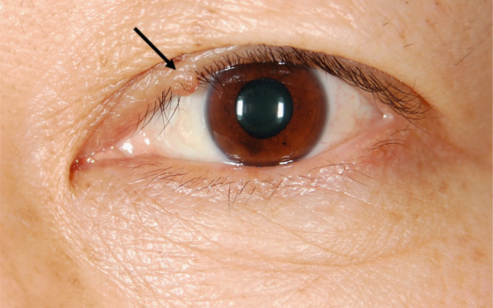
Figure 2.
Intradermal nevus. (A) A pigmented melanocytic nevus at the lower eyelid margin in a 34-year-old man (black arrow). (B) A nonpigmented melanocytic nevus of lower eyelid in a 69-year-old woman (black arrow).

Figure 3.
Squamous papilloma. A Slightly elevated, pigmented mass located on the lower eyelid in a 52-year-old woman (black arrow).
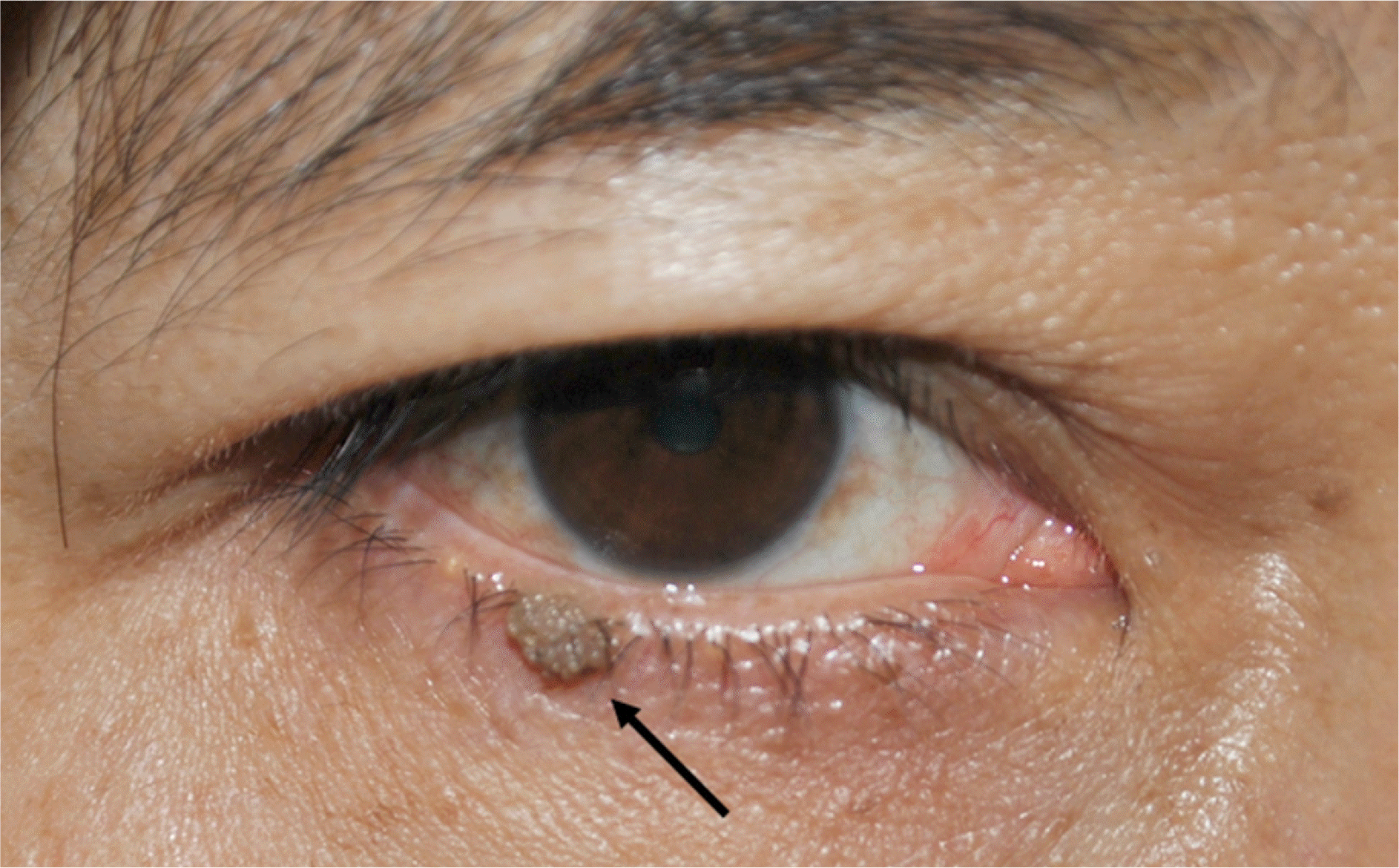
Figure 4.
Seborrheic keratosis. An elevated brown-colored mass located on the upper eyelid in a 47-year-old woman (black arrow).
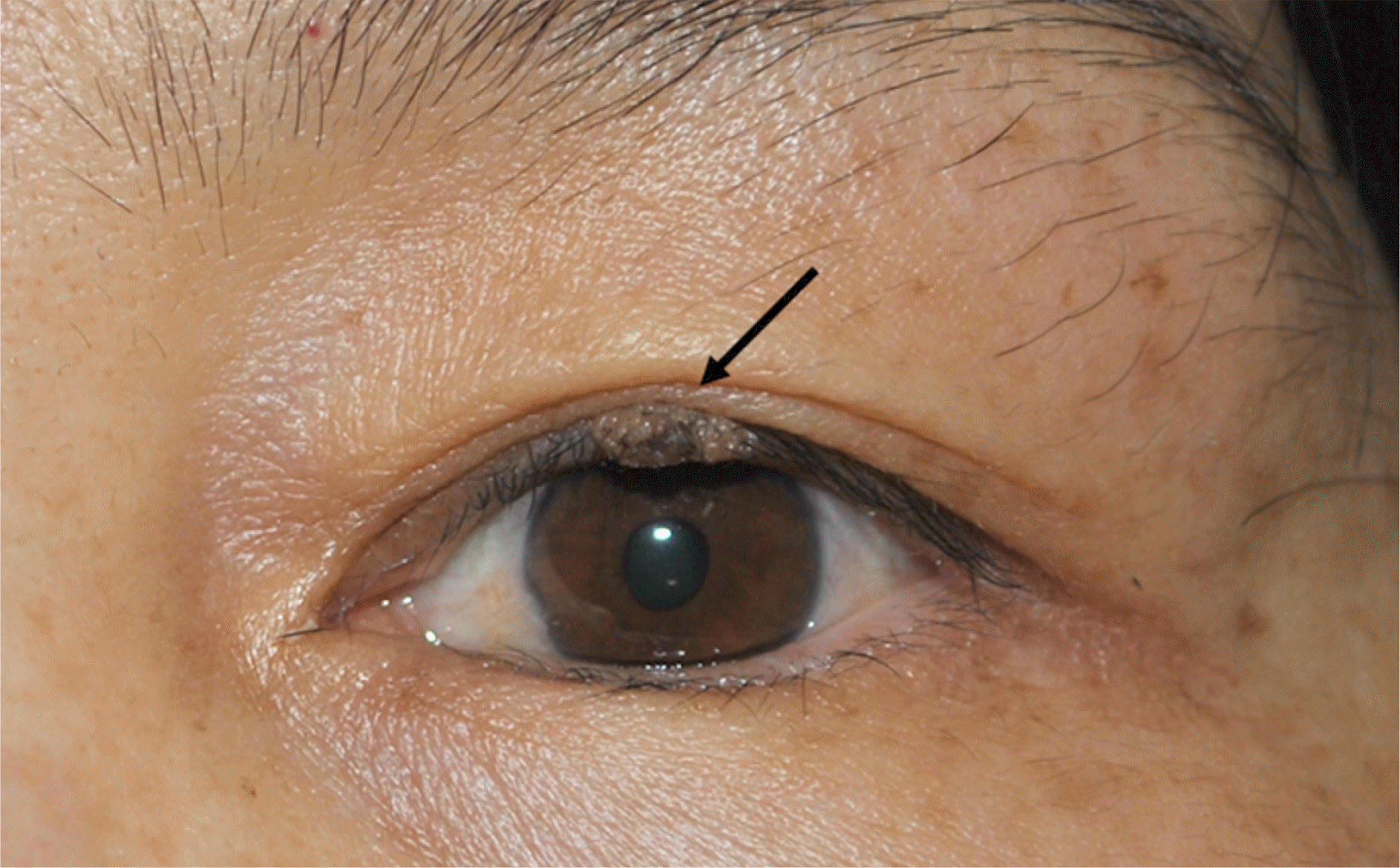
Figure 5.
Epidermal cyst. A smooth, soft, yellowish ruptured epidermal cyst located on the upper eyelid in a 27-year-old man (black arrow).
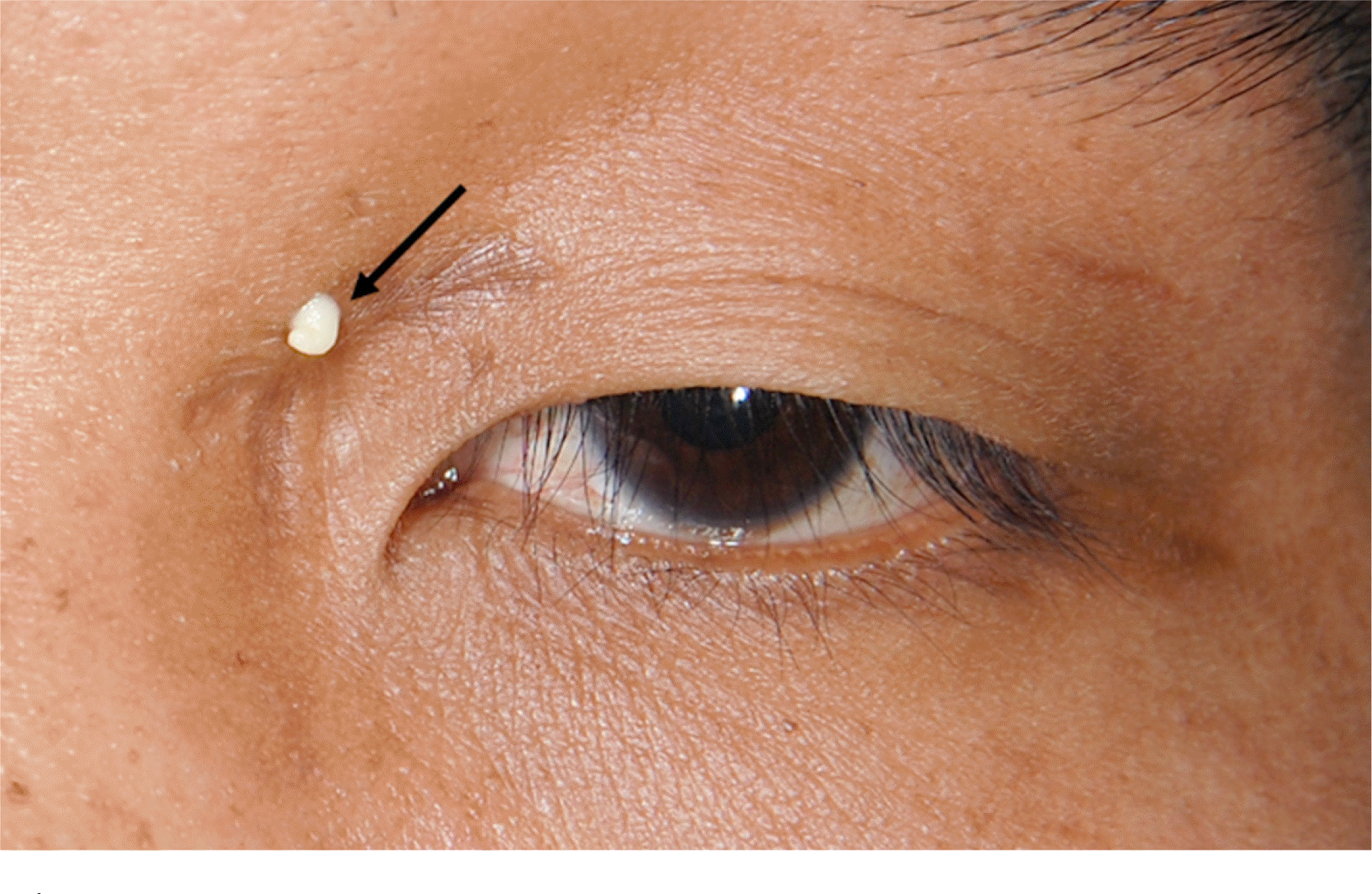
Figure 6.
Dermoid cyst. A smooth, subcutaneous mass in the lateral aspect of the eyebrow in a 27-year-old woman (black arrow).
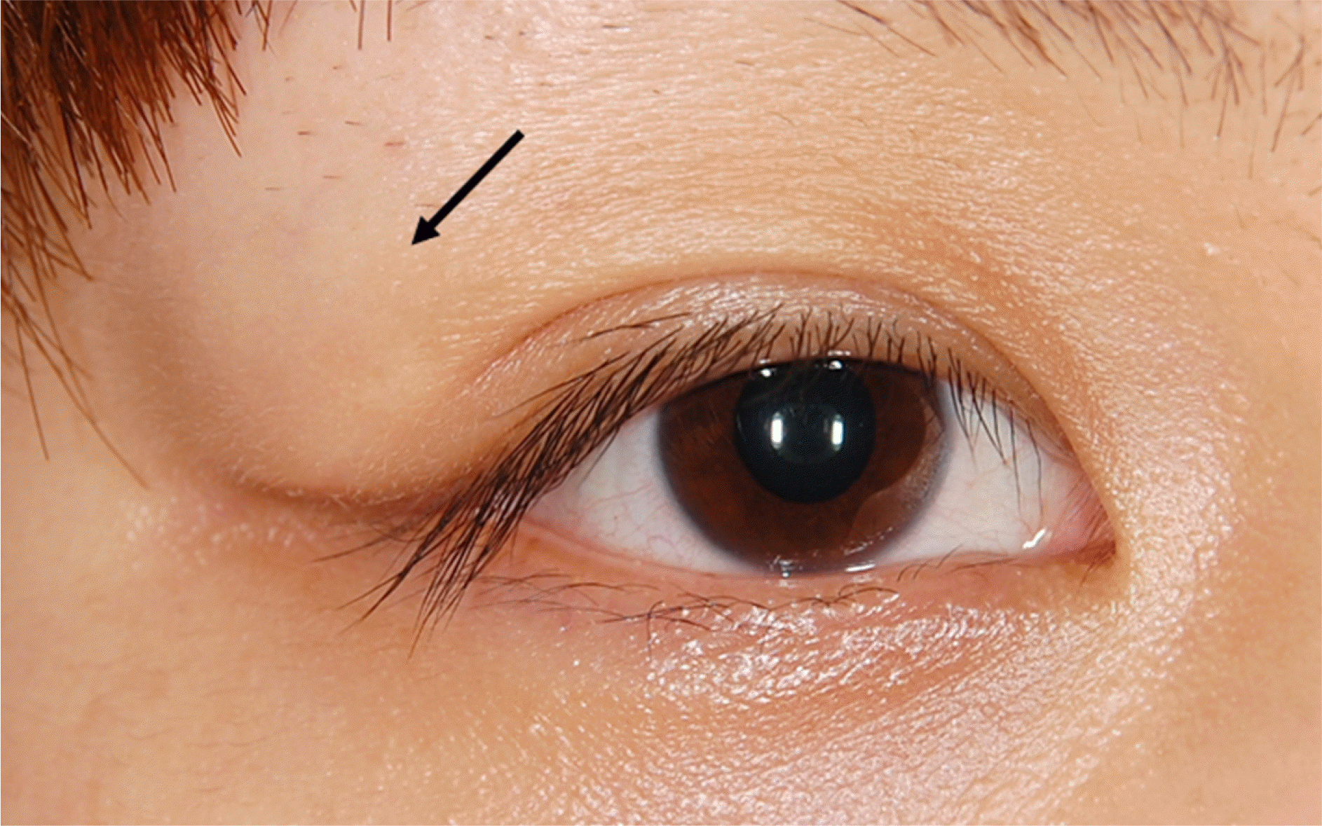
Figure 7.
Pilomatrixoma. (A) A fairly well circumscribed mass located on the upper eyelid in a 3-year-old child (black arrow). (B) When he closed his eye, the lesion is well demarcated (black arrow).

Table 1.
Sex and age distribution of benign eyelid tumors
Table 2.
Benign eyelid tumors


 PDF
PDF ePub
ePub Citation
 Print
Print


 XML Download