Abstract
Cavernous sinus syndrome (CSS) is defined as the involvement of two or more of the third, fourth, fifth (V1, V2) or sixth cranial nerves or involvement of only one of them in combination with a neuroimaging-confirmed lesion in the cavernous sinus. Some cases of CSS are attributed to Tolosa-Hunt syndrome (THS), an idiopathic inflammatory disease of the cavernous sinus. THS is characterized by painful ophthalmoplegia due to granulomatous inflammation in the cavernous sinus. THS is a diagnosis of exclusion that requires a vigorous series of differential diagnoses, and corticosteroid therapy is known to dramatically resolve clinical findings of THS. We report a case of a patient with painful ophthalmoplegia associated with vision loss, which was suspected to be THS. This patient followed a relatively typical clinical course of THS on steroid pulse therapy. We emphasize the differential diagnosis of THS, its presentation, and treatment.
References
1). Kang IG, Jung JH, Kim ST, Cha HE. A Case of Tolosa-Hunt Syndrome. J Rhinol. 2009; 16(1):65–7.
2). Taylor EJ, Anders UM, Martel JR, Martel JB. Tolosa-Hunt syndrome masquerading as a carotid artery dissection. Clin Ophthalmol. 2014; 8:707–10.
3). Nieri A, Bazan R, Almeida L, Rocha FC, Raffin CN, Bigal ME, et al. Bilateral painful idiopathic ophthalmoplegia: a case report. Headache. 2007; 47(6):848–51.

4). Kang H, Park KJ, Son S, Choi DS, Ryoo JW, Kwon OY, et al. MRI in Tolosa-Hunt syndrome associated with facial nerve palsy. Headache. 2006; 46(2):336–9.

5). Headache Classification Committee of the International Headache S. The International Classification of Headache Disorders, 3rd edition (beta version). Cephalalgia. 2013; 33(9):629–808.

6). Forderreuther S, Straube A. The criteria of the International Headache Society for Tolosa-Hunt syndrome need to be revised. J Neurol. 1999; 246(5):371–7.
7). Beckham S, Kim H, Truong A. Painful ophthalmoplegia of the right eye in a 20-year-old man. J Emerg Med. 2013; 44(2):e231–4.

8). Rotstein DL, Tyndel FJ, Tang-Wai DF. A case report of cavernous sinus syndrome in a patient with Takayasu's arteritis. Headache. 2014; 54(8):1371–5.

9). Slattery E, White AJ, Gauthier M, Linscott L, Hirose K. Tolosa-Hunt syndrome masquerading as Gradenigo syndrome in a teenager. Int J Pediatr Otorhinolaryngol. 2013; 77(7):1219–21.

10). Hung CH, Chang KH, Chu CC, Liao MF, Chang HS, Lyu RK, et al. Painful ophthalmoplegia with normal cranial imaging. BMC Neurol. 2014; 14:7.

11). Sanchez Vallejo R, Lopez-Rueda A, Olarte AM, San Roman L. MRI findings in Tolosa-Hunt syndrome (THS). BMJ Case Rep. 2014; 2014.

12). Çakirer S. MRI findings in Tolosa-Hunt syndrome before and after systemic corticosteroid therapy. European Journal of Radiology. 2003; 45(2):83–90.

13). Abdelghany M, Orozco D, Fink W, Begley C. Probable Tolosa-Hunt syndrome with a normal MRI. Cephalalgia;. 2014.

14). Kim IT, Park KY, Kim YM, Yang KH, Park YM. Lateral Sinus Thrombophlebitis Caused by Isolated Sphenoid Sinusitis. J Rhinol. 1997; 4(1):68–70.
Fig. 1.
On pre-op evaluation, ptosis of the left eye, left-superior fixation of the eye ball on resting state, and limitation of the left eye on left gaze was observed, indicating weakness of the left 3rd and 6th cranial nerves.

Fig. 2.
On pre-op nasal endoscopic examination, the left maxillary osteomeatal unit was intact, and no lesions were observable in the ethmoid complex.
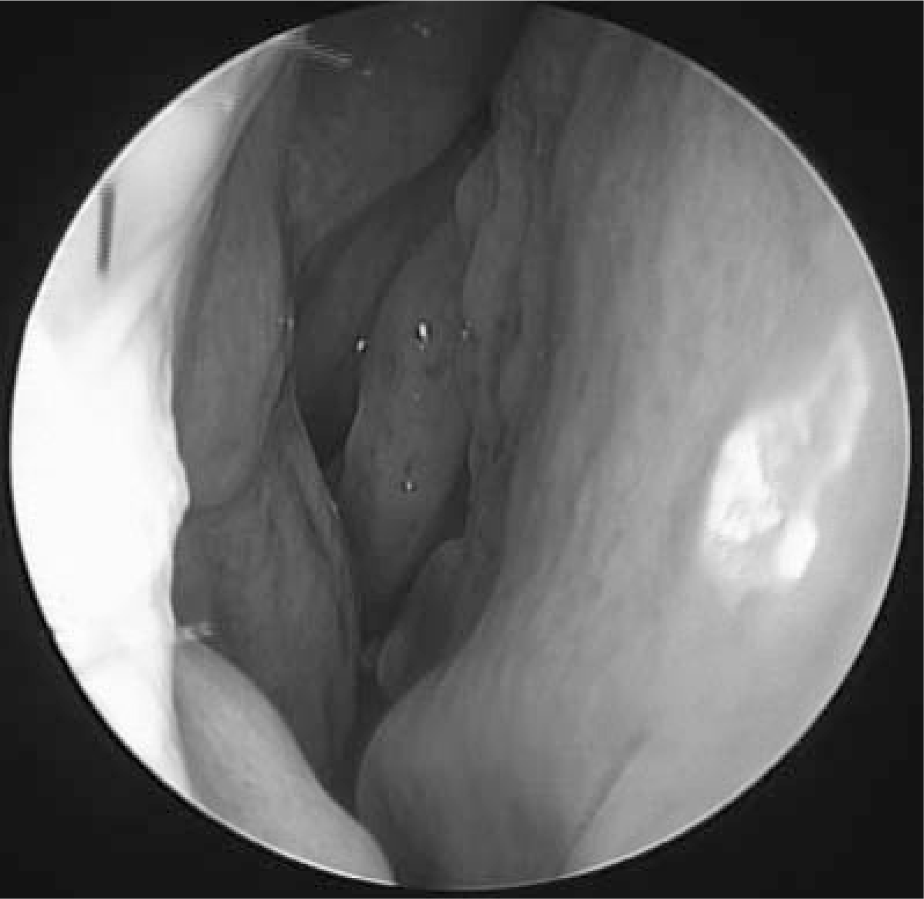
Fig. 3.
On pre-op computer tomo-grphy, soft tissue density indicating mucosal thickening of the left sphenoid sinus was observed (A, white arrow), soft tissue was not enhanced on contrast (B, white arrow).
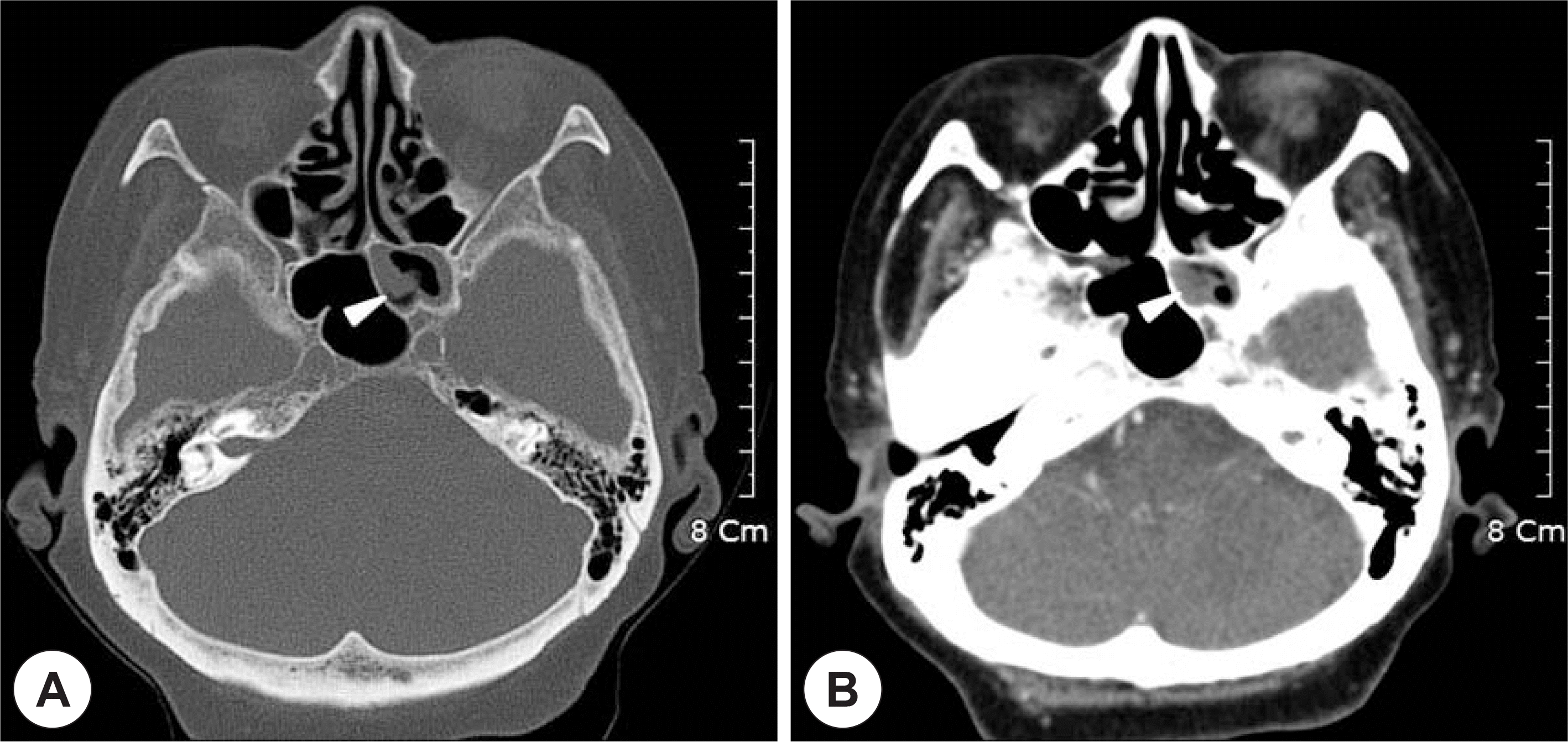
Fig. 4.
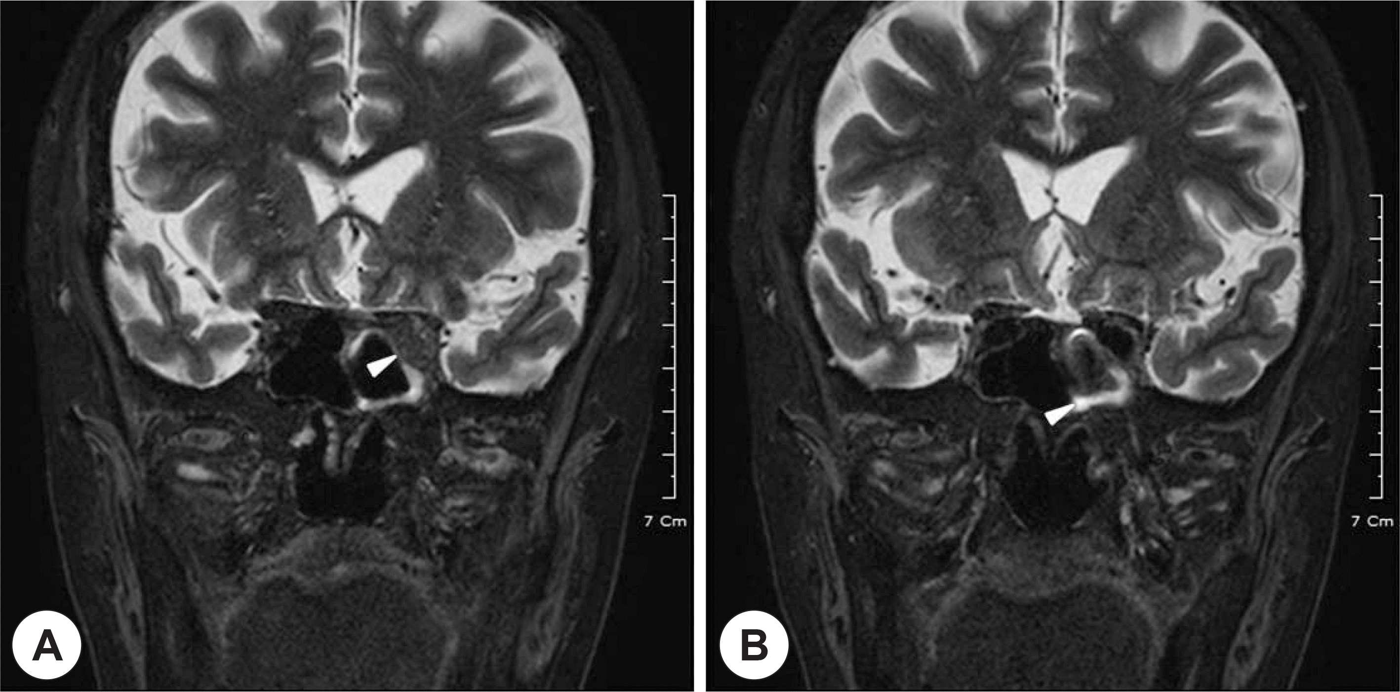
Fat-saturated T2 weighted view on MRI shows infiltrative mass in the left orbital apex (A, white arrow), including the inferior orbital fissure, extending to the left cavernous sinus (B, white arrow).
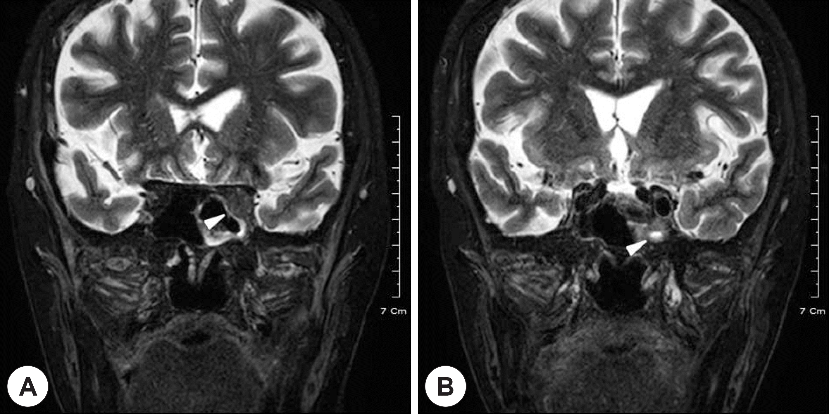
Fig. 5.
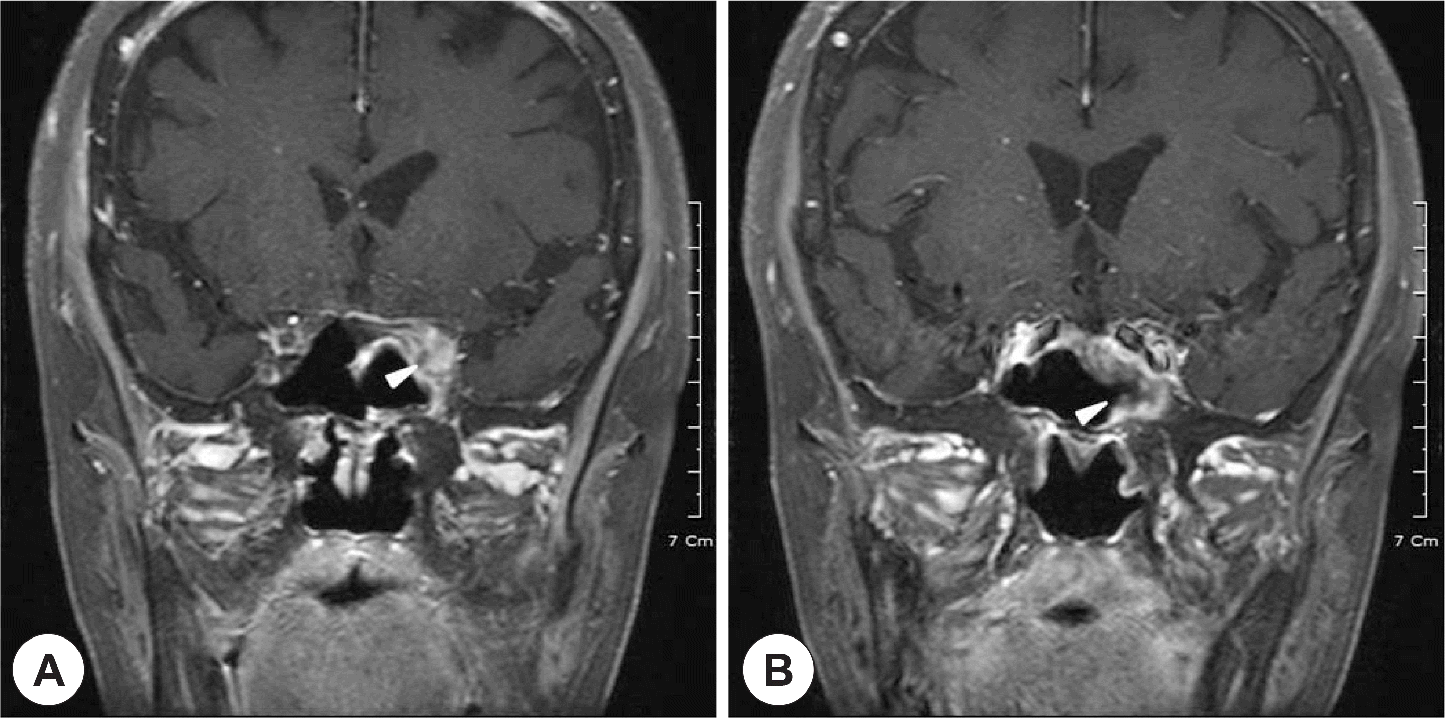
Gadolinium enhanced T1 weighted MR images show intense but inhomogeneous enhancement in the left orbital apex (A, white arrow), and cavernous sinus (B, white arrow).
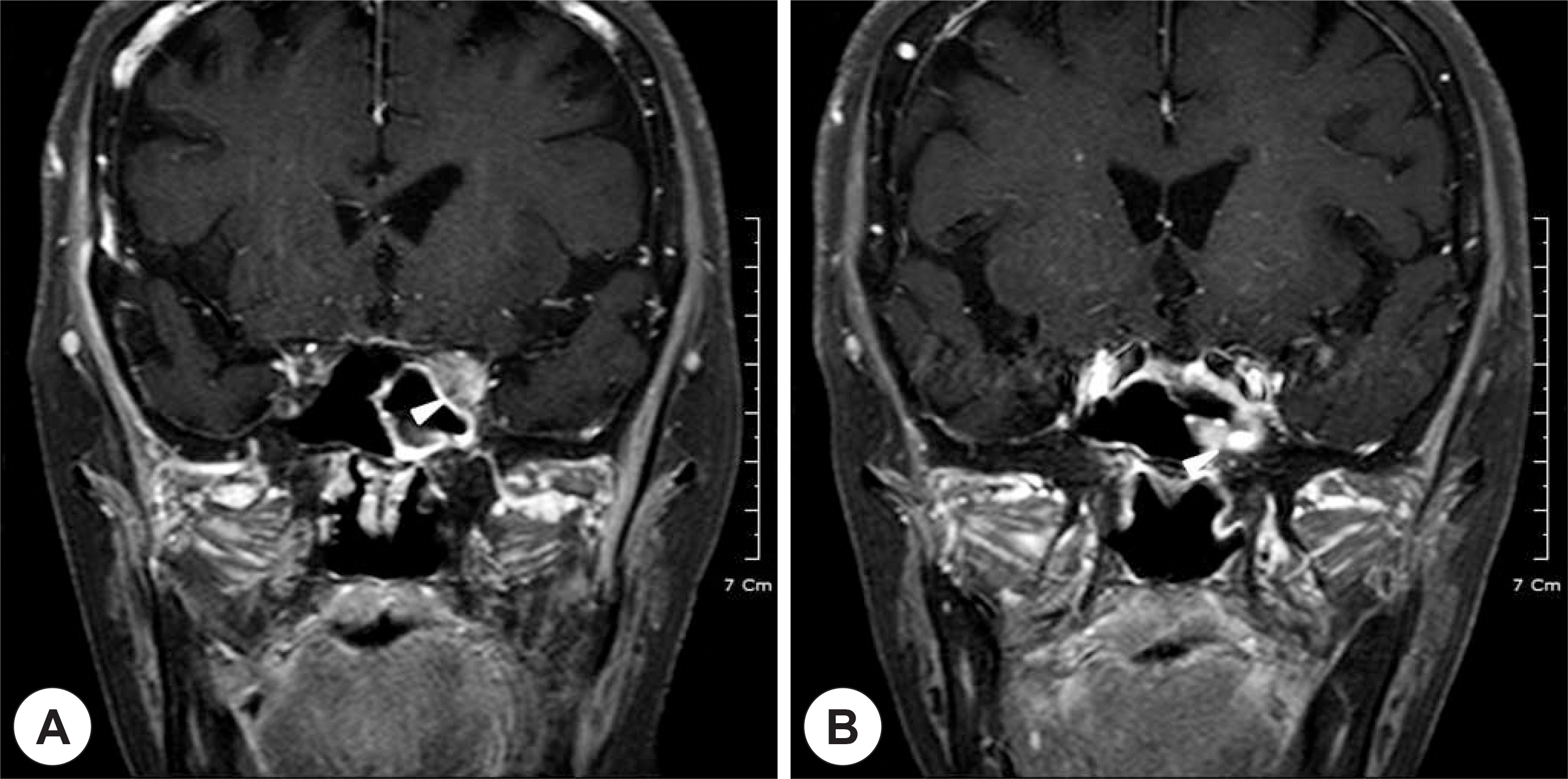
Fig. 6.
Intraoperative endoscopy showed no lesions of sphenoid sinus, other than mild mucosal thickening (NS: nasal septum, SS: sphenoid sinus, MT: middle turbinate).
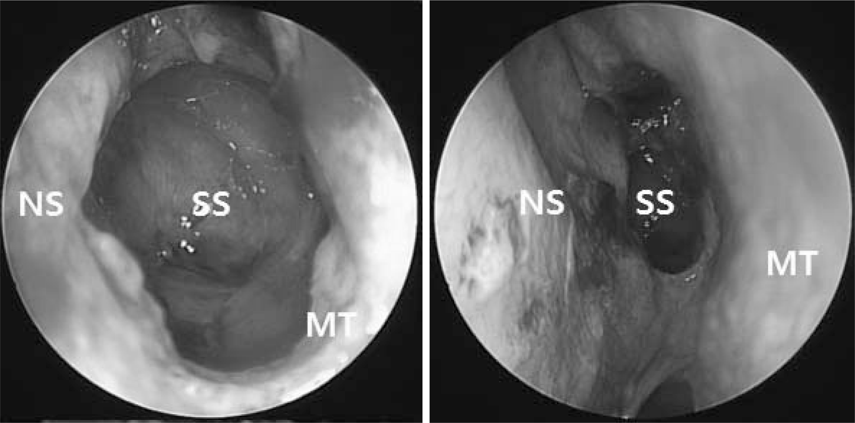
Fig. 7.
On physical examination of eye movements 1 week after endoscopic drainage and biopsy, and 10 days of pulse steroid therapy, no ptosis or left eye movement limitation was observed.


 PDF
PDF ePub
ePub Citation
 Print
Print


 XML Download