

 PDF
PDF ePub
ePub Citation
 Print
Print


INTRODUCTION
Brain metastasis (BM) develops in approximately 10% to 30% of all cancer patients and it is a common neurologic complication of lung cancer, breast cancer, and melanoma [123]. As for gynecologic cancer, the most common metastatic sites are liver, lung, bones, and lymph nodes [4]. However, BM originating from gynecologic cancer is extremely rare, with the exception of choriocarcinoma, and considered as a late manifestation [5]. The incidence of BM from ovarian, endometrial, and cervical cancer has been reported to be 0.3–2.2%, 0.4–1.2%, and 0.3–0.9%, respectively [12]. However, the occurrence of BM in gynecologic malignancies appears to have increased in recent years, due to advances in neuroimaging such as computed tomography (CT) and magnetic resonance imaging (MRI), together with the prolonged survival of patients [126].
Because of its rarity, only a few papers about outcome and prognostic factors of gynecologic cancer have been published, and a consensus on therapeutic guidelines has not been established [6]. Although the prognosis of BM differs according to primary tumor, general treatment principle has been alike regardless of primary cancer. Therfore, the treatment options for BM from gynecologic cancer include surgery, radiosurgery, or whole brain radiation therapy (WBRT) depending on the condition of the patients. However, the increasing incidence of BM of gynecologic cancer calls for special attention since the treatment strategy clearly affects patient prognosis [789]. In this study, we reviewed our experience with 20 patients who underwent treatment for BM from gynecologic cancer. We reviewed the clinical characteristics of patients, and analyzed their outcomes by primary origin and primary treatment.
MATERIALS AND METHODS
Patient populations
A retrospective review of the medical records of 951 BM patients who were treated at the Neurosurgical Department of our institution from July 2003 to February 2016 (patients who underwent treatment or management in other departments were not included) identified 20 (2%) patients originating from gynecologic cancer. BM patients with primary choriocarcinoma were excluded to create a more uniform patient population.
The medical and radiological records of those 20 patients were reviewed. Clinical variables were the type of primary cancer, therapeutic modalities, overall survival time, progression-free survival time, and the Karnofsky Performance Status (KPS) score. Each case of gynecologic cancer was staged based on the International Federation of Obstetricians and Gynecologists Staging System [10]. Outcomes according to the therapeutic modalities for the BM, that is, Gamma Knife surgery (GKS), surgical resection followed by WBRT, or WBRT only were analyzed. The study was approved by the Institutional Review Boards of Bundang Seoul National University Hospital (B-1708-417-108). The informed consent was obtained from all patients for collection of clinical data.
Selection of therapeutic modalities
Among a total of 20 patients, 6 (30%) patients underwent surgical resection followed by WBRT at initial diagnosis of BM, 11 (55%) patients received GKS, and the remaining 3 (15%) patients had WBRT only.
Clinical indications for surgery followed by WBRT in this study included single lesion, non-eloquent location of the lesion, and symptoms and signs of intractable intracranial hypertension, intractable seizures, a reduced level of consciousness, or progressive neurologic deficit. Basically, neurosurgical candidates were expected to survive more than 6 months.
When the location of the lesion was critical for surgical resection (located in eloquent area) or the patient's general condition was too poor to undergo major surgery, and the number of lesions was less than 10, GKS was selected as the treatment modality. When the lesion recurred after initial treatment, and if the patient previously underwent WBRT so that the patient was unable to take another WBRT, GKS was the only treatment of choice as the second therapy.
When there were more than 10 lesions or the general condition of the patient was too poor, WBRT was selected as the initial treatment modality. And even if the number of the lesions were less than 10, when the size of the lesion was too large to take GKS (larger than 3 cm in maximum diameter), WBRT was selected for the optimal treatment modality.
Statistical analysis
Statistical analyses were conducted using SSPS 20.0 (IBM Corp., Armonk, NY, USA). The origin of the primary cancer, time interval between the diagnosis of primary cancer and BM, KPS score, and therapeutic modalities were regarded as candidate prognostic factors. The overall survival and progression- free survival were estimated using the Kaplan-Meier method and analyzed based on the log-rank test. A p value of less than 0.05 was considered statistically significant.
RESULTS
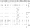
The characteristics of 20 patients with a primary gynecologic malignancy and BM are summarized in Table 1. The mean age at time of BM diagnosis was 54.4 years (range from 28 to 76 years). The median interval time from the diagnosis of the primary gynecologic cancer to the development of BM was 28 months (range from 0 to 99 months). Fourteen (70%) patients had primary ovarian cancer, 4 (20%) had uterine cancer, and 2 (10%) patients had cervical cancer. Median follow up period of over all patients was 13 months.
Those patients who underwent surgical resection followed by WBRT were all alive during the follow-up periods. The maximal size of the lesions ranged 2.7 cm to 5.3 cm, and the average was 4.8 cm in this group. There were no major, but only minor complications such as headache or vomiting. However, 2 out of 6 patients experienced local recurrences, and the second treatments for recurred BM were also surgical resection in both cases, as there was no evidence of systemic progression.
In those patients who underwent GKS as initial treatment, the number of lesion was up to 8. Two out of 11 patients had lesions involving the eloquent areas. The maximal size of the lesions ranged 0.9 cm to 4 cm, and the average was 3 cm in this group. The prescription doses of each lesion are listed in Table 1, and average of all doses was 19.2 Gy. The clinical manifestations after GKS were minor and transient: confusion, headache, dysmetria, or seizure. During the follow-up periods after GKS, 6 out of 11 patients had local recurrences, and repeated GKS were performed in 3 cases, surgical resection followed by WBRT in one, and WBRT in two, depending on the number of lesions and clinical manifestations. Two patients were lost to follow up, and 3 patients died during the follow-up periods. The causes of death were systemic aggravation (one patient with pulmonary effusion and the other with hepatic failure due to the progression of primary cancer) rather than intracranial problems.
Out of 3 patients who had WBRT only as the primary treatment, two patients had 2 lesions involving the eloquent area, and one patient had more than 10 lesions at the time of BM diagnosis and some of the lesions were near the eloquent area. The former 2 patients initially treated WBRT rather than taking GKS despite the number of lesions was only 2, because the patients decided WBRT considering their poor general condition and economic problem. In this group, all 3 patients had local recurrences eventually, and they all underwent GKS as the second treatment. One patient was lost to follow up and another patient died during follow up periods.
The information about prescribed radiation doses and fractionation is summarized in Table 1, and there were two groupspostoperative radiation group (6 patients) and WBRT only group (3 patients). Overall doses seemed higher in postoperative radiation group, but the radiation doses in that group included boost radiation to the operation site. The radiation doses which cover whole brains were almost similar between two groups.
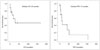
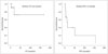
The median overall survival time after the diagnosis of brain metastases was 28 months, and the progression-free survival time was 15 months (Fig. 1). For patients with ovarian cancer, median overall survival time did not reach during follow-up periods and the progression-free survival time was 15 months (Fig. 2). For uterus and cervix cancer patients, the number of patients was too small to analyze statistically.
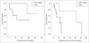
According to therapeutic modality, the median overall survival time of the 11 patients who received GKS as the initial treatment was 17 months and that of the 6 patients who underwent surgical resection followed by WBRT was 37.3 months (p=0.16). The median progression-free survival time for patients who received GKS as the primary treatment was 12 months and that for patients who underwent surgical resection along with WBRT was 42 months (p=0.042) (Fig. 3). The number of patient who underwent WBRT only as the initial treatment (3 patients) was too small to analyze statistically
DISCUSSION
BM originating from gynecologic cancer has been described as being rare in the literature. In our study, we also found the rarity: twenty out of 951 (2%) BM patients had gynecological cancer as their primary cancer. However, with increased survival times and regular screening programs, it has in fact become relatively more common than a decade ago [11].
According to the previous reports, the median overall survival time after a diagnosis of BM from ovarian cancer was 6 to 7 months [101213], that from endometrial cancer was 1 to 2 months [8], and that from cervical cancer was 9.9 months [4]. However, in our study, the median overall survival time after BM diagnosis from all gynecologic cancer was 28 months. Our study showed relatively good outcomes compared with the published reports. It might be due to the use of effective chemotherapeutics and the improvement of surgical techniques, but there was also patient selection bias [11]. Increased diagnostic sensitivity resulting from improved cerebral imaging technologies also made it possible to detect small intracranial lesions and early diagnosis during the course of disease recurrence [46]. In our study, surgical resection followed by WBRT is a treatment modality significantly associated with improved survivals. This finding is consistent with a previous study that aggressive and multimodality treatment methods such as neurosurgery and combination chemoradiotherapy increased the survival time for patients with gynecologic cancer and BMs.
However, these results must be interpreted carefully, because the characteristics of the patients are different among the groups by treatment modalities (Table 1). In most cases, the number and the location of the lesions determined the choice of primary treatment modality. If there were too many lesions or the location of the lesions was risky to be surgically resected (e.g., the basal ganglia, pons, and so on), GKS or WBRT were considered as the initial treatment modality.
In case of old patient, oligometastases, or critical location of the lesion, surgical resection is not usually indicated and GKS could be an alternative treatment option. The outcome of GKS treatment in our study (median overall survival time of 17 months) was better than that of a previous study. In most recent study, the median overall survival time after GKS for gynecological cancer BM patients was 9.5 months [35]. Moreover, in our data, there were no severe complications after GKS.
Preoperative performance status (which measured in KPS score) was also related to treatment outcome. As shown in Table 1, in mortality cases, patients showed lower KPS score (which ranged 50 to 80 and the average was 65) when brain metastases were diagnosed. When the performance status of the patients at the time of diagnosis of brain metastases were not poor, that is KPS scores are 70 or more, we could choose more aggressive treatment like surgery, which led good outcomes.
There are several limitations to consider when interpreting our findings. First, there were biases in the selection of patient and primary treatment modality for BM, given the retrospective nature of this study. Second, the extracranial metastases were not considered, which could influence the outcomes. Lastly, small number of patients is not appropriate to make a generalized consensus.
Despite these limitations, our study is worthwhile considering the rarity of BM patient from gynecologic cancer. Our study revealed that prognosis of brain metastases from gynecologic cancer is not always poor. Surgical resection followed by WBRT might be a treatment modality significantly associated with a longer survival when indicated. Further studies with a larger sample size at the multi-center or national level are necessary to provide a more comprehensive and comparative analysis.
 XML Download