

 PDF
PDF ePub
ePub Citation
 Print
Print


Metatarsus adductus is a deformity characterized by adduction of the lesser metatarsals. Concomitant metatarsus adductus is prevalent in about 30% of patients undergoing hallux valgus surgery.1) Metatarsus adductus is a risk factor for postoperative recurrence as well as a cause of hallux valgus.2) Moreover, it is difficult to correct hallux valgus in the presence of metatarsus adductus because there is insufficient space between the first and second metatarsals for lateral translation of the first metatarsal, due to varus position of the second metatarsal.3) In addition, there is an increased risk of recurrence after surgery for hallux valgus without correction of metatarsus adductus, because medial translation force by the second metatarsal is constantly applied at the first metatarsal head.
Although the importance of metatarsus adductus has been highlighted in hallux valgus surgery, few studies have discussed management of hallux valgus with metatarsus adductus. Some methods were already introduced to correct metatarsus adductus using distal oblique osteotomy4) or lateral closing base wedge osteotomy of the lesser metatarsals.5) However, these are extensive procedures with a risk of complications associated with the shortening and malposition of the lesser metatarsals. Although some authors proposed another method to correct hallux valgus using a stable osteotomy such as a scarf osteotomy,6) or a stable fixation device such as a locking plate7) for prevention of recurrence, these methods cannot obtain sufficient space for translation of the first metatarsal because of the lack of correction of metatarsus adductus.
The technique described here is believed to be easier to perform and has several advantages over the above techniques. The aim of this study was to introduce this simple and effective method for modified proximal chevron osteotomy for hallux valgus with metatarsus adductus. The present case was approved by our Institutional Review Board and informed consent was obtained from the patient.
TECHNIQUE
The procedure was performed under regional anesthesia using sciatic and femoral blocks in the supine position. A tourniquet using an elastic bandage was applied at the thigh.
A 7-cm medial transverse incision was made from 1 cm distal to the first metatarsophalangeal joint, continuing proximally along the inferior margin of the first metatarsal. After exposure, medial capsular resection was performed. The medial eminence of the first metatarsal head was removed about 2 mm medial to the sagittal sulcus, in line with the medial cortex of the metatarsal shaft, using a micro-oscillating saw. Lateral soft tissue release was performed using transarticular approach. The lateral capsular structures were exposed using manual traction of the great toe. First, the metatarsosesamoid ligament was released along the lateral sesamoid with a number-15 blade. Then, the capsule of the metatarsophalangeal joint and the adductor hallucis tendon was released. Varus stress to the metatarsophalangeal joint was applied to complete the release of the adductor hallucis tendon.
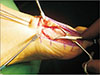
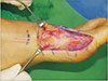
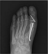
Proximal chevron osteotomy was performed through the same medial incision. The apex of the chevron was proximally located 7 mm distal to the metatarsocuneiform joint. The angle of each arm of the chevron was made at 30° to the first metatarsal longitudinal axis. Before osteotomy, Kirschner wire of 1.6-mm diameter was placed at the apex of the chevron to avoid extension of the osteotomy into the joint. After completion of the osteotomy, a small curette was placed on the lateral side of the dorsum of the proximal fragment to lever the proximal fragment medially as much as possible, while the distal fragment was angulated laterally. At this point, the distal fragment was displaced medially using a towel clip placed on the proximal portion of the distal fragment (Fig. 1), in contrast with previously reported proximal chevron osteotomy technique. After correction of the deformity, 3 or 4 Kirschner wires of 1.6-mm diameter were inserted from proximal to distal into the metatarsal head. The correction was then checked using C-arm fluoroscopy (Fig. 2). The Kirschner wires were buried under skin and a bone graft was prepared using resected bone, and was placed in the gap of the osteotomy site (Fig. 3). The medial joint capsule was repaired using absorbable sutures, and correction was rechecked using C-arm fluoroscopy after wound closure (Fig. 4).
The feet are placed in splints for 1 week after surgery. Weight-bearing on the heels and lateral border of the foot was permitted on the day after surgery. Weightbearing on the first ray was allowed at 6 weeks after surgery, and the Kirschner wires were removed at 12 weeks after surgery.
DISCUSSION
Metatarsus adductus is a structural deformity occurring at the tarsometatarsal joints, with the metatarsals being deviated medially with reference to the lesser tarsus.2) Some studies have identified a relationship between hallux valgus and metatarsus adductus.18) It is suggested that metatarsus adductus may cause the development and postoperative recurrence of hallux valgus.2) However, satisfactory correction of the deformity is challenging because the lesser metatarsals have adductive deviation and occupy the first and second intermetatarsal space. In addition, with postoperative weightbearing, the second metatarsal head pushes the first metatarsal medially, so the recurrence rate is increased.2)
Although most surgeons have recognized the significance of metatarsus adductus in hallux valgus surgery for a long time, few reports have presented a surgical technique to overcome this problem. One method uses lateral closing base wedge osteotomy of the lesser metatarsals for correction of metatarsus adductus.5) Another method uses multiple distal4) or middle9) oblique osteotomy of the lesser metatarsals to obtain space to translate the first metatarsal. These methods use fundamental approaches to treat metatarsus adductus; however, they are extensive procedures with an inherent risk of shortening and malposition of the sagittal plane of the lesser metatarsals. In addition, they require a longer healing time and lengthy immobilization, and may not be suitable for young patients with high functional demands.4) Correction of hallux valgus using a stable osteotomy such as a scarf osteotomy6) or stable fixation device7) could be an option to prevent recurrence after surgery. These methods can reduce complications associated with surgery of the lesser metatarsals; however, they cannot obtain sufficient space for translation of the first metatarsal due to adduction of the second and third metatarsals, which would impinge on further reduction.10)
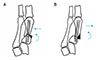
Our new technique of proximal chevron osteotomy and medial translation of the distal fragment is a simple and effective procedure for hallux valgus deformity with severe metatarsus adductus. When performing proximal chevron osteotomy, reduction was commonly performed with lateral angulation and lateral translation of the distal fragment to reduce the first and second intermetatarsal spaces. Our technique is similar to proximal chevron osteotomy, except for medial translation of the distal fragment (Fig. 5). In our technique, sufficient space between the first and second metatarsals can be obtained by medial translation of the distal fragment, thereby reducing the risk of recurrence after surgery. In addition, compared with previously reported methods, our technique can provide shorter healing times and reduce the risks associated with extended operative times and multiple procedures. We believe that our new, simple technique can be an option for correcting hallux valgus deformity with severe metatarsus adductus.

 XML Download