Abstract
Purpose:
Diabetic foot gangrene has a high morbidity rate and a great influence on the quality of life. Amputation is an appropriate treatment if conservative treatment is impossible according to the severity of gangrene and infection. The purpose of this study was to evaluate the usefulness of preoperative percutaneous transluminal angioplasty for the postoperative outcome.
Materials and Methods:
From February 2013 to April 2016, among 55 patients with diabetic foot gangrene, who require surgical treatment, percutaneous transluminal angioplasty was performed on patients with an ankle brachial index (0.9 and stenosis) 50% on angiographic computed tomography. The study subjects were 49 patients, comprised of 37 males (75.5%) and 12 females (24.5%). The mean age of the patients was 70.0±9.6 years. The treatment results were followed up according to the position and length of the lesion and the changes during the follow-up period.
Results:
As a result of angiography, there were 13 cases of atherosclerotic lesions in the proximal part, 11 cases in the distal part and 25 cases in both the proximal and distal parts. As a result of the follow-up after angiography, in 13 patients, the operation was not performed and only follow-up and dressing were performed around the wound. Sixteen patients underwent debridement for severe gangrene lesions and 20 patients, in whom the gangrene could not be treated, underwent amputation (ray amputation or metatarsal amputation, below knee amputation).
REFERENCES
1.Reiber GE., Lemaster JW. Epidemiology and economic impact of foot ulcers and amputations in people with diabetes. Bowker JH, Pfeifer MA, editors. editors.Levin and O’Neal’s the diabetic foot. 7th ed.Philadelphia: Mosby;2008. p. 3–22.

2.LoGerfo FW., Gibbons GW., Pomposelli FB Jr., Campbell DR., Miller A., Freeman DV, et al. Trends in the care of the diabetic foot. Expanded role of arterial reconstruction. Arch Surg. 1992. 127:617–20. discussion 620-1.
3.Rosenblum BI., Pomposelli FB Jr., Giurini JM., Gibbons GW., Freeman DV., Chrzan JS, et al. Maximizing foot salvage by a combined approach to foot ischemia and neuropathic ulceration in patients with diabetes. A 5-year experience. Diabetes Care. 1994. 17:983–7.

4.Faglia E., Favales F., Quarantiello A., Calia P., Brambilla G., Ram-poldi A, et al. Feasibility and effectiveness of peripheral percutaneous transluminal balloon angioplasty in diabetic subjects with foot ulcers. Diabetes Care. 1996. 19:1261–4.

5.Hanna GP., Fujise K., Kjellgren O., Feld S., Fife C., Schroth G, et al. Infrapopliteal transcatheter interventions for limb salvage in diabetic patients: importance of aggressive interventional ap- proach and role of transcutaneous oximetry. J Am Coll Cardiol. 1997. 30:664–9.
6.Boulton AJ., Vileikyte L., Ragnarson-Tennvall G., Apelqvist J. The global burden of diabetic foot disease. Lancet. 2005. 366:1719–24.

7.Beach KW., Bedford GR., Bergelin RO., Martin DC., Vandenberghe N., Zaccardi M, et al. Progression of lower-extremity arterial occlusive disease in type II diabetes mellitus. Diabetes Care. 1988. 11:464–72.

9.Gaspar L., Komornikova A., Kruzliak P., Rodrigo L., Gabbasov Z., Staffa R. Contribution of prostaglandin E1 treatment in patients with critical limb ischemia. Int J Clin Exp Med. 2016. 9:3227–31.
10.Flynn MD. The diabetic foot. Tooke JE, editor. editor.Diabetic angiopathy. London: Arnold;1999. p. 92–8.
11.Caputo GM., Cavanagh PR., Ulbrecht JS., Gibbons GW., Karchmer AW. Assessment and management of foot disease in patients with diabetes. N Engl J Med. 1994. 331:854–60.

12.Ouriel K., Fiore WM., Geary JE. Limb-threatening ischemia in the medically compromised patient: amputation or revascularization? Surgery. 1988. 104:667–72.
13.Choi D., Pyun WB., Yoon YS., Jang Y., Shim WH. Frequency of combined atherosclerotic disease of the coronary, periphery, and carotid arteries found by angiography. Korean Circ J. 1999. 29:883–90.

14.Wolosker N., Nakano L., Duarte FH., De Lucia N., Leao PP. Peroneal artery approach for angioplasty of the superficial femoral artery: a case report. Vasc Endovascular Surg. 2003. 37:129–33.
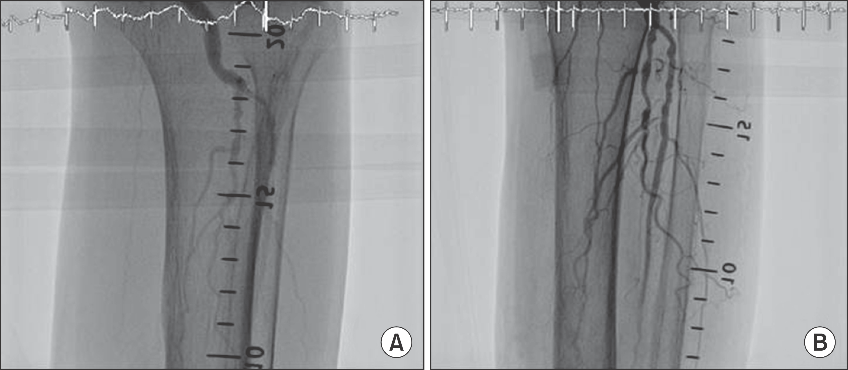
Figure 1.
Angiography before (A) and after (B) intervention of anterior tibial artery and superficial femoral arteries. Blood flow was restored after the intervention.

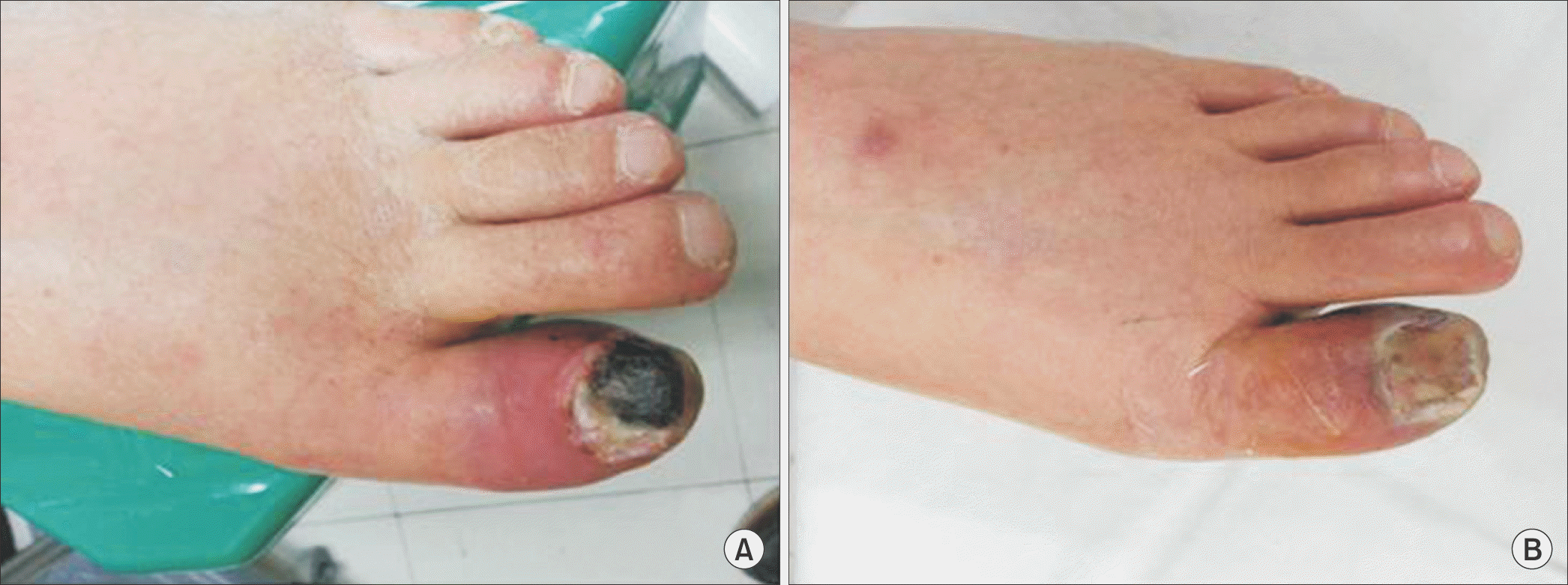
Figure 2.
(A) Diabetic gangrene with ischemia and infection. (B) Fourteen days after balloon dilatation of peripheral artery.

Table 1.
Wagner’s Diabetes Mellitus Foot Classification
Table 2.
Stenosis Location by Angiography
| Location of stenosis | No. of patients | |
|---|---|---|
| Proximal | Superficial femoral artery | 10 |
| Popliteal artery | 1 | |
| ATA | 21 | |
| PTA | 6 | |
| Distal | Distal DPA | 17 |
| Distal PTA | 19 |
Table 3.
Final Treatment of Diabetes Mellitus Foot after Percutaneous Transluminal Angioplasty
| Treatment | No. of patients (%) |
|---|---|
| Conservative management | 13 (26.5) |
| Debridement | 16 (32.7) |
| Amputation | 20 (40.8) |
Table 4.
Results of Arterial Territory according to the Different Regions of Artery
| Conservative | Debridement | Amputation | p-value | |
|---|---|---|---|---|
| SFA stenosis | 2 | 7 | 1 | 0.68 |
| PA stenosis | 0 | 0 | 1 | 0.74 |
| ATA stenosis | 11 | 7 | 3 | 0.78 |
| PTA stenosis | 1 | 2 | 3 | 0.17 |
| DPA stenosis | 6 | 4 | 7 | 0.47 |
| dPTA stenosis | 3 | 4 | 12 | 0.68 |
Table 5.
Final Treatments after Angioplasty according to the Lengths of the Proximal ATA & PTA Stenosis


 PDF
PDF ePub
ePub Citation
 Print
Print


 XML Download