

 PDF
PDF ePub
ePub Citation
 Print
Print


INTRODUCTION
Atopic eczema (AE, or atopic dermatitis) is a chronic, relapsing, pruritic, inflammatory eczematous eruption that usually starts in early life.1 The causes of AE remain unclear, but are likely to be multifactorial in nature, involving genetic, socioeconomic, and environmental factors.23 For example, mutations in FLG are found to be associated with AE development.4 Recently, the prevalence of AE is increasing,5 and the reason for this is still not clear. Some studies suggest that environmental factors influence the increase in the prevalence of AE. Small family size, increased income, education, migration from rural to urban environments, and increased use of antibiotics may all be associated with the rise in AE.6 Recent reports demonstrated that indoor air pollution, outdoor exposure to allergens, and environmental tobacco smoke are considered to be some of the environmental factors.37 However, the association between serum vitamin D levels or obesity and AE has still been controversial.3
AE is a major global public health problem, affecting 1%-20% of people worldwide. The prevalence of AE in adults is about 1%-3%, and 10%-20%, in children.8910 AE is the most common form of eczema in childhood. Since 1960s, the prevalence of AE has increased more than 3-fold.11 The reasons for the rising prevalence are as yet unclear. We suggest that the basis for this increase in prevalence, as well as the causes of AE, involve an interaction between genetic and environmental factors. The International Study of Asthma and Allergies in Childhood (ISAAC) is a survey designed to investigate the prevalence of AE through the use of standardized epidemiologic tools.12 In ISAAC Phase I (1992-1997), about 715,033 children from 154 centers in 56 countries were recruited to estimate the prevalence of AE. In ISAAC Phase I, the prevalence of AE was found to be approximately 0.6%-20.5% of the population.9 In 2009, the ISAAC Phase III (1999-2004) study was published, which included data from 143-230 centers in 60-96 countries (1,049,109 children).8 By comparing ISAAC phase I and III, we can clearly see that the prevalence of AE is rising. Notably, the global prevalence in the age group of 6-7 years in ISAAC Phase III (7.9%) was higher than that in ISAAC Phase I (6.1%).813 It was suggested that environmental factors or genetic-environmental interactions might have played an important role in disease expression. In ISSAC Phase III, Odhiambo et al.8 observed that disease prevalence in 6-7 year-old children from 143 centers in 60 countries ranged from 0.9% in India to 22.5% in Ecuador. For the age group of 13-14 years from 230 centers in 96 countries, disease prevalence ranged from 0.2% in China to 24.6% in Colombia. Another study conducted by the European Community Respiratory Health Survey reported that the 12-month prevalence of AE was 2.4% among adults age 27-56 years.14 In children, the rate was 6% in the United States, 9.2% in Switzerland.1516 In a recent national survey of the U.S., AE prevalence was 10.7% in children under 17 years.17 In Japan, the prevalence of AE was estimated to be 11.8% for 6-7 years old and 10.5% for 11-12 years old in 2001-2002, whereas the rate in elementary school children increased to 12.1% in 2007-2008.1819
In Korea, according to ISSAC in 1995, the prevalence of AE was 7.3% and 3.9% in age groups of 6-12 years and 12-15 years, respectively.20 In 2000, the prevalence of AE increased by 10.7% in 6-12 years and 6.1% in 12-15 years. The epidemiologic study showed that parents' allergic diseases including AE might affect the development of AE in their offspring.21 Taken together, results from the conducted in Korea were similar to the forementioned studies performed worldwide.
AE is a worldwide public health concern with significant financial burden. In addition, AE affects the quality of life of families as well as victims themselves. Treatment for AE depends on/varies by the severity, distribution, and extent of the condition as well as patients' age. Therefore, AE remains a challenging disease for physicians and patients. In this review, we proposed recommendations for the general treatment of AE based on the recent literature.
TREATMENT OF AE
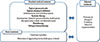
AE is a chronic skin inflammation and its symptoms wax and wane with various manifestations. Individualized therapy for the patient should be implemented according to patients' age, severity and extent of AE, and distribution of the lesion. To control AE, in addition to main pharmacologic therapy, other measures such as cutaneous hydration, identification and elimination of aggravating factors, relief of pruritus, and patient education should be considered.22 Flares need to be prevented by multiple systemic approaches. We categorize the treatment options into 3: basic, standard medical, and adjuvant treatments (Figure).
Basic treatment
Reduction in dryness with emollient often relieves pruritus. Aggravating factors should be avoided by individualized evaluation.
Cutaneous hydration
AE is characterized by an impaired skin barrier with xerosis, which needs to be strictly controlled by cutaneous hydration. FLG gene mutations are commonly shown in AE patients, which decrease the natural moisturizing factors. Furthermore, the correlation between FLG gene mutations and AE severity is well known.23 Cutaneous hydration can help the skin retain water, improve barrier function, and relieve itch sensations.24 Emollients can reduce flare-ups of AE and the need for topical steroid use.2526 Taking baths with warm water for about five to ten minutes are recommended for hydrating the stratum corneum and eliminating scale, crust, sweat, irritants, and allergens.27 When patients with AE take a shower or bath, using nonirritating, mild acidic soaps is desirable, and scrubbing should be avoided. Importantly, emollients should be applied within 3 minutes after showers or baths, as the skin can become dry otherwise. Emollients need to be applied on the skin at least twice a day, including the unaffected skin. The National Institute for Health and Clinical Excellence (NICE) guideline recommends to use emollients more than 250 g weekly.25 There are several types of moisturizers, including lotions, creams, and ointments. The most appropriate moisturizer is decided depending on the season, patient's preferences, and symptoms. For example, in the summer season, lotions are nmostly preferred to ointments. In addition, preservatives or fragrances that may aggravate the skin condition should be considered. In our previous study, we proved that AE patients, in general, did not use the proper amount of emollients.28 Therefore, we propose that education about proper cutaneous hydration methods needs to be provided for all patients. Wet dressing can promote the transepidermal penetration of topical glucocorticoids with skin barrier recovery, especially for acute oozing lesions.29 After removing a wet gauze, the skin needs to be immediately hydrated with emollients. In cases involving oozing lesions, dressing with wet gauze can reduce the chances of infection, and the drying effect due to evaporation can be beneficial to oozing lesions. Dressing is also effective to protect the skin from scratching.30
Identification and elimination of aggravating factors
When assessing patients with AE, physicians need to identify aggravating factors, through a detailed history intake/detailed interview, careful evaluation of clinical manifestations, and allergy tests with clinical relevance. In the management of AE, many aggravating factors have to be considered and identified on a patient-by-patient basis. AE should be properly managed based on a detailed assessment of any potential aggravating factors.
General Considerations
Commonly, individuals with AE have more sensitive skin than the general population. The first recommendation is to avoid known irritants, such as soaps or detergents, chemicals, wool or nylon clothing, abnormal temperature/humidity, or sudden temperature changes. Perfumed fabric softeners can also cause irritation. Wool can cause irritation, and nylon cannot absorb sweat. Smooth clothing such as cotton is preferred to minimize skin irritation. New clothing should ideally be laundered before use. Double rinsing is helpful for the removal of detergents. AE patients should always make sure to maintain a pleasant temperature and humidity level in their environment. Mild sports activities or swimming is good to relieve stress, although sports that may induce intense perspiration or heat should be restricted. Sunscreen is good for preventing sunburn, but as it can also lead to skin irritation, thereby patients should always choose nonirritating products.
Specific allergens
There are numerous triggering and exacerbating factors in AE. The cornerstone in AE treatment is an individualized recommendation on aggravating and/or triggering factors. Food and inhalant allergens can aggravate the symptoms of AE. In children in particular, food allergens can exacerbate AE, although this correlation is still controversial. In cases of severe AE, which can be suddenly aggravated after therapy is discontinued, food allergens are often considered triggering factors. Tests for evaluating food allergies include skin prick testing, serum-specific IgE level checks, radioallergosorbent tests, and immunoCAP tests. Negative test results are helpful to rule out suspected allergens. Positive results require clinical correlation and confirmation by scrutinizing and eliminating foods that are suspected to be the cause. After avoiding the suspected food for 4 to 6 weeks, an oral food challenge should be performed to confirm whether it is indeed a cause of AE flares for the patient. The double-blind placebo-controlled food challenge is considered the gold standard for diagnosing food allergies.31 Keeping a food diary for recording intake and symptoms can be helpful in identifying food allergens. The avoidance of food allergens is the best therapy, although the imprudent elimination of food can cause nutritional deficiencies. Therefore, maintaining a good diet is also very important. Food-related AE can often resolve with time; intermittent rechallenging should be undertaken every 6 to 12 months. The National Institute of Allergy and Infectious Diseases Food Allergy Expert Panel suggest that food allergens, such as cows' milk, eggs, wheat, soy, and peanuts, should be considered to be restricted in patients under five years of age with moderate to severe AE.32 In cases of patients with peanut allergy, the symptoms can be serious and may continue throughout their lifetime.
In contrast to food allergens, positivity to aeroallergens increases with age. Sensitivity to inhalant allergens, such as dust mites, pollen, animal danders, and fungi, is more common in moderate-to-severe AE patients.33 These allergens can aggravate the symptoms of AE. Dust mites are the most common allergen among patients with AE, and avoiding this allergen is helpful to patients.34 Physicians should recommend AE patients with a sensitivity to dust mites to encase their pillow and mattress, and also wash beddings in hot water weekly and vacuum frequently.3536 Minimizing carpeting, curtains, and drapes is good for controlling AE. Maintaining proper humidity and temperature levels through ventilation is recommended. The prevalence of allergic contact dermatitis is increasing in AE patients. The most common contact allergens are nickel, neomycin, fragrances, formaldehyde, lanolin, and rubber chemicals.37 Patch tests should be considered for patients with AE who have symptoms consistent with allergic contact dermatitis. Because there are many triggers contributing to flares of AE, attention needs to be paid to identifying and controlling factors that contribute to flares on a patient-by-patient basis.
Psychological support
Psychological problems, such as anxiety, depression, and attention deficit hyperactivity disorder, can occur in AE patients. Emotional stress can provoke itching and scratching, and thus exacerbate AE. Therefore, AE patients with emotional or psychological problems should try to relax, and counseling can be helpful to break the itch-scratch cycle, especially in adolescents and young adults.3839 In some instances, scratching is simply habitual. Relaxation, behavioral modification, or biofeedback may also be of benefit, especially in patients with habitual scratching.30
Standard medical treatment
For patients with mild AE symptoms, topical medications are the primary choice of treatment. However, if the basic and topical therapies fail, a systemic approach may be necessary. Cyclosporin and short-term systemic glucocorticoids are commonly used. Alternative therapies include phototherapy, antimetabolites, interferon-gamma, allergen immunotherapy, andbiologics, depending on cases. The effectiveness of new biologics on AE is being reported in recent years, suggesting that biologics can be a promising targeted therapy for AE in the future.
Topical glucocorticoids
Topical glucocorticoids are a mainstay therapeutic agent for AE. They are known to be one of the most effective pharmaceutics in controlling AE symptoms, such as itchiness and inflammation, although their application can accompany a number of side effects. Potential adverse effects include the development of striae, skin atrophy, perioral dermatitis, acne rosacea, and adrenal suppression.
Glucocorticoids are categorized into seven classes by potency based on vasoconstrictor assays. Physicians should recommend topical steroids of the appropriate potency considering the patient's age, disease severity, and extent and, distribution of lesions. Very potent steroids are effective intreating AE flares; however, as improvement is observed, providers need to use steroids of lower potency and reduce the frequency of applications. In addition, physicians should educate patients to limit the use of high potency steroids to severe or lichenified lesions only. The use of high potency steroids should be avoided on thin skin, such as face or skin folds. Also, it is not recommended to use them for more than 2 weeks in a row. There have been reports that the long-term use of low potency steroids such as fluticasone 1 to 2 times a week on AE lesions, including those already healed, can prevent the aggravation of AE.4041 This method of applying steroids to residual lesions, as well as unaffected skin, is called proactive therapy.42
A finger tip unit (FTU), a practical measure of the amount of ointment, is defined as the amount of 0.5 g cream or ointment expressed from a tube with a 5 mm diameter nozzle, applied from the distal skin-crease to the tip of the index finger. The appropriate amount of cream or ointment should be determined in FTUs based on the area of lesions requiring treatment.
Factors that influence the effectiveness of topical steroids are as follows: (1) potency, amount, and vehicle of agents, (2) duration of application, (3) whether using occlusive dressing, (4) host factors such as type and the total area of lesion, (5) existence of infection or allergic reaction, and (6) patients' compliance to therapy.
Topical calcineurin inhibitors
Topical immunomodulators, tacrolimus and pimecrolimus, are nonsteroidal, topical calcineurin inhibitors.43 They bind to FK-binding protein and inhibit the production of cytokines from activated T cells and inflammatory cells. Tacrolimus and pimecrolimus appear to have an anti-inflammatory potential similar or slightly less than that of midpotency corticosteroids.44 Tacrolimus ointment 0.03% and pimecrolimus cream 1% is approved for the treatment of patients ≥2 years old. The use of tacrolimus ointment 0.1% is approved for adults only. Transient burning or itching sensations are its only adverse effects. No complications such as skin atrophy are associated with its long-term use.45 These agents may be favored for the treatment of facial and eyelid AE. Like fluticasone, topical calcineurin inhibitors are also good for proactive therapy when applied 2 to 3 times a week.46 Recent studies have reported that it is safe to use tacrolimus or pimecrolimus for up to 2 and 4 years, respectively.4748 We were not able to find well-designed, established randomized, controlled studies or prospective studies concerning the safety of long-term use of topical calcineurin inhibitors. However, 1 case-control study reported no increased risk of lymphoma in AE patients who used topical calcineurin inhibitors for a long period of time, despite the existing concern about the development of lymphoma with the chronic use of the agent.49
Antihistamines
Oral antihistamines relieve histamine-induced itching sensations by blocking H1 receptors. Although frequently prescribed to patients with AE, their efficacy has not been proven by controlled clinical trials.5051 Pruritus can be caused by various mediators in addition to histamine, so many patients cannot be treated by antihistamines alone. For AE patients with concomitant urticaria or concurrent allergic rhinitis, oral antihistamines may be effective.52 In addition, sedating antihistamines, such as hydroxyzine or chlorpheniramine, can prevent scratching during sleep and may offer symptomatic relief for itching that worsens at night. However, they can impair adults' driving ability or children's learning ability.
Anti-infectious agents
Infections of bacteria, viruses, and fungi can frequently accompany AE, and symptoms of AE can be aggravated following an infection. Any secondary infection should be controlled. Staphylococcus aureus is known to play a significant role in AE. Toxins from S. aureus act as superantigens and are known to exacerbate AE.53 In cases of local infection, especially in impetiginized features, the application of fusidic acid or mupirocin ointment is helpful. In patients with extensive superinfection without resistant S. aureus strains, cephalosporins, or penicillinase-resistant penicillins (dicloxacillin, oxacillin, or cloxacillin) are effective.54 Physicians should be careful in the use of antibiotics that can lead to the growth of resistant organisms, especially macrolide antibiotics or topical antibiotics.55 Among resistant organisms, methicillin-resistant S. aureus in particular, is a growing problem in AE patients. Therefore, empirical antistaphylococcal antibiotics should be used with caution in patients without any signs of infection. The choice of antibiotics should be based on the sensitivity of bacteria as determined by bacteria culture. Baths with dilute sodium hypochlorite (bleach) may also benefit AE patients infected with bacteria.56
Herpes simplex virus can also cause AE flares. Eczema herpeticum is difficult to be ruled out clinically. However, the development of particular signs such as the punched-out erosions, vesicles, and/or oozing crusts suggests secondary infection with herpes simplex virus and can be differentiated by Tzanck test, direct immunofluorescence assay, polymerase chain reaction, and culture. When herpes simplex virus in AE patients spread to other parts, a systemic antiviral therapy needs to be promptly provided.57
Systemic therapy
Systemic therapies using pharmaceutical drugs, such as glucocorticoids, cyclosporin, methotrexate, mycophenolate, azathioprine, are generally reserved for patients with severe and refractory AE. These agents must be used carefully, with consideration of their potential adverse effects.
Cyclosporine
Cyclosporin is an immunomodulator which primarily acts on T cells. It binds intracellular cyclophilin and inhibits cytokine transcription. Cyclosporin is effective for severe, refractory AE in both children and adults. The optimal doses of cyclosporine ranges from 3-6 mg·kg·d.61 However, rapid relapse is a concern after abrupt discontinuation of cyclosporin treatment. Patients receiving cyclosporin should be monitored for potential adverse effects, such as kidney or liver function impairment and hypertension.
Systemic glucocorticoids
Although systemic glucocorticoids are frequently used, they have shown to only temporarily suppress AE. Moreover, they are rarely indicated for the treatment of chronic AE. Still, short courses of oral corticosteroids are sometimes necessary to control atopic flares. Once symptoms improve, systemic steroids should be tapered or discontinued while keeping maintenance therapy such as cutaneous hydration and the use of topical agents. If systemic corticosteroids are abruptly stopped, a disease flare or a rebound phenomenon can occur.
Antimetabolites
Methotrexate, azathioprine, and mycophenolate mofetil are recommended as systemic agents for the treatment of refractory AE. Methotrexate is a folate antagonist, which inhibits inflammatory cytokine synthesis and cell chemotaxis and acts as an antimetabolite. It is frequently used for treatment-resistant adult AE patients as well as patients with psoriasis.62 Azathioprine is a purine analog with anti-inflammatory and antiproliferative effects. It has been used for severe adult and child AE patients.5051 It can cause significant side effects such as myelosuppression.
Mycophenolate mofetil is a purine biosynthesis inhibitor. Once ingested, it undergoes ester hydrolysis to its active form, mycophenolic acid. It is effective for treatment-resistant adult AE at doses of 2 g daily. Patients undergoing treatment with this agent need to be monitored for herpes retinitis and dose-related bone marrow suppression.63
Interferon-γ
IFN-γ acts to reduce IgE levels and downregulates Th2 cells. There have been reports that treatment with IFN-γ can lead to clinical improvements.646566 In those studies, there are significant correlations between reducd clinical severity and decreased total eosinophil count. However, some short-term side effects similar to influenza-like symptoms, such as fever and headache, may occur in the early stage of treatment. Further studies regarding the long-term side effects of the therapy are needed.
Phototherapy
Natural sunlight may be beneficial to some patients with AE, while it can also induce sweating and itching sensations. Phototherapy can be a secondary therapeutic option for patients with AE. In general, UVA-1 (340 to 400 nm) is used for acute severe lesions, and narrow-band UVB (311 nm) is used for chronic AE.67 Epidermal Langerhans cells and eosinophils may be targets of UVA phototherapy. UVB exerts immunosuppressive effects by blocking the function of antigen-presenting lymphocytes and by altering keratinocyte cytokine production.68 Phototherapy needs to be carefully considered for cutaneous malignancies as well as erythema, skin pain, pruritus, and pigmentation.69
Allergen immunotherapy
Subcutaneous and sublingual specific immunotherapies (SITs; e.g. against house dust mites, pollen, or cow's milk protein) are another approach to the treatment of AE. Immunotherapy with inhalant allergens is useful for allergic rhinitis and asthma. However, it has not been proven effective for AE. Reports have been published suggesting that SITs may play a role in the subgroup of AE patients who have become sensitized to dust mite allergens.707172 Recently, SIT is effective as much as evidence B in AE patients with sensitized inhalant allergen from a meta-analysis.72 However, the complex procedures and long durations (≥ 1-2 years) involved with this therapy can lead to poor compliance.73
Anti-CD20 therapy
Rituximab is an antibody against CD20 which depletes B cells. Treatment with Rituximab improved skin symptoms in patients with severe AE, suggesting its potential role for B-cells in the pathogenesis of AE.74
Anti-IgE
Omalizumab is a monoclonal antibody which binds and neutralizes IgE. Some AE patients have shown clinical improvement with anti-IgE therapy, but others have experienced no response or even aggravation of their symptoms.757677 Further studies are needed to determine whether omalizumab deserves a place in routine AE therapy, or whether its costs or side effects outweigh possible benefits.
Anti-IL-4 receptor therapy
It is well known that Th 2 cytokine plays an important role in atopy. Pitrakinra, an inhibitor of IL-4 receptor signaling, hinders the binding of interleukin 4 and interleukin 13 to interleukin-4 alpha receptor complexes. The biologic substance was suggested to improve the symptoms of asthma.78 Interestingly, another anti-IL-4 receptor antibody, dupilumab, was proved to be effective in the treatment of some AE cases from a multicenter, randomized, double-blind study.79
Anti-TNFα therapy
A pilot study with a TNF antagonist, infliximab, was conducted in 9 patients with moderate or severe AE. Treatment with infliximab improved clinical symptoms, but the effect was not continued through the maintenance therapy.80
Anti-IL-5 therapy
IL-5 is also another important cytokine produced by Th2 cells. Treatment with mepolizumab, a humanized monoclonal antibody, which binds to IL-5, did not induce clinical improvement in patients with AE, despite a significant decrease in peripheral blood eosinophils.81 However, a recent double-blind study showed that mepolizumab had a significant glucocorticoid-sparing effect in patients with severe eosinophilic asthma.82 Further studies are required to determine whether anti-IL5 therapy may be used to treat AE.
Anti-IL-6 receptor therapy
Tocilizumab or atlizumab is a humanized monoclonal antibody against the IL-6 receptor which is used mainly for the treatment of rheumatoid arthritis. A recent study showed the potential effectiveness of interrupting IL-6-receptor signaling in patients with AE.83 However, bacterial superinfections were also reported to be associated with the therapy. Further studies are needed to investigate the efficacy and safety of IL-6 receptor antagonists.
Anti-IL-31 therapy
IL-31 is primarily produced by type 2 helper T cells (Th2). The structure of IL-31 places it in the IL-6 family of cytokines. IL-31 serum levels correlate with disease activity and Th2 cytokine levels in children with AE.84 Anti-IL 31 monoclonal antibody is under investigation in a phase I clinical trial (clinicaltrials.gov).
Anti-TSLP therapy
TSLP is an epithelial-cell-derived cytokine, which plays a key role in the maturation of T cell populations through activation of antigen presenting cells. TSLP production may initiate allergic inflammation. AMG 157 is a human anti-TSLP monoclonal immunoglobulin G2λ that binds human TSLP and prevents receptor interaction. Treatment with anti-TSLP antibody decreased allergen-induced early and late asthmatic responses in patients with mild allergic asthma.85 A phase I clinical trial with anti-TSLP antibody is being conducted in healthy subjects and subjects with moderate to severe AE (clinicaltrials.gov).
Adjuvant treatment
Adjuvant therapy should be considered if AE symptoms are uncontrolled by adequate basic treatment.
Primrose oil
Evening primrose oil has been thought to be beneficial to AE patients because of its gamma-linoleic acid content. Although a recent meta-analysis has shown that primrose oil failed to significantly improve AE, it is still considered an adjuvant therapy with a small risk of side effects.86
Omega-3
Recently, it has been suggested that the increasing prevalence of AE may be associated with changes in the Western diet: reduced consumption of omega-3 polyunsaturated fatty acids (PUFA) and increased omega-6 PUFA intake. A pilot study demonstrated that dietary omega-3 PUFA intake may have a therapeutic effect on the symptoms of AE.87
Probiotics
Probiotics are live microbial food supplements. Prebiotics are indigestible carbohydrates that stimulate the growth of probiotic bacteria in the intestine. Both are known to act on and modulate T helper cell cytokine activation as immunomodulators.88 Baquerizo Noel et al.89 recently stated that probiotics and prebiotics appear to be effective in reducing the incidence of AE in infants. However, their role in AE treatment is still controversial. In a Cochrane review, probiotic treatment was shown to carry only a small risk of adverse events.90 Further studies are needed to determine whether these agents can play a role in AE treatment by modulating immunological dysfunction of AE.
Chinese herbal medicines
Chinese herbal medicine has been increasingly used for AE. It has been reported that 42% of AE patients have tried alternative medicine at some point, such as homeopathy and herbal remedies.91 Our previous study showed that Korean red ginseng ameliorated and prevented AE-like skin lesions in NC/Nga mice.92 Although there are many interesting studies on traditional Chinese herbal therapies, the temporary nature of its effect and possible complications need to be noted. The potential side effects include hepatic toxicity, cardiac side effects, and idiosyncratic reactions. Further well-designed, randomized, double-blind trials are needed to evaluate the long-term efficacy and safety of Chinese herbal therapies in the treatment of AE.
Oral vitamin D
There is controversy regarding vitamin D supplementation for the treatment of AE. A previous report demonstrated an inverse relationship between the severity of AE and the serum vitamin D level.93 Therefore, vitamin D supplementation can be helpful for patients deficient in vitamin D or whose symptoms aggravate/worsen during winter.94
CONCLUSIONS
The prevalence of AE is steadily increasing, currently ranging 1%-20% of the general population. AE may be caused by genetic factors and may be influenced by environmental factors. Most AE patients have a chronic, relapsing disease course characterized by remission and intermittent flares. Therefore, controlling symptoms of chronic AE is still challenging. The treatment options for AE includes (1) standard medical treatment with pharmaceutical agents such as topical steroids and topical immunomodulators, (2) adjuvant therapy, and (3) the following basic treatment such as using emollients. Moreover, avoidance of triggering and/or aggravating factors that exacerbate itching, such as woolen clothes, emotional stress, and uncomfortable climatic conditions, is essential at all times. Furthermore, phototherapy or systemic immunosuppressants, such as cyclosporine, azathioprine, and mycophenolate mofetil, should be considered if conventional methods fail to control AE symptoms properly.
In the future, with better understanding of the pathogenesis of AE, revolutionary treatment options, including targeted biologic therapies, are being introduced. In particular, anti-IL-4 receptor therapy is expected to be a promising choice for refractory AE.
 XML Download