



 PDF
 Citation
 Print


Introduction
Accurate measurement of urinary bladder volume is essential in diverse clinical settings, including assessment of postoperative urinary retention, monitoring of neurogenic bladder, and evaluation of lower urinary tract symptoms. Reliable volumetry guides catheterization decisions and enables longitudinal monitoring in both inpatient and outpatient care [1–3]. Ultrasound is the most widely used noninvasive modality for bladder assessment because it is safe, portable, and rapidly deployable. In current practice, bladder volume estimation typically involves acquisition of orthogonal transverse and sagittal planes [4–6], bladder segmentation, and application of an empirical shape coefficient, commonly 0.52–0.72, to the product of three linear dimensions [7–9].
Although effective in many cases, this workflow has several inherent limitations. First, it requires near-simultaneous acquisition of two orthogonal planes, which increases workflow complexity and makes the measurement susceptible to interplane misregistration. Second, the use of a fixed, non–patient-specific shape coefficient imposes a heuristic anatomical assumption that may not reflect actual bladder geometry and may therefore introduce systematic error. Third, identifying the “maximal” caliper positions is often ambiguous and morphology dependent, reducing reproducibility. These limitations have motivated reduced-acquisition strategies, which generally fall into three categories: (1) limited-beam sagittal methods that estimate volume from sparse beam samples and hand-tuned formulas [10,11] but remain sensitive to probe angle and shape variability; (2) A-mode, single-transducer designs intended for wearable or portable systems [12], which sacrifice rich two-dimensional context for simple one-dimensional echoes; and (3) early artificial intelligence-based approaches that continue to rely on multiplanar acquisition, segmentation, or shape-factor scaling and have limited generalizability beyond internal validation cohorts [12–14]. Consequently, misregistration between planes, dependence on heuristic shape factors, and underuse of perivesical echotexture remain unresolved challenges.
To address these limitations, the authors propose a deep learning-based approach that directly estimates bladder volume from a single B-mode sagittal ultrasound image and its corresponding binary bladder mask. The central premise is that sagittal bladder appearance may encode features associated with overall bladder geometry and thereby support volume estimation from a single plane. Prior anatomical studies support this premise: sagittal and transverse bladder shapes are correlated [15], and perivesical soft-tissue patterns may constrain three-dimensional morphology. In magnetic resonance imaging, the detrusor muscle forms a distinct hypointense band relative to surrounding fat, creating high-contrast boundary layers [16]. Ex vivo micro-computed tomography studies have similarly demonstrated nonuniform bladder wall thickness and filling-dependent geometric variation [17]. Although these observations come from modalities other than ultrasound, they suggest that boundary compliance and perivesical context may carry information relevant to bladder filling and overall geometry, a principle that can be leveraged by modern deep learning models applied to ultrasound images.
Building on this anatomical rationale, the present method jointly learns from the sagittal bladder mask and the full-image echotexture to capture cues from both the bladder and adjacent perivesical tissues. A hybrid architecture integrated convolutional neural network features with self-attention mechanisms, allowing the model to encode local detail while also leveraging long-range contextual relationships within a single plane.
This study developed and evaluated a model designed to learn a patient-specific implicit mapping between single-plane sagittal image features and bladder volume from full-resolution two-dimensional ultrasound data, eliminating the need for explicit orthogonal-plane acquisition. By performing end-to-end volume regression, the method avoids reliance on fixed heuristic shape factors and simplifies acquisition, with the potential to improve robustness compared with traditional two-plane, coefficient-based approaches. A single-plane solution may be especially valuable in settings where orthogonal views are difficult to obtain, such as emergency departments, outpatient clinics, and handheld or portable ultrasound workflows.
Materials and Methods
Compliance with Ethical Standards
All procedures followed the principles of Good Clinical Practice and were approved by the Severance Hospital Institutional Review Board (IRB Nos. 1-2022-0076 and 1-2025-0025). Written informed consent was obtained from all participants before enrollment. Distinct Institutional Review Board approvals and nonoverlapping enrollment periods were used to maintain clear separation between the training and test groups.
Deep Learning Model Development
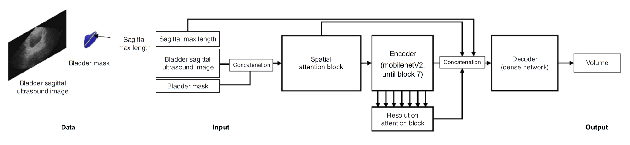
This study developed a single-stage deep learning network for bladder volume regression that operates on a sagittal B-mode ultrasound image and its corresponding ground-truth bladder mask, as shown in Fig. 1. The ground-truth mask used for model input and sagittal length computation was generated independently by two expert sonographers, each with more than 10 years of experience, who worked in a blinded manner. A urologist with at least 10 years of clinical experience adjudicated the annotations and finalized the consensus segmentation. The ground-truth bladder volume reference was obtained from Foley catheter drainage measured immediately after scanning.
Model inputs
The network receives a channel-wise concatenation of the sagittal ultrasound image (330×570 pixels) and its ground-truth mask. The precomputed maximum sagittal bladder length is provided as an auxiliary scalar input to the prediction head.
Spatial self-attention module
To model long-range echotextural dependencies that are not captured by local convolutions, a spatial self-attention block was incorporated directly into the concatenated image–mask input. The input was divided into nonoverlapping 30×30 patches, forming an 11×19 grid of 209 tokens. Each patch was embedded into a 64-dimensional token representation, and multihead self-attention was applied across tokens. This mechanism highlights texture-informative regions and provides global contextual cues analogous to patch-token representations in vision transformers.
Backbone encoder and cross-scale attention
The primary convolutional encoder consisted of MobileNetV2 blocks 1–7, initialized with pretrained weights to leverage efficient inverted residuals with linear bottlenecks [18]. To capture multiresolution information, a resolution self-attention block was introduced: batch-normalization activations from encoder blocks 1 through 7 were extracted, projected into 64-dimensional tokens, and processed with self-attention across scales [19–22]. The resulting cross-scale representation was concatenated with the spatial attention features and auxiliary sagittal length.
Prediction head and training
The fused representation was passed to a lightweight fully connected prediction head to produce the final bladder volume estimate. The model was trained using mean squared error loss on an NVIDIA RTX A6000 GPU, with a learning rate of 1×10⁻³. Fig. 1 illustrates the overall architecture of the proposed single-plane estimator.
Clinical Data Acquisition
Data were collected at Severance Hospital for model development and evaluation. The training dataset was acquired between January 2023 and September 2023, and the test dataset was acquired between June 2025 and March 2026, ensuring strict temporal separation between cohorts.
All ultrasound scans were obtained by trained clinicians under stable anesthetic conditions. During acquisition, operators visually confirmed the sagittal bladder view in real time and positioned the probe at a consistent infraumbilical landmark. To promote operator heterogeneity and improve model generalizability, scans were performed by multiple clinicians with varying levels of experience across the Institutional Review Board-governed studies. Participants were adult patients undergoing elective abdominal or urologic surgery that required general anesthesia and Foley catheter placement as part of routine perioperative care. These participants included healthy kidney donors and patients undergoing surgery for renal or prostate conditions. Foley catheterization was not performed solely for research purposes. After ultrasound acquisition, catheter drainage was performed immediately to obtain the reference bladder volume. Patients were excluded if Foley catheter placement was not clinically feasible or if suprapubic ultrasound scanning could not be performed because of severe skin injury or other local conditions at the scanning site.
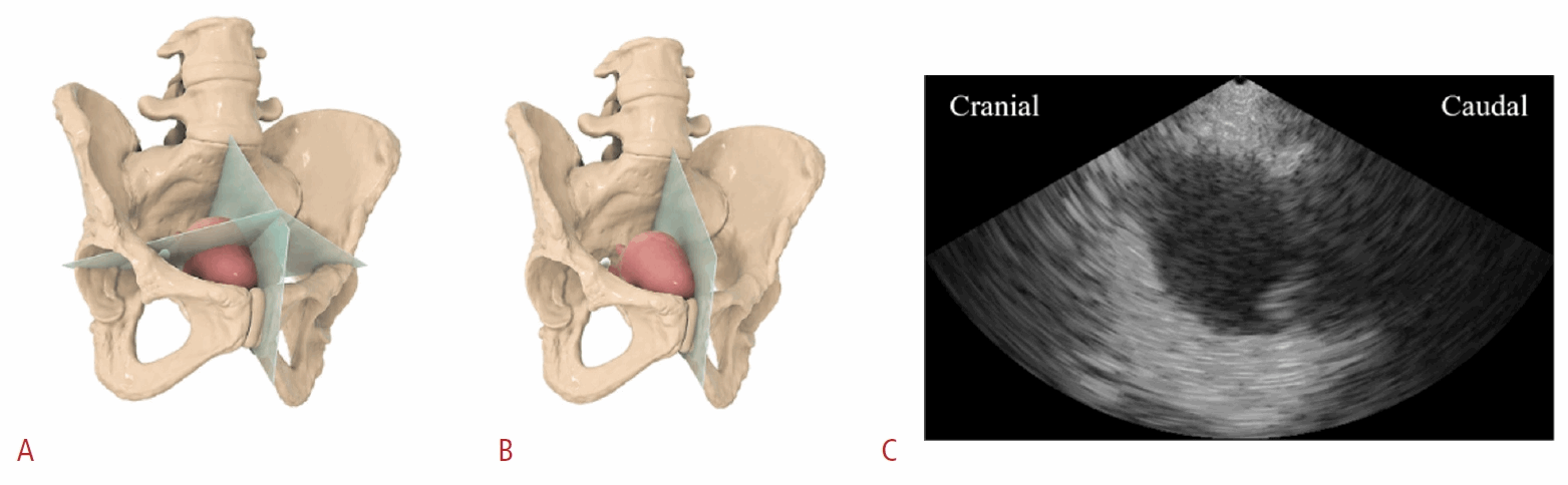
A total of 315 participants were enrolled, including 262 participants in the training cohort and 53 participants in the test cohort. Training images were collected using the EdgeFlow UH10 bladder scanner (Edgecare Inc., Seoul, Korea), whereas test images were captured using the EdgeFlow UW20 wearable ultrasound system (Edgecare Inc.). Table 1 summarizes the demographic characteristics of the study participants. Fig. 2 illustrates the conceptual difference between conventional multiplanar bladder assessment and the proposed single-plane acquisition strategy. In Fig. 2A, orthogonal imaging planes represent conventional cross-sectional assessment requiring multiple views. In Fig. 2B, only the sagittal imaging plane is retained, corresponding to the proposed single-sagittal-plane approach used for volume estimation in this study. Fig. 2C shows a representative sagittal B-mode ultrasound image, illustrating the cranial-to-caudal orientation and typical bladder appearance in the sagittal plane.
Reference Standard and Labeling
The reference standard for bladder volume was Foley catheter drainage measured immediately after ultrasound acquisition. Catheter drainage is widely accepted as the clinical reference standard for assessing postvoid residual volume and was therefore used as the direct benchmark for model evaluation. Bladder segmentation masks were generated to support both model input and computation of maximum sagittal bladder length. Two sonographers, each with more than 10 years of experience in genitourinary ultrasound, independently annotated anonymized sagittal images using the computer vision annotation tool (CVAT) annotation platform in a blinded manner. An experienced urologist with at least 10 years of clinical experience, who was not involved in the initial annotation process, reviewed both sets of masks and adjudicated a final consensus segmentation for each image. The finalized ground-truth masks were used for two purposes: (1) model input, as a binary mask channel alongside the B-mode image, and (2) sagittal length estimation, to compute the maximum sagittal bladder length incorporated as an auxiliary scalar feature. In this proof-of-concept study, expert-annotated ground-truth masks were provided during both model development and held-out evaluation because the study focused on the volume-estimation component rather than the segmentation model. To enhance transparency and reduce potential bias, reference-standard determination was separated from model prediction. Image annotation was performed by clinical experts who were blinded to model outputs, and discrepant segmentation results were adjudicated by an experienced urologist independent of the initial annotation process. Foley catheter drainage, rather than ultrasound-derived volume estimation, was used as the reference standard for bladder volume. Company-affiliated authors contributed to algorithm development, data processing, and manuscript preparation based on the provided dataset, but they were not involved in device operation or data acquisition. The clinical reference standard and final segmentation adjudication were determined through predefined clinical procedures.
Results
Visual Inspection
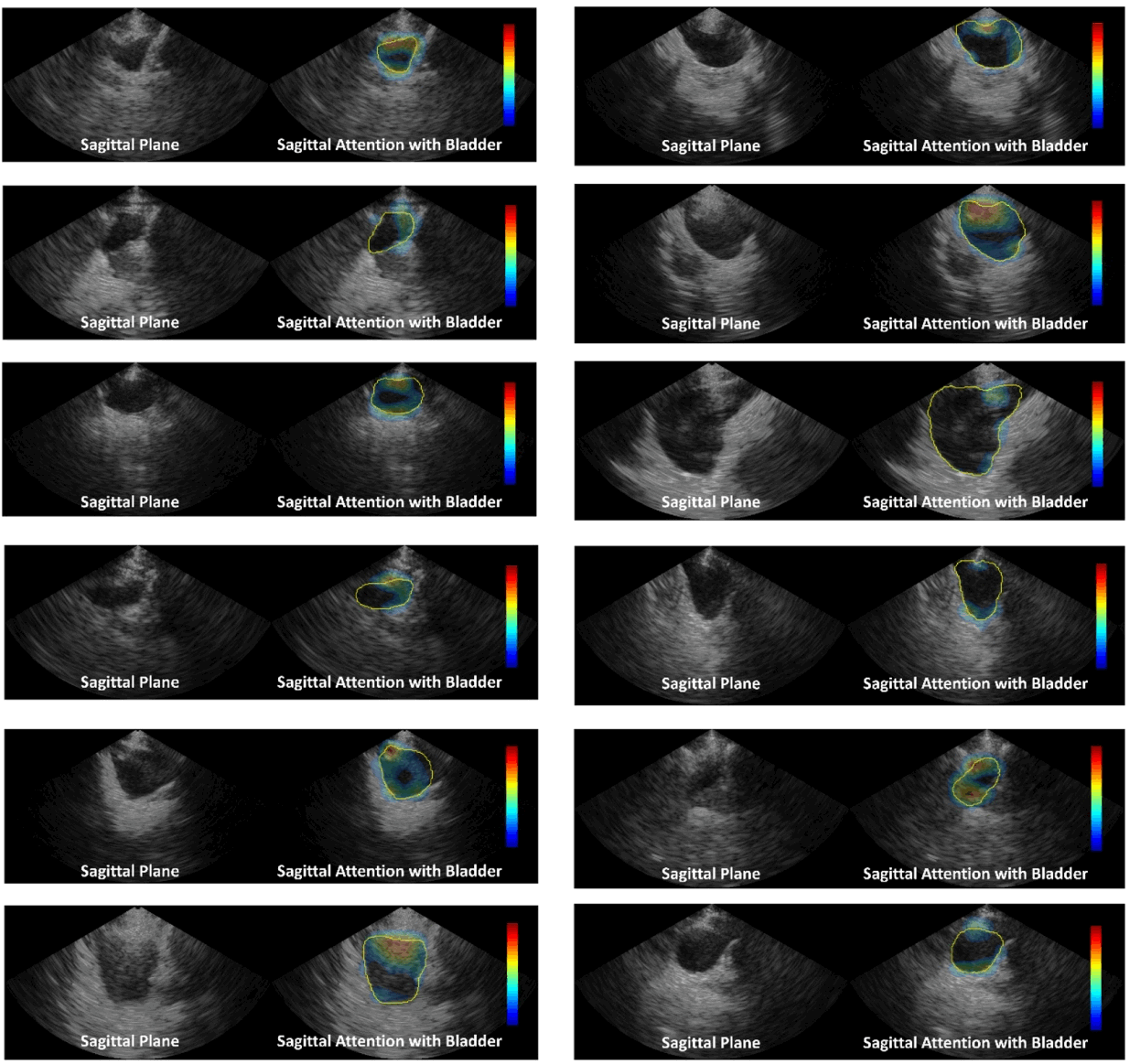
For visualization, attention maps were obtained from the spatial self-attention module applied to the concatenated ultrasound image and bladder mask input. The attention output was mapped at the patch level according to the nonoverlapping 30×30 patch structure used in the model, corresponding to an 11×19 spatial grid. The resulting patch-level attention map was resized to the original ultrasound image size and overlaid on the grayscale B-mode image. Color intensity represents the relative attention weight within each image, with warmer colors indicating regions that received greater model attention. Each map was independently normalized using within-image min–max scaling for display; therefore, color intensity represents relative spatial weighting within each case rather than absolute attention magnitude across cases.
Across representative test cases, the proposed model’s spatial self-attention consistently highlighted the bladder wall and adjacent perivesical soft tissues while largely suppressing the anechoic bladder lumen. These attention patterns were concentrated around boundary curvature and adjacent echotextural bands near the bladder wall, suggesting that the network used contextual soft-tissue cues and surrounding morphology, rather than the binary bladder mask alone, to extract image features relevant to volume estimation. The persistent concentration of attention along the bladder interface and adjacent perivesical tissues is consistent with the hypothesis that sagittal morphology and contextual echotexture contain cues relevant to overall bladder volume. Anatomically, the bladder wall comprises the mucosa, submucosa, detrusor muscle, and outer perivesical layers; the generated overlays qualitatively suggest attention overlap with these boundary regions. Representative attention maps from several test cases are shown in Fig. 3 and illustrate consistent localization of salient regions across patients.
Statistical Analysis
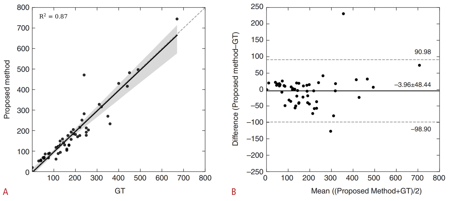
In the held-out test set (n=53; reference standard, Foley catheter drainage), the proposed single-plane model demonstrated a strong linear association with the reference volume, yielding R²=0.87 (Fig. 4A). Bland-Altman analysis of the model-derived volume minus the reference volume showed a mean bias of −3.96 mL with a standard deviation (SD) of 48.44 mL, corresponding to 95% limits of agreement from −98.90 to +90.98 mL (Fig. 4B).
These values are summarized in Table 2: bias, −3.96 ± 48.44 mL; limits of agreement, −98.90 to +90.98 mL; and R²=0.87. In stratified analyses, the 0–100 mL cohort (n=13) yielded a mean absolute error of 14.15 mL, whereas the ≥100 mL cohort (n=40) yielded a mean absolute percentage error of 16.64%. Percentage error was reported only for volumes ≥100 mL to avoid instability from near-zero denominators.
Compared with previous work using the same reference standard, the proposed single sagittal-plane model achieved agreement metrics within the range reported for two-plane approaches. Relative to Jeong et al. [23], the magnitude of bias in this study was 39.1% smaller than that of their artificial intelligence method (6.5±50.4 mL) and 73.6% smaller than that of their conventional calculation (15.0±50.9 mL). The present study’s limits-of-agreement width (189.88 mL) was marginally narrower than the 197.7–199.6 mL reported for two-plane methods, consistent with the slightly smaller residual SD herein (48.44 mL vs. approximately 50 mL). Correlation was also comparable (R²=0.87 vs. R²=0.91). Alpert et al. [24] reported narrower limits of agreement (−52.7 to +86.9 mL) and a stronger correlation (approximately 0.94), although their reference was manual human estimation rather than catheter drainage, limiting direct numeric comparability. Overall, despite relying on only a single sagittal acquisition, this method achieved agreement approaching that of established two-plane techniques while substantially reducing the acquisition burden.
Discussion
Clinical bladder volumetry using ultrasound typically multiplies three orthogonal lengths—longitudinal length, transverse width, and anteroposterior height—and applies a shape coefficient, commonly 0.52 for a prolate ellipsoid, although other coefficients are also used. This workflow requires acquisition of two orthogonal planes and depends on a fixed, non–patient-specific factor, which can misrepresent anatomy and is susceptible to misregistration and caliper-placement variability. The present study investigated whether a single sagittal plane can support accurate volume estimation by learning the relationship among sagittal morphology, surrounding echotexture, and image features associated with bladder filling and overall geometry.
The single-plane model developed in this study closely tracked the reference standard (R²=0.87). Bland-Altman analysis of the model-derived volume minus the reference volume showed a small negative bias of −3.96 mL with an SD of 48.44 mL, yielding 95% limits of agreement from −98.90 to +90.98 mL. Stratified error analysis showed a mean absolute error of 14.15 mL for volumes of 0–100 mL (n=13) and a mean absolute percentage error of 16.64% for volumes ≥100 mL (n=40), with percentage error restricted to volumes ≥100 mL to avoid instability near zero.
Visual inspection of the model’s spatial attention showed consistent focus on the bladder wall and adjacent perivesical soft tissues, with suppression of the anechoic lumen across a range of bladder sizes and probe positions. This pattern suggests that the network used contextual echotexture, rather than the binary mask alone, to infer surrogate shape cues relevant to volume estimation. However, consistent with prior cautions and ongoing debate in the explainability literature, this study treated attention maps as diagnostic visualizations rather than causal mechanistic evidence; they support biological plausibility but do not prove the underlying computation.
A single-sagittal-plane approach offers several practical benefits: it eliminates the need for near-simultaneous orthogonal acquisitions, reduces opportunities for misregistration, and removes reliance on fixed shape coefficients by generating patient-specific predictions directly from image content. These features align with broader efforts to improve reproducibility and streamline ultrasound workflow while maintaining accuracy.
These findings should be interpreted in light of several limitations. First, the data do not establish that sagittal echotexture is deterministically linked to transverse shape in all patients; rather, they demonstrate predictive utility compatible with such a relationship. Second, attention visualizations are not causal explanations, and complementary attribution analyses, such as gradient-based methods or attention rollout, would strengthen interpretability. Third, sagittal maximal length was derived from ground-truth masks, and all data were collected at a single center under protocol-governed acquisition conditions, which may limit generalizability. Finally, validation in larger cohorts is warranted to refine the precision of the Bland-Altman limits of agreement and support broader generalizability.
Despite these limitations, the present results demonstrate the technical feasibility of estimating bladder volume from a single sagittal ultrasound plane. This capability may be particularly valuable when orthogonal views are impractical, such as in emergency departments, outpatient clinics, or portable and handheld ultrasound settings. In these contexts, a single-plane estimator may serve as a screening or adjunctive tool rather than a full replacement for multiplane or three-dimensional methods. Within clinical workflows, the proposed formulation achieved catheter-referenced performance approaching that of two-plane approaches while requiring only one acquisition, supporting its potential practicality in time-limited or handheld scenarios.
This feasibility study demonstrated that a deep learning model operating on a single sagittal ultrasound image estimated bladder volume with accuracy approaching that of conventional two-plane methods referenced to Foley catheter drainage. By leveraging both bladder morphology and perivesical echotexture, the proposed framework avoids the need for orthogonal-plane acquisition and eliminates dependence on fixed heuristic shape coefficients. These characteristics may offer practical advantages for continuous bladder monitoring when consistent transverse views are difficult to obtain. Although these findings support the technical viability of single-plane, patient-specific volumetry, further work is needed to establish broader clinical applicability. Future studies may incorporate extended attribution analyses to better characterize the model’s decision pathways. Nevertheless, the present results suggest that reduced-acquisition ultrasound strategies may streamline bladder assessment without compromising accuracy, providing a foundation for future integration into continuous bladder monitoring applications.
 XML Download