



 PDF
 Citation
 Print


INTRODUCTION
Extrapulmonary tuberculosis (EPTB) accounts for 15%~20% of all active TB cases, with estimates ranging from 10% to 40% depending on the geographic region and host immune status [1]. Within EPTB, musculoskeletal TB is prevalent only in 1%~11% of cases, most commonly presenting as arthritis or osteomyelitis [2]. Skeletal muscle involvement, first described in 1886, is extremely rare and reported in only 0.6%~1.8% of TB cases worldwide [3]. Majority of such cases are reported from endemic regions such as Asia and Africa. Multifocal muscle involvement is an even rarer subset, accounting for less than 40% of all skeletal muscle TB cases [4].
These cases were therefore selected to illustrate the spectrum, diagnostic pitfalls, and management considerations of multifocal tuberculous myositis within the broader musculoskeletal TB cohort. This study presents a case series of patients with multifocal tuberculous myositis reported in tertiary care settings in India.
Given the protean manifestations and overlap with autoimmune myositis, a high index of clinical suspicion and maintained clinical vigilance are essential for timely identification of multifocal tuberculous myositis in TB-endemic settings.
MATERIALS AND METHODS
The retrospective and multi-centre observational study was conducted at two tertiary care centres in India. The study reviewed and collected the data of patients diagnosed with TB who presented to the Rheumatology or Infectious Disease outpatient or inpatient departments between October 2019 and January 2025. Patients were included if they had evidence of muscle involvement at two or more non-contiguous sites concurrent with a TB diagnosis. Ethics approval was obtained from the respective Institutional Review Boards of Manipal Hospitals, Bengaluru and King George’s Medical University, Lucknow (protocol numbers: ECR/34/Inst/KA/2013/RR-24 and 3110/Ethics/2025). The requirement for informed consent was waived owing to the retrospective design of the study.
TB was confirmed either microbiologically, via acid fast bacilli (AFB) staining, GeneXpert mycobacterium TB (MTB), or culture, or histopathologically, based on the presence of caseating granulomatous inflammation with exclusion of other causes. Muscular involvement was defined as radiological evidence of muscle inflammation on computed tomography (CT), magnetic resonance imaging (MRI), ultrasound, or fluorodeoxyglucose-positron emission tomography (FDG-PET), with or without elevated muscle enzymes or clinical muscle weakness.
Data were extracted from patient records using a standardized proforma, gathering information on disease onset, clinical features, diagnostic evaluation, sites of TB involvement, immunosuppressive treatments, anti-tubercular treatment (ATT) regimens, treatment duration, and outcomes. All patients received ATT as per the National Tuberculosis Elimination Program (NTEP) guidelines. For cases with persistent symptoms or inflammation despite ATT, treatment was escalated to conventional synthetic disease-modifying antirheumatic drugs such as hydroxychloroquine (HCQ), methotrexate (MTX), sulfasalazine (SSZ), or mycophenolate mofetil (MMF) or intravenous immunoglobulin (IVIg).
To support the discussion, a comprehensive review of English-language literature was conducted. A systematic search across databases—PubMed, Web of Science, Scopus, and the Cochrane Library—was performed using terms such as “tuberculous myositis,” “tuberculous pyomyositis,” and “muscular tuberculosis.” Relevant articles were selected and integrated into the discussion. Twelve case reports of TB myositis, including seven reports of multifocal involvement, six series, one study, and one meta-analysis were analysed and compared to the findings of our study.
RESULTS
A total of 74 patients with musculoskeletal TB were evaluated, of which six (8.1%) had multifocal muscle involvement i.e., two or more non-contiguous sites and had a clinical presentation that mimicked inflammatory myopathy or other rheumatic musculoskeletal disorders (details of the 74 cases are included in the Supplementary Table 1).
The median age of selected patients was 42.5 years (range: 21~61), with an equal sex distribution (male-to-female ratio: 1:1). The mean diagnostic delay was 13.4±10.4 months. Inflammatory markers were significantly elevated, with a mean erythrocyte sedimentation rate of 51.2 mm/hr (reference: 0~20 mm/hr) and mean C-reactive protein of 93 mg/L (reference: <5 mg/L). Muscle enzymes were within or slightly above the upper normal limit, with a mean creatine phosphokinase (CPK) of 127.8 U/L (2.13 µkat/L) (reference: 21~215 U/L; 0.17~1.50 µkat/L) and lactate dehydrogenase of 333.8 U/L (5.56 µkat/L) (reference: 135~225 U/L; 1.67~3.17 µkat/L).
Muscle biopsy was performed in three patients (50.0%) and three underwent intramuscular pus aspiration (50.0%). The histopathology of the muscle samples yielded necrotizing granulomatous inflammation (Case 1), myonecrosis and inflammation (Case 2), and non-specific inflammation in Case 3. GeneXpert MTB was positive in 2/3 of the biopsy specimens (Cases 1 and 2). Among the intramuscular pus aspirated, all three patients were GeneXpert MTB positive without Rifampicin resistance (Cases 4, 5, and 6), AFB staining positive in one (Case 6), MTB culture was positive without drug resistance in two (Cases 5 and 6). All six samples were tested for GeneXpert MTB, AFB, and Mycobacteria growth Indicator Tube culture. Case 3 had positivity for Gene Xpert on lymph node biopsy.
Imaging was performed as per the discretion of the treating clinician. PET/CT was performed in 4/6 patients as there was a clinical suspicion of malignancy/infection in those patients. MRI was performed in 3/6 patients and ultrasound was done to locate the intramuscular abscess in one case.
One patient (16.7%) had isolated muscle disease. Concomitant organ involvement included pulmonary TB in two patients (33.3%), generalized lymphadenopathy in four patients (66.7%), and skeletal TB in three patients (50.0%), with osseous lesions occurring both adjacent to and distant from affected muscles.
Two patients had underlying connective tissue diseases, one with inflammatory myositis and another with undifferentiated connective tissue disease. Three patients (50.0%) had received immunomodulatory therapy prior to TB diagnosis, including corticosteroids, MTX, and HCQ; one patient had been on high-dose corticosteroids (>0.5 mg/kg/day prednisolone equivalent). All patients were human immunodeficiency virus (HIV)-negative, with no ATT resistance detected. The clinical presentation, organ involvement, diagnostic modalities, and follow-up are summarized in Table 1.
Case summaries
1) Case 1
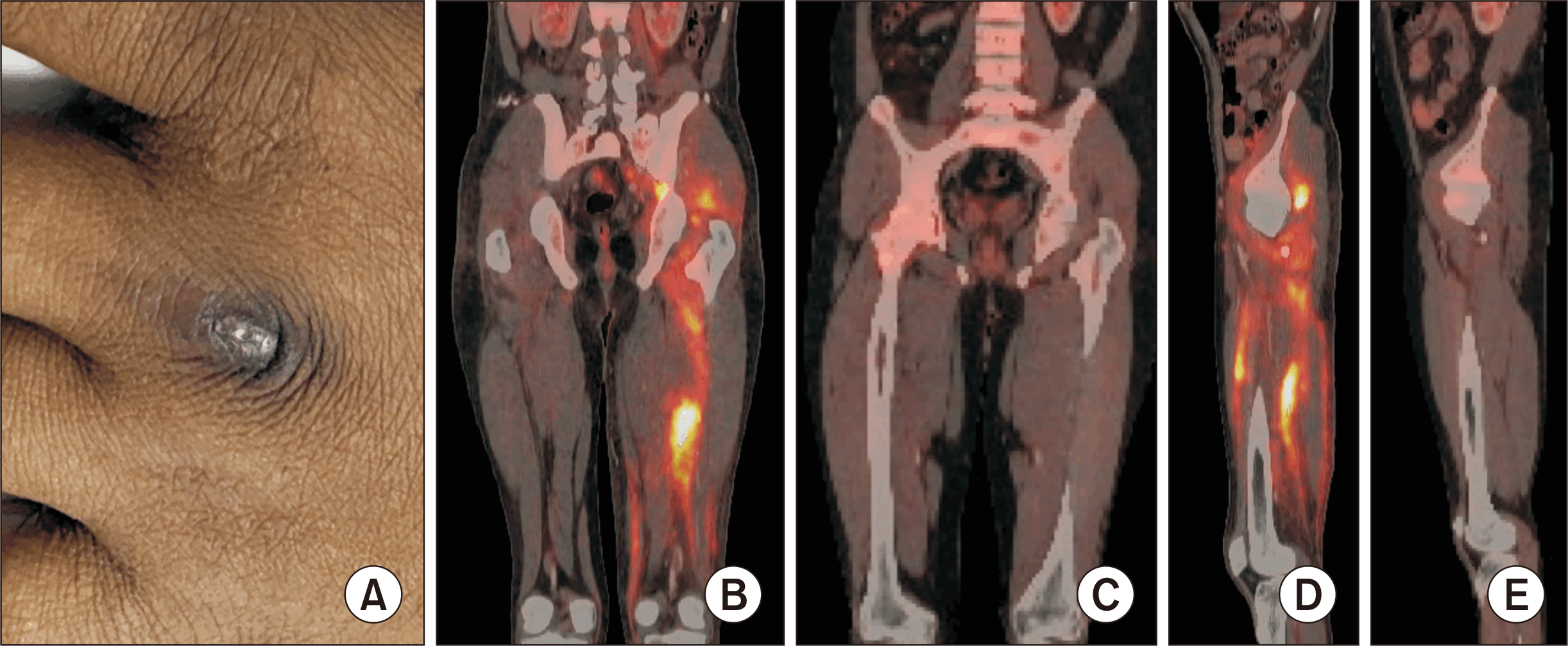
A 27-year-old male presented with an 18-month history of arthralgia, skin rashes, muscle weakness, fever, and a 10-kg weight loss. He had initially been treated for dermatomyositis with corticosteroids, HCQ, and MTX, but showed minimal improvement. On examination, there was a malar rash, Gottrón-like lesions (Figure 1A), and proximal muscle weakness (Manual muscle testing-8 score: 131/150). PET/CT indicated generalized lymphadenopathy, and FDG uptake was noted in multiple lower limb muscles (Figure 1), and bilateral lung consolidation.
Gluteus medius muscle biopsy demonstrated necrotizing granulomatous inflammation, and GeneXpert MTB testing was positive. The patient was treated with a 9-month course of ATT along with tapering corticosteroids, which resulted in complete clinical and radiological resolution. There was no relapse during a 3-year follow-up period.
2) Case 2
A 56-year-old female presented with a 2-month history of symmetrical proximal muscle pain, weakness, and panniculitis lesions. She was initially diagnosed with inflammatory myositis based on elevated CPK levels, endomysial inflammation in muscle biopsy, and a partial response to corticosteroids.
Her symptoms worsened after 6 weeks. MRI revealed bulky thigh muscles with T1 and T2 hyperintensities. A repeat muscle biopsy confirmed myonecrosis with inflammation, and GeneXpert MTB testing was positive. She was treated with IVIg (2 g/kg), a 9-month course of ATT, and corticosteroids, followed by MMF (1.5 g/day) for persistent myositis. Follow-up at 5 years of follow-up showed that she was stable on MMF.
3) Case 3
A 60-year-old female presented with a 15-day history of arthralgia and myalgia. Examination revealed muscle tenderness, weakness, and arthritis of the ankles, knees, and feet. PET/CT demonstrated generalized necrotic lymphadenopathy, bilateral lung consolidations, generalised FDG-avid muscle uptake, a subperiosteal abscess over the left lateral malleolus, and osteomyelitis of the right second metatarsus.
Lymph node biopsy showed necrotizing granulomatous inflammation, and GeneXpert MTB testing was positive. She was treated with a 9-month course of ATT, resulting in complete resolution of symptoms.
4) Case 4
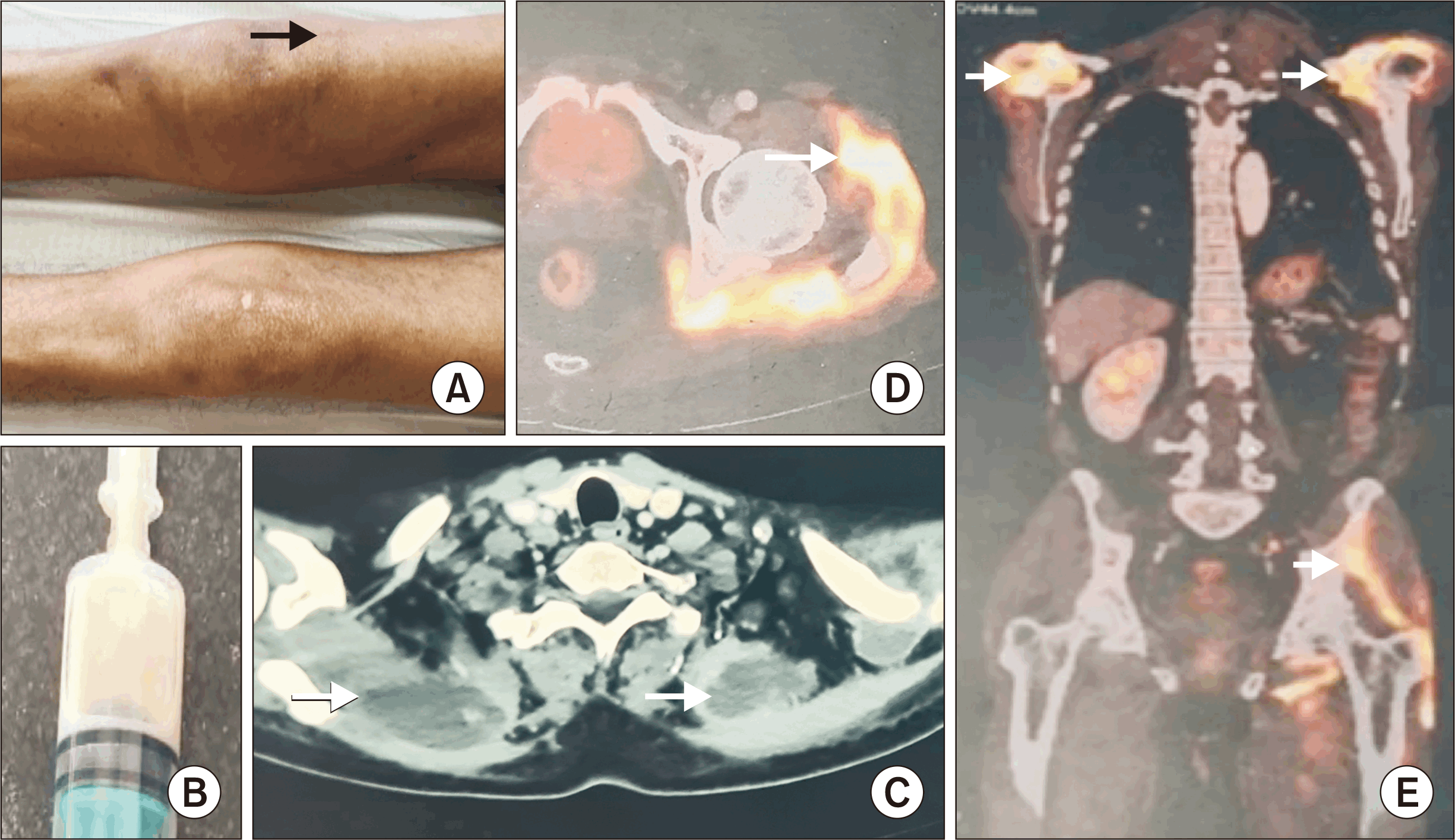
A 61-year-old male presented with a 1-year history of inflammatory polyarthritis, diffuse myalgia, weight loss (11 kg), and bed-bound status. Electromyography demonstrated myopathic changes. PET/CT indicated multifocal intramuscular FDG uptake, a gluteal collection (Figure 2), generalized lymphadenopathy, and polyarthritis. Intramuscular aspiration tested positive for MTB on GeneXpert. He was treated with a 6-month course of ATT, which led to resolution of muscle symptoms and stabilisation of weight. SSZ was initiated for persistent arthritis.
5) Case 5
A 29-year-old male presented with a 2-year history of lower back, gluteal, and knee pain, fever, and weight loss (15 kg). He had previously been treated for inflammatory arthritis with steroids and SSZ. PET/CT revealed multiple FDG-avid intermuscular collections and necrotic para-aortic lymphadenopathy. Gluteal abscess aspiration tested positive on GeneXpert MTB. A 9-month course of ATT contributed to his complete recovery.
6) Case 6
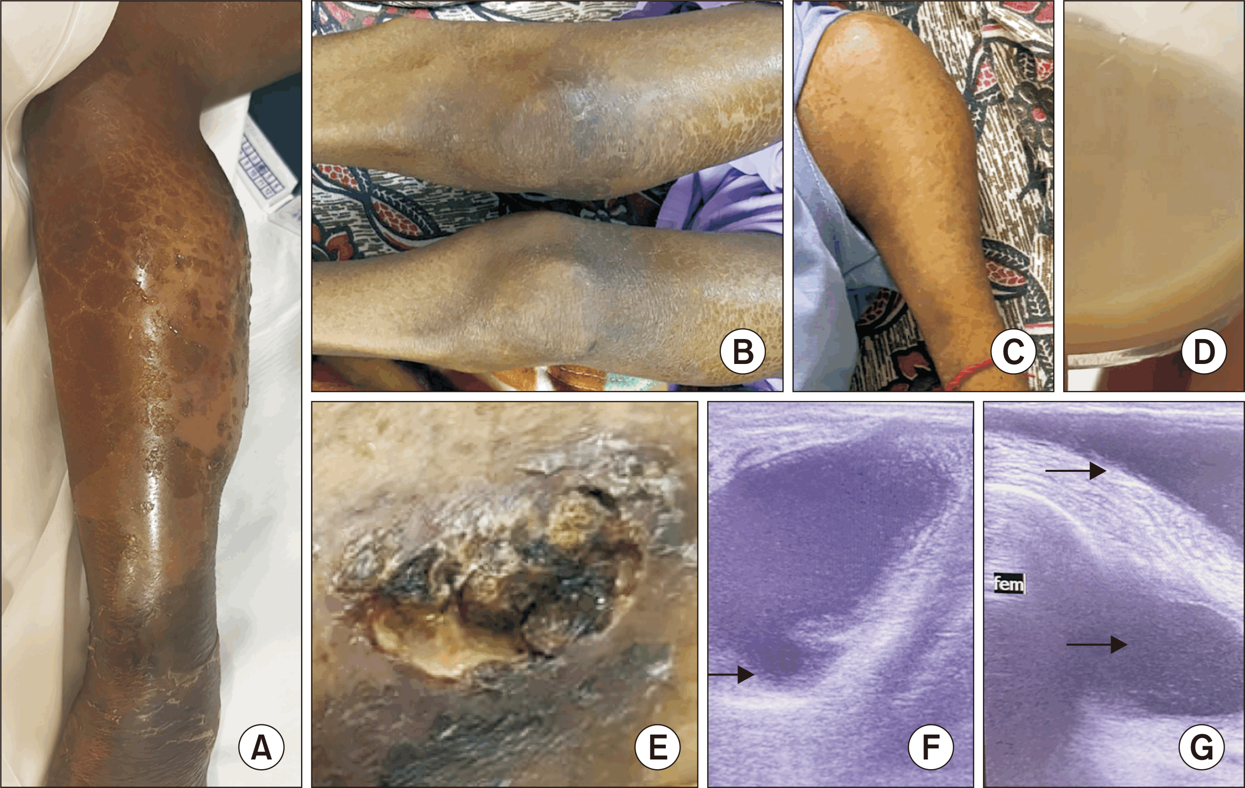
A 21-year-old female presented with a 1-year history of fever, weight loss (15 kg), myalgia, weakness, and bed-bound status. Examination revealed generalized lymphadenopathy, scrofuloderma in the inguinal region, muscle wasting, and multiple tender limb swellings. Ultrasound demonstrated multiple intramuscular and subcutaneous abscesses (Figure 3). Laboratory findings showed severe anaemia (haemoglobin 5 g/dL), Raynaud’s phenomenon, and positivity for multiple autoantibodies. Intramuscular aspirate tested positive on GeneXpert MTB, AFB staining and TB culture.
She was treated with ATT for 9 months, oral prednisolone (20 mg) for probable TB-associated immune reconstitution inflammatory syndrome, HCQ 5 mg/kg for the underlying rheumatological disease, abscess drainage, and one dose of intralesional streptomycin injection into two major abscesses. After 6 months of treatment, she demonstrated symptomatic improvement and a resolution of abscesses and muscle inflammation.
DISCUSSION
Unlike the classical presentations of TB, the varied clinical patterns noted in tuberculous myositis indicate diverse underlying mechanisms of bacillary spread [5]. These manifestations often mimic autoimmune rheumatic disorders, causing diagnostic delays, inappropriate use of immunosuppressive therapy, and associated complications.
The present series demonstrated equal sex distribution and a broad age range among affected individuals, findings that align with previously reported case series [3].
Pathogenesis
Skeletal muscle is generally considered as an unfavorable site for MTB survival and proliferation, and this could explain the rarity of its involvement. High lactic acid content, absence of lymphatic and reticuloendothelial tissue, a highly differentiated cellular state, and enriched vascular supply are reported as the proposed protective factors [6]. However, these natural defenses may be compromised by trauma, prior local inflammation, immunosuppressive states, or systemic comorbidities such as HIV/AIDS and malnutrition [7].
Disease or medication-related immunosuppression is a major risk factor and may account for the multifocal pattern and increased morbidity seen in some patients [3]. Autoimmune diseases such as systemic lupus erythematosus (SLE) and inflammatory myositis further predispose to tuberculous myositis and the probable risk factors include inflammation-induced microtrauma, immunomodulatory therapy, and masking of constitutional features. Prior studies have reported prevalence of underlying SLE in 2.8%~8.2% and inflammatory myositis in 20% of patients with tuberculous myositis [3,7]. The absolute incidence remains low, with only one case of tuberculous pyomyositis (0.6%) among 167 inflammatory myositis patients followed for 4 years. Gupta et al. [8] reported a higher risk for active TB in idiopathic inflammatory myositis (IIM) compared with SLE (68-fold vs. 30.4-fold), especially among patients receiving immunosuppressive therapy.
Clinical stages and mimics
The progression of infectious myositis occurs through three major stages, namely invasive, suppurative, and late [9]. In TB, an additional pre-suppurative inflammatory phase may occur, particularly in multifocal or hematogenous spread. This stage frequently mimics IIM. The wider adoption of muscle biopsy has enhanced diagnostic precision and underscored the need to consider tuberculous myositis in the differential diagnosis of IIM. TB myositis, once described as a “polymyositis-like reaction” is now understood to involve direct bacillary invasion of muscle tissue rather than an autoimmune process. This distinction is further supported by microbiological confirmation of MTB on muscle samples [10].
Spread and distribution
The clinical phenotypes reflect the route of spread in tuberculous myositis. Contiguous extension from adjacent skeletal structures or lymph nodes is most common, whereas multifocal or bilateral involvement usually indicates hematogenous dissemination [3]. While axial muscles, particularly iliopsoas and chest wall, are classically implicated, more recent reports describe the predominant involvement of gluteal and thigh muscles, with occasional calf and upper limb disease [11,12].
Concomitant pulmonary TB, which is not universal, has been reported only in 45%~51% of previous series and 33% in the present cohort [3]. This highlights the limitation of relying solely on chest imaging for screening.
Clinical features
Clinical manifestations of tuberculous myositis are heterogeneous, ranging from focal or disseminated pyomyositis to multifocal inflammatory edema without suppuration. In a review of 30 cases by Zhu et al. [13], pain was the most frequently reported feature (67%), followed by swelling (63%) and nodular masses (23%). Abscesses are reported in 45%~80% of cases, though not universally noted. Compared with bacterial pyomyositis, tuberculous myositis is characterized by a more indolent onset, and constitutional symptoms such as fever and weight loss are inconsistently reported (23%~50%) [7,14].
The findings in our series are consistent with earlier reports, with predominant local pain, swelling, and palpable nodules, and less pronounced muscle weakness. This relative preservation of strength may serve as a distinguishing feature for tuberculous myositis from inflammatory myopathies. Erythema of the overlying skin is also observed [15].
A comparison of clinical features of our cohort with a series by Wang et al. [3] and Ahmad et al. [16] is described in Table 2. The cohort described by Wang et al. [3] included 35 patients of muscle culture or histologically proven muscle disease by MTB, supplemented by CT/MRI, both unifocal and multifocal. Five patients in their cohort were noted to have multifocal muscle involvement. The difference noted was a smaller mean disease duration i.e., 2 months compared to 13 months in our series, a distinct clinical pattern of muscle involvement, with involvement of chest wall muscles, which was not seen in our series, a higher rate of surgical intervention (82.9% vs. 16.7%), and a higher mortality rate (14.3% vs. 0%).
Ahmad et al. [16] included 14 patients in their series of tuberculous myositis, all 14 were unifocal, showed lower rates of underlying immunosuppression/comorbidities compared to our series (14.3% vs. 50.0%) and showed a more distal limb distribution (50.0%) compared to our series (16.7%).
A study on MRI findings of 65 patients of TB pyomyositis demonstrated psoas muscle to be the most commonly involved in 60%, paravertebral muscles in 52%, pelvic iliacus muscles in 39%, and gluteal in 29%. Multiple muscles were involved in 97% of patients, and abscesses in 60% of patients, compared to 50% in our series. Underlying comorbidities or immunosuppression was seen in 40% in their study vs. 67% in our series. Spinal involvement was more common in their cohort (76%) vs. 17% in our series [17].
Diagnosis
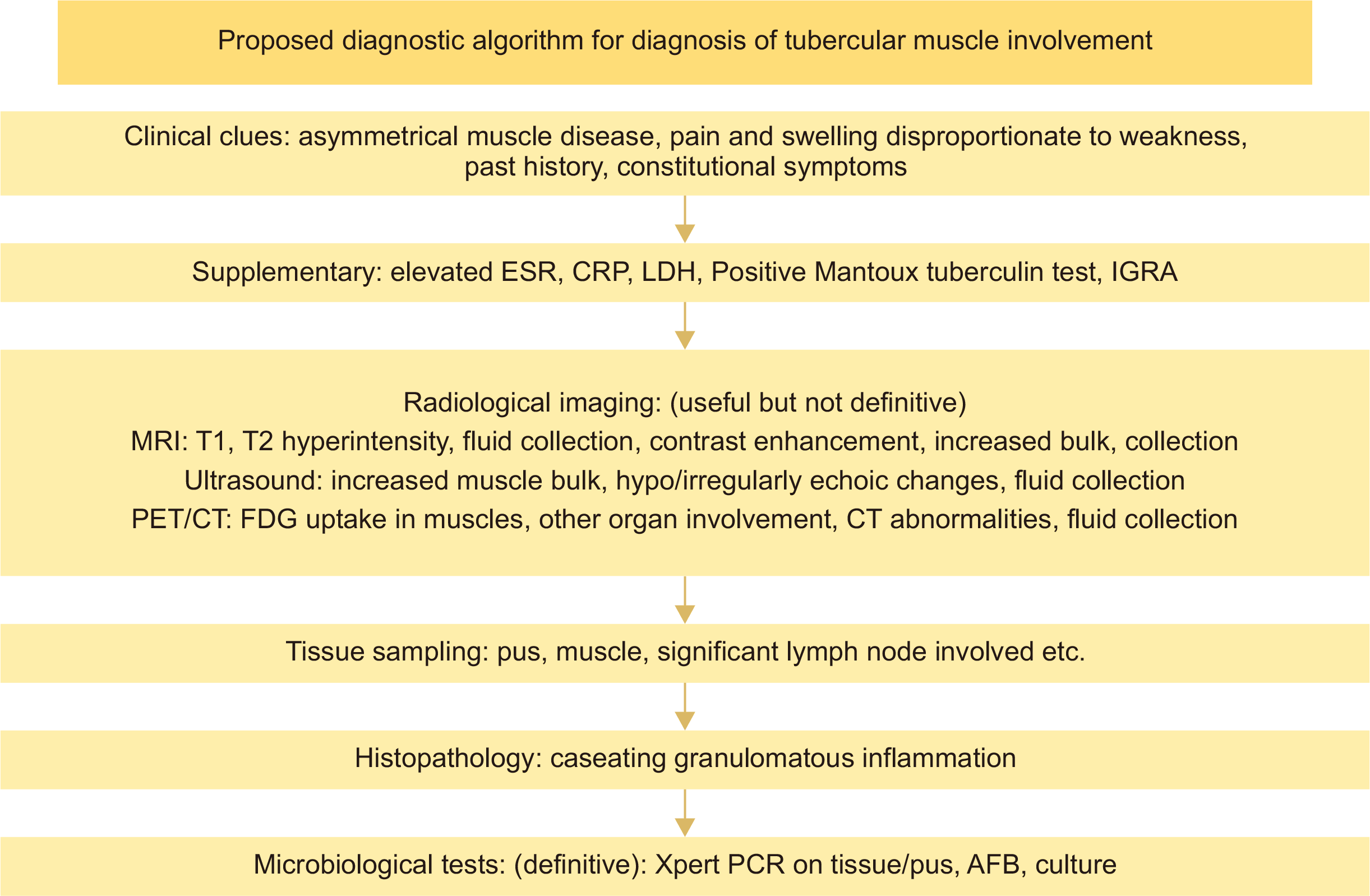
As with TB of other organs, the diagnostic cornerstone of muscular TB is tissue evidence, supported by imaging, biochemical markers, and tests for latent TB (Figure 4). Muscle biopsy or aspiration is warranted in cases lacking evidence of systemic TB. Although culture is the gold standard, its limited sensitivity and delayed results restrict its utility. PCR-based methods such as GeneXpert offer higher yield and rapid results from both tissue and pus, and they are superior to the AFB smear.
Comparison of the present case series with a meta-analysis by Zhu et al. [13] demonstrated that both cohorts showed relatively high detection rates with GeneXpert MTB from muscle biopsy or pus (83% vs. 89%). A substantial proportion of patients in both series showed tuberculin test/ interferon gamma release assay positivity (50% vs. 69%). AFB and TB culture on muscle biopsy/pus rates were 33% vs. 41% and 50% vs. 74% respectively in the two cohorts. These findings underscore the significance of combining microbiological methods for confirming muscular TB, emphasizing on the usage of GeneXpert MTB on all suspicious tissue/pus samples, especially in TB-endemic countries.
Caseating granulomas are typically noted in histopathology. In the current case series, myositis-specific autoantibodies were negative, while myositis-associated antibodies were occasionally positive in high titers, highlighting the need for cautious interpretation in patients with suspected IIM.
MRI, marked by features such as multifocal involvement, bulk enlargement, T2 hyperintensity, and occasional abscesses, is recognized as the radiological modality of choice [17]. However, these findings can overlap with those of IIM. Specific MRI features can help differentiate tuberculous from inflammatory myositis, such as a T2 hypointense abscess rim, focal edema, and possible asymmetry in TB myositis. The MRI findings of the 2 are compared in Table 3.
A study on MRI findings of 136 patients of pyomyositis, including 65 patients of tuberculous pyomyositis showed enlarged muscle contour in 80%, isointense muscle signals on T1 weighted imaging in 88% and hyperintense signals on T2 weighted images in 100% without any significant difference between tubercular and bacterial pyomyositis. Adjacent spinal and psoas muscle involvement was significantly more common in TB pyomyositis than in bacterial pyomyositis [17].
CT and ultrasound findings are supportive, particularly for guiding tissue sampling. PET/CT may demonstrate FDG uptake patterns suggestive of TB; however, in the absence of suppuration, such findings are nonspecific [18].
Treatment and outcomes
Standard musculoskeletal TB regimens are recommended, typically with isoniazid, rifampicin, pyrazinamide, and ethambutol for 9~12 months [19]. Immunocompromised states or multifocal disease may warrant extended regimens. Surgical intervention is generally limited to cases with diagnostic uncertainty, abscess drainage, or compressive symptoms. Alternative approaches such as percutaneous aspiration with intracavitary streptomycin have also been described [20,21].
In the present cohort, four patients achieved complete remission with ATT, while two required long-term immunosuppression due to underlying autoimmune disease. No deaths were reported. These outcomes are in line with prior reports showing response rates of 84%~92% [11,13], where adverse outcomes were more frequent in the context of corticosteroid use, high bacillary load, multifocal involvement, or disseminated disease [3].
Strengths, limitations, and future implications
The present case series holds significant relevance in the Indian context, where TB remains highly prevalent and presents with diverse clinical manifestations. The diagnostic delay of more than one year noted in the present cohort reflects real-world challenges within Indian settings, where atypical presentations of TB are often overlooked. With the wider recognition of autoimmune diseases in India, the coexistence of TB and underlying rheumatologic conditions further complicates clinical decision-making, particularly when immunosuppressive therapy is initiated without considering TB as a differential diagnosis. By documenting these cases, the study provides valuable regional data to the limited global literature, highlights the need for clinical vigilance in TB-endemic regions, and underscores the importance of integrating NTEP guidelines into routine rheumatology and medical practice. However, the rarity of this condition and the small sample size limit the generalizability of the findings. Future multicenter studies are warranted to better define risk factors, imaging correlates, and treatment outcomes.
CONCLUSION
Multifocal muscular TB is a rare but clinically important form of EPTB that can closely mimic autoimmune myopathies. In TB-endemic regions, it should be considered in the differential diagnosis of patients presenting with atypical or treatment-refractory myositis. Muscle inflammation without overt abscess formation may indicate an early phenotype of multifocal involvement. Early recognition, appropriate use of imaging and microbiological tools, and timely initiation of ATT are crucial to avoid misdiagnosis, inappropriate immunosuppression, and adverse outcomes.
SUPPLEMENTARY DATA
Supplementary data can be found with this article online at https://doi.org/10.4078/jrd.2025.0128
 XML Download