Journal List > J Rheum Dis > v.33(3) > 1516096075

Chaisrimaneepan, Khoruamklang, Sodsri, and Jakramonpreeya: Non-specific cutaneous lupus erythematosus and clinical significance: a literature review

Abstract

Cutaneous lupus erythematosus (CLE) is classified into LE-specific cutaneous lesions and LE-non-specific cutaneous lesions. LE-specific lesions, which include acute cutaneous LE, sub-acute cutaneous LE, and chronic cutaneous LE have been well-described and studied regarding their association with systemic lupus erythematosus (SLE) and disease activity. Non-specific CLE is divided into thrombogenic (vascular), neutrophilic, or uncertain pathogenesis. Some features such as Raynaud, vasculitis, and alopecia have been well-documented with reported association with other clinical features of SLE and its disease activity. Still, some are under-recognized and not well-studied for their correlation with SLE disease activity, or clinical significance concerning SLE. This review aims to encompass each non-specific CLE and its clinical relevance in SLE. However, the literature was limited in many non-specific LE conditions with the lack of statistical evidence to prove its correlation to SLE or other signs and symptoms of SLE, including morbidity and mortality. Future research can provide predictions of the disease based on non-specific CLE.

INTRODUCTION

Systemic lupus erythematosus (SLE) is an autoimmune disease with a wide variety of clinical presentations, ranging from limited disease to major organ involvement. Cutaneous manifestations are the second most common presentation in SLE patients, involving 70%~80% of the cases. Cutaneous LE represents either an exclusive cutaneous disease of lupus features or a component of SLE [1]. Gilliam and Sontheimer first proposed the classification of cutaneous LE cutaneous lesions into specific and non-specific lesions. The specific cutaneous LE lesions are well-established and defined by the presence of LE features, which include interface dermatitis, with or without SLE. They are divided into 3 categories based on morphology and clinical characteristics, not chronology–acute cutaneous LE (ACLE), sub-acute cutaneous LE (SCLE), and chronic cutaneous LE (CCLE). However, the classification contains some limitations [2]. A new classification was later proposed by Lipsker, based on clinical characteristics and histopathological findings, dermo-epidermal, dermal, or hypodermic involvement regardless of interface dermatitis [3]. The non-specific cutaneous lesions comprise any other cutaneous manifestations associated with LE and may be present in other conditions. They are divided into vascular (thrombogenic), neutrophilic, or uncertain pathogenesis [3], modified and summarized in Table 1.
Specific cutaneous LE is known to be associated with SLE and SLE activity at different degrees. In contrast, the relationship between non-specific cutaneous LE and SLE is vague. This review aims to explore non-specific LE and its clinical significance in SLE.
For this type of study, ethics approval is not required. Written consents obtained for learning and publication were obtained from patients whose figures were demonstrated in this manuscript without identifiable details of the patients.

VASCULAR (THROMBOGENIC)

Vasculitis

Cutaneous vasculitis (CV) is not uncommon in SLE, reported to be approximately 30%~70% [4,5]. It may be presented as palpable purpura, punctate lesions, urticaria-like, ulcers, papules erythematous macules/plaques, or necrosis, depending on the types of vessels involved.
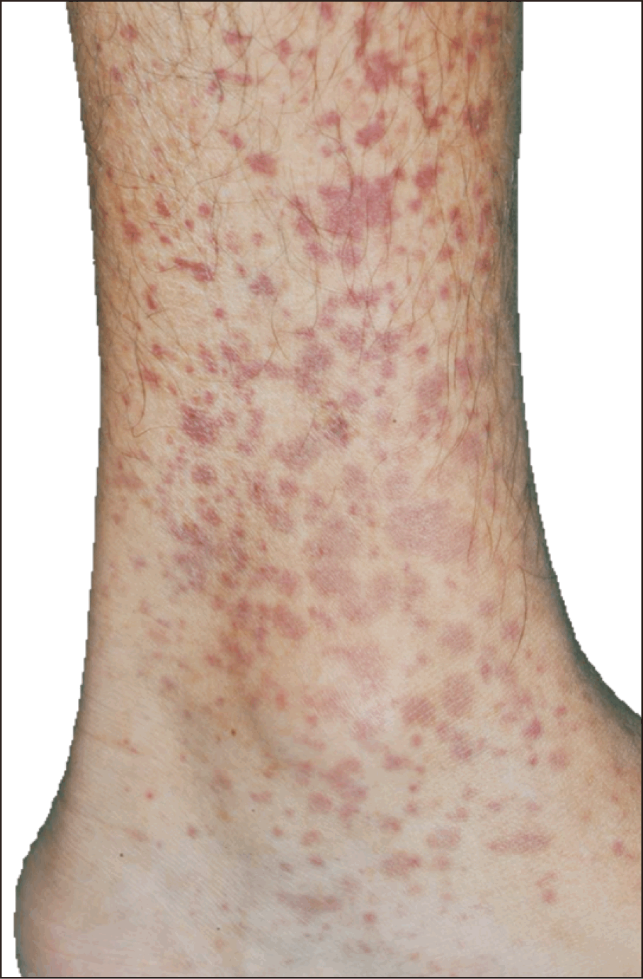
Palpable purpura (Figure 1), a form of leukocytoclastic vasculitis (LCV), is the most frequently observed manifestation of small vessel vasculitis (SVV) [6]. It is characterized by raised hemorrhagic papules or plaques with diameters up to several centimeters [7]. The lesion is most found on the lower legs and buttocks. LCV is caused by immune-complex deposition in small vessels, particularly cutaneous dermal capillaries and venules. This leads to complement activation, neutrophil recruitment, and subsequently vascular injury. Histopathology reveals neutrophil infiltration, leukocytoclasia or the disintegration of neutrophil nuclei, fibrinoid necrosis, and vascular damage [8]. Palpable purpura, as a presentation of CV, is found in 25% of SLE patients with vasculitis [9]. To our knowledge, there is no known study about the association between palpable purpura itself and SLE disease activity. However, studies show a higher SLE Disease Activity Index (SLEDAI) and SLICC/ACR (Systemic Lupus International Collaborating Clinics/American College of Rheumatology) damage index in SLE patients with CV compared to those without [10,11].
Race and genetic factors play a role in the occurrence of CV; the highest prevalence is seen in Hispanic and African-American [11]. Asian descents with CV-associated SLE were at increased risk of renal and neuropsychiatric involvement [10]. Multiple observational studies have described the correlation of the presence of CV with other systemic clinical manifestations or antibodies.
Musculoskeletal manifestations and lupus nephritis (LN) are increased in CV patients [12,13]. Antiphospholipid syndrome (APS) also appears to occur more frequently in patients with CV but with no significant difference [4]. The first cutaneous manifestation of CV has been reported to develop APS subsequently after SLE diagnosis [14]. Notably, hypocomplementemia was associated with CV compared to non-CV SLE patients and high disease activity [4]. Cryoglobulinemic vasculitis, which results in palpable purpura, can be associated with SLE, other than SVV from SLE itself [9].
In one study, CV in juvenile SLE (jSLE) is associated with more seizures and granular cast while adult SLE (aSLE) is associated with Raynaud’s phenomenon (RP), musculoskeletal manifestations, myositis, or no major organ involvement [4,10,15]. However, a study by Kallas et al. [11] demonstrated that the presence of CV is associated with an increased risk of organ damage. Myositis, RP, hematological manifestation, discoid lupus erythematosus (DLE), anti-Smith, and anti-ribonucleoprotein (RNP) were predictors of the development of later cutaneous SVV [11]. Anti-Ro antibody is significantly associated with CV in SLE [16]. In general, CV is one of the markers of high SLE disease activity. It is included in many scores, including the SELENA-SLEDAI, the SELENA Flare Index, the European Consensus Lupus Activity Measurement, and the British Isles Lupus Assessment Group [17-19]. The earlier the vasculitis is treated, the better the prognosis of SLE [20].

Urticarial vasculitis

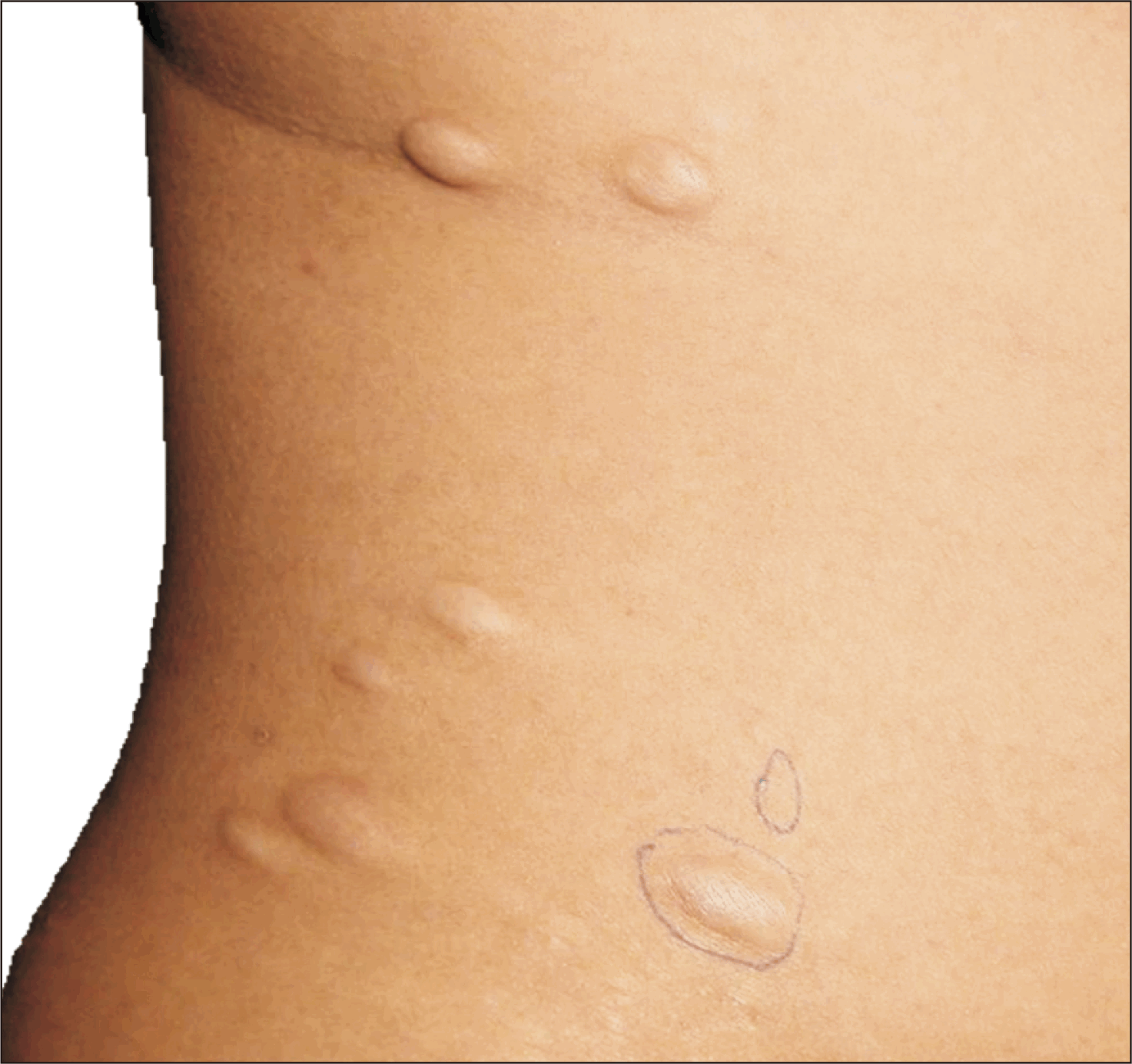
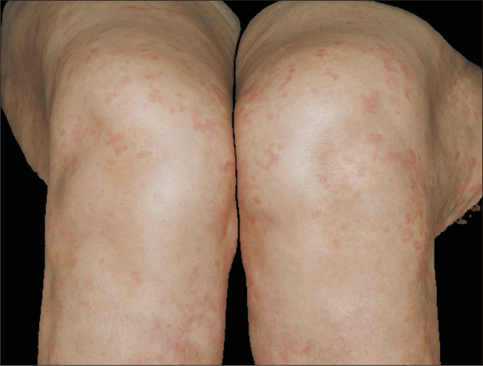
Urticarial vasculitis (UV), a variant of cutaneous SVV, is characterized by recurrent urticaria, an eruption of erythematous wheels (Figure 2), and evidence of LCV in biopsy. UV is commonly painful, nonpruritic, and persists for more than 24 hours, which helps distinguish it from urticaria. The lesion usually resolves with post-inflammatory hyperpigmentation or purpura [21]. There are two main subtypes of UV: normo-complementemic UV group and hypo-complementemic urticarial vasculitis (HUV) [22]. Some studies extrapolated that HUV is associated with SLE due to their clinical and immunological resemblance. C1q antibodies act as a diagnostic marker of HUV and are also found in about 30% of SLE patients [23]. The prevalence of HUV in SLE patients is about 7%~8%. HUV could be a presenting symptom of SLE or a flare-up of SLE, and 54% of HUV cases are diagnosed with SLE during follow-up, leading to the conclusion that HUV is a subtype of SLE in some studies [24]. Anti-double-stranded DNA (anti-dsDNA) antibodies and rheumatoid factor (RF) were more frequently detected in UV with SLE patients than in non-SLE patients [25].
However, there is still no specific study for UV regarding its relationship with SLE disease activity due to the limited number of cases to perform statistical analysis [26].

VASCULOPATHY

Livedoid vasculopathy

Livedoid vasculopathy (LV) is characterized by painful focal petechiae or purpura and progresses to recurrent leg ulcers (Figure 3). In SLE, similar lesions can be observed on the upper extremities and were associated with other organ involvement [27]. Hypercoagulability and autoimmune processes attributed to the pathogenesis of LV, including SLE with or without the presence of APS [28]. Histopathological findings demonstrate segmental hyalinization, intravascular fibrin, and endothelial proliferation, denoting thrombotic pathogenesis without features of true vasculitis [29,30].
Unlike vasculitis, studies indicating strong correlation of LV with SLE are lacking. A study by Yasue [27] observed that 16.7% of SLE patients develop LV. Among these, 90.9% eventually developed central nervous system (CNS) lupus, and 11.5% developed LN. With CNS or renal involvement, corticosteroids were used in such cases.
No treatment guidelines are available for LV. Hydroxychloroquine (HCQ), steroids, methotrexate (MTX), intravenous immunoglobulin (IVIg), and antiplatelet (ASA)/anticoagulation have been used in LV associated with SLE [31-33].

Livedo reticularis

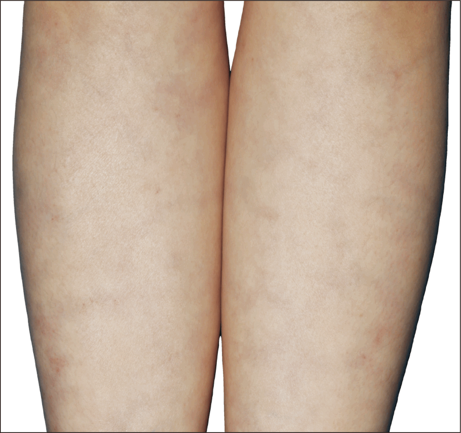
Livedo reticularis (LR) is a distinctive cutaneous manifestation characterized by a violaceous, net-like reticulated pattern, affecting the lower extremities, buttocks, and arms (Figure 4). It results from altered blood flow dynamics within the dermal microvasculature, leading to deoxygenation and subsequent discoloration of venous blood [34,35]. LR in SLE may be a clinical marker of underlying vascular pathology, immune-mediated endothelial dysfunction, and increased thrombotic risk in these conditions [36,37].
The development of LR is closely linked to the microvascular anatomy of the skin. Histological findings reveal varying degrees of perivascular inflammatory infiltrates, endothelial cell swelling, and fibrin thrombi within small vessels, particularly in APS-associated cases. Notably, in cases of deep-seated thrombogenic vasculopathy, skin biopsies may show only superficial telangiectasia unless deeper vascular pathology is specifically sampled [38,39].
LR is frequently observed in patients with SLE and APS, with a prevalence ranging from 11.7% to 25.5% in different cohorts. It is more commonly associated with systemic disease involvement (56.5% vs. 24.7%, p=0.007) and may indicate an increased risk of thrombotic complications [2]. Among the different subtypes of LE, LR is more frequently seen in ACLE (9.9%) compared to CCLE (2.8%) or intermittent cutaneous lupus erythematosus (1.5%). It is also significantly correlated with anti-phospholipid antibodies (aPL), including anticardiolipin (aCL) and lupus anticoagulant [40,41]. These autoantibodies are associated with hypercoagulability, subjected to arterial and venous thrombotic risk. Additionally, LR is more prevalent in SLE patients with RP, photosensitivity, and diffuse alopecia, suggesting a broader immunopathogenic interplay [41,42].
The presence of LR in patients with SLE warrants thorough clinical evaluation as it may serve as an early indicator of systemic vascular involvement. Several key clinical implications include increased SLE disease activity, particularly in cases with concurrent vasculitis or APS, prompt screening for aPL to assess thrombotic risk in SLE patients, and monitoring for ongoing vascular damage, immune-mediated endothelial dysfunction, and susceptibility to ischemic complications.

Raynaud’s phenomenon

RP is one of the most common vascular manifestations in SLE. It is characterized by the discoloration of fingers and/or toes with cold exposure in sequence of blanching, cyanosis and erythema, as a result of reversible vasospasm. RP is classified into primary and secondary in accordance to the absence or presence of an associated disease, especially connective tissue disease like SLE, systemic sclerosis (SSc) or Sjogren syndrome [43]. The incidence was estimated to be 10%~45% in both aSLE and jSLE [44,45]. The presence of RP is higher in positive SSc-specific autoantibody SLE patients, especially anti-centromere [46].
RP in SLE was more frequently observed among anti-phosphatidylserine (aPS) immunoglobulin M (IgM) positive patients [47,48] while a study by Caccavo et al. [49] demonstrated no association between RP and aPS and aPL. A higher prevalence of aCL IgG was also found in RP with SLE [49,50]. Anti-RNP, anti-Sm antibodies, and RF were reportedly related to the development of pulmonary hypertension (PH) in SLE patients with RP [51-53]. However, an observational study did not report any significant association of RP and autoantibodies [54]. There was a higher frequency of positive dsDNA antibodies associated with RP in children with SLE [45].
RP has been reportedly associated with higher incidence of PH [55-57], peripheral neuropathy in SLE [44,58], and alopecia [44,59], but lower glomerulonephritis [52,60,61] and hemolytic anemia [62]. The occurrence of PH in SLE was postulated to be due to vasospasm in pulmonary arteries as a result of endothelial dysfunction and the increased vasoconstrictors [63]. Although patients with RP have a higher risk of PH, they have a more favorable outcome [64]. Dimant et al. [52], also found that patients with RP had significantly higher incidence of arthritis, malar rash and photosensitivity, and were less likely to have severe disease, consistent with few other studies [60,65,66]. Likewise, RP was less frequently observed in patients with unfavorable prognosis [67]. In jSLE, the presence of RP was not suggestive of less severe disease or better prognosis [68].
Symptoms of PH in SLE are non-specific. Therefore, RP can be an important clue to PH in SLE patients. The high prevalence of RP in SLE patients with PH supports the assumption that RP could be a marker for the disease activity [59,69].

Erythromelalgia

Erythromelalgia is an uncommon disorder recognized by the triad of erythema, burning pain and localized warmth on extremities. Secondary erythromelalgia, being less common than primary, is a unique presentation in SLE [70]. The exact incidence is unknown, but seemingly rare. Only case reports and case series have reported the coexistence of erythromelalgia and SLE. It can precede SLE as long as 4 years, present as the only clinical manifestation of SLE or occur following the diagnosis of SLE [71-74].
Erythromelalgia is clinically diagnosed and does not require laboratory confirmation; its presence in association with systemic symptoms should raise suspicion for SLE. The treatment goals are symptomatic relief and to prevent long-term complications such as ulcers, gangrene and infections which may develop secondarily to symptom-relieving behaviors like long duration clod water immersion. Non-pharmacological measures include avoiding heat, maintaining cool environment and limb elevation. Several pharmacological drugs have been used such as ASA, vasodilators, antidepressants, etc. [75,76].

Degos disease-like lesions

Degos disease or malignant atrophic papulosis is a vaso-occlusive disorder, characterized by porcelain-white centers and raised erythematous telangiectatic rims. It can be divided into two subtypes; the first involves the skin and internal organs (malignant form) and the second type (benign form), so-called Degos-like lesions, involves only skin and is associated with connective tissue diseases [77-79].
Only case reports and case series were documented. Whether to Degos disease-like lesions are strongly related to SLE or mere co-incidence remain elusive. Up to date, less than 15 cases of Degos-like lesions with SLE have been reported [80,81]. Degos-like lesions in SLE had reported internal organ involvement (renal and CNS) [82]. Cutaneous lesions of both types cannot be differentiated, suggesting a shared pathogenesis of vasculopathy [80].
CCLE associated with Degos-like lesions was reported in a case [83]. Endothelial proliferation, subintimal thickening, and fibrous tissue deposition between endothelium and intima can be seen histologically and are indistinguishable from classic Degos disease [84].
Given the suggestive microangiopathic occlusive pathogenesis, HCQ and ASA have been reported in the treatment of Degos-like disease in SLE. HCQ was found to have an anti-thrombogenic effect, inhibit platelet aggregation, and interfere with aPL production, which may be appropriate for aPL-positive lupus [83]. However, corticosteroids may worsen skin lesions [84].

Periungual telangiectasias

Periungual telangiectasias represent a significant microvascular abnormality observed in various autoimmune diseases, mainly SLE, SSc, and dermatomyositis [40]. They are characterized by small, tortuous, and dilated blood vessels measuring approximately 0.5 to 1 millimeter in diameter. They appear as erythematous or purplish capillary loops that become more prominent upon nail fold capillaroscopy, a non-invasive diagnostic imaging technique. They are significantly associated with SLE (p<0.001) [41].
The overall prevalence of SLE is approximately 2.4%~15% of cases [34,38,41,85]. The prevalence of periungual telangiectasias varies across different lupus subtypes, with notable differences in systemic involvement. The prevalence of periungual telangiectasias in ACLE, SCLE, and CCLE are 6.2%, 2.1%, and 1.8%, respectively [34]. The prevalence is increased in patients with active lupus manifestations, including CV and RP [34].
The underlying vascular pathology is endothelial cell dysfunction secondary to persistent immune activation and microvascular inflammation due to chronic exposure to pro-inflammatory cytokines leading to vascular dilation and architectural disorganization [86]. Autoantibodies targeting vascular structures, such as anti-U1 ribonucleoprotein (U1RNP) and anti-dsDNA antibodies, contribute to immune complex deposition [35,41]. The observed capillary dropout and neoangiogenesis reflect progressive vascular damage, correlating with disease severity.
Periungual telangiectasias is not just a diagnostic finding but a significant marker with necessary diagnostic and prognostic implications in LE and SLE. These lesions were identified in 76% of patients with SLE and concurrent DLE, while no cases were observed in patients with exclusive DLE [2,42]. These firm evidenced statistical analysis from observational studies indicated that it provides crucial insights into active and systemic disease involvement, ongoing vascular injury, and immune-mediated endothelial dysfunction, particularly in moderate to severe SLE cases. Periungual telangiectasias also serve as an essential clinical marker of systemic involvement and disease activity.
They were more frequently observed in patients with significant internal organ involvement (80.0% vs. 24.5%) [41] and strongly linked to Raynaud’s phenomenon, reflecting underlying microvascular dysfunction [35,87]. Their presence correlates with anti-U1RNP antibodies, which are indicative of systemic immune activation [35].
By reflecting chronic vascular inflammation, they serve as a warning sign to clinicians for the potential risk of microvascular ischemia and tissue damage. This awareness can prompt early intervention to improve patient outcomes, enhancing the audience’s vigilance and commitment to patient care.

Thrombophlebitis

Thrombophlebitis is rarely reported, in contrast to major thrombosis. It was reported in 3%~5% of the study population [88]. Two cases reported thrombophlebitis of cerebral vessel SLE patients resulting in multiple hemorrhagic infarcts, herniation, and death [89].
For all thrombotic or vasculopathy conditions, they serve as a warning sign to clinicians for the potential risk of microvascular ischemia and tissue damage. From our perspective, the association of thrombophlebitis with SLE may have been under reported. This awareness can prompt early intervention to improve patient outcomes, enhancing the audience’s vigilance and commitment to patient care.

Cutis laxa/anetoderma

Cutis laxa (CL) is a rare heterogenous dermatosis with several etiologies. It can be inherited or acquired, associated with preceding cutaneous inflammation (SLE, urticaria, amyloidosis) in adult-onset disease [90,91]. Acquired CL has two forms affecting dermal elastin. Type I can be localized or generalized with internal organ involvement while type II involves localized post-inflammatory elastolysis without internal organ involvement [92]. The immune-mediated mechanism was presumed to be the cause of CL in which elastase produced by activated neutrophils breaks down collagen resulting in elastolysis [93,94].
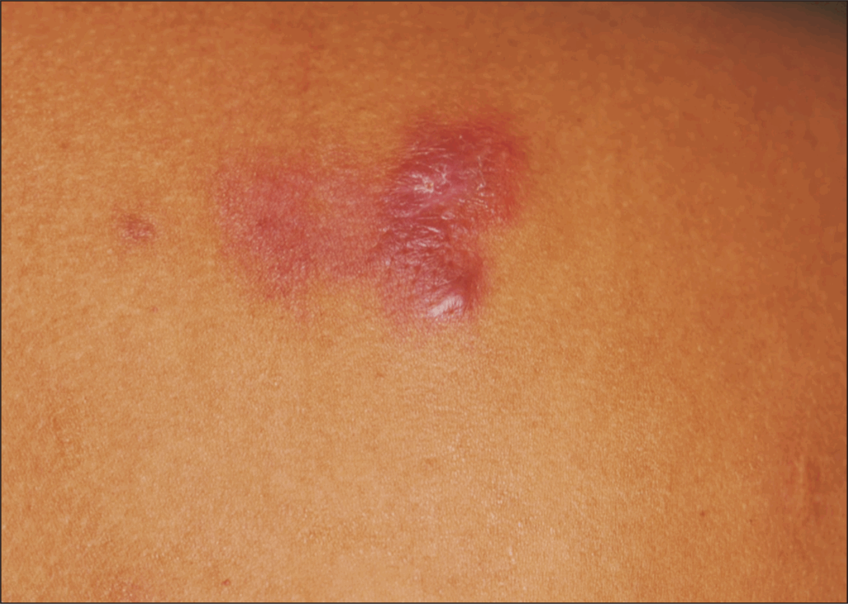
Anetoderma is characterized by slack skin due to focal loss of elastic tissue caused by infection or autoimmune processes (Figure 5) [95,96]. Co-occurrence of SLE and anetoderma has shown to be associated with aPL [97], which may also suggest that it could be a manifestation of APS. It was believed that dermal tissue ischemia may have triggered elastic tissue degeneration [98], which is evidenced by microthrombosis observed in some skin biopsies [96,99]. Due to the rarity of CL/anetoderma itself, currently, less than 15 cases of SLE and secondary anetoderma have been reported [95]. Among these, several patients have anetoderma and discoid LE [95] and lupus profundus concurrently [100]. Anetoderma can precede an autoimmune disorder for many years; however, the temporal relation with SLE is unknown [101,102]. Further studies are required to determine whether there is a temporal correlation between these skin conditions and SLE as current evidence is based on case series and case reports only.

NEUTROPHILIC

SLE-associated neutrophilic dermatosis

Neutrophilic dermatosis (ND), a rare manifestation in SLE, refers to cutaneous disorders that are predominantly infiltrated with mature neutrophils in the dermis on histopathology with or without vasculitis [103,104]. The reported cases of SLE-associated ND include but are not limited to Sweet syndrome (Figure 6), pyoderma gangrenosum (PG), palisaded neutrophilic and granulomatous dermatitis (PNGD), neutrophilic eccrine hidradenitis, amicrobial pustulosis of the fold [105,106].
Non-bullous neutrophilic lupus has also been reported but is believed to be a variant of Sweet’s syndrome or the early form of bullous LE [107,108].
The skin lesions are typically erythematous papules and plaques. Important clues to the diagnosis of SLE-associated ND in some patients were the presence of interface dermatitis and dermal mucin [105,109]. A study by Lee et al. [110] showed that neutrophilic urticarial dermatosis (NUD) in SLE patients was not associated with systemic signs and SLE flaring, while 80% of Sweet-like neutrophilic dermatosis (SLND) experienced SLE flaring and 60% developed SLND as a presenting symptom of SLE. SLND can be distinguished from Sweet syndrome by distribution which is more predominant in sun-exposed areas and chronicity of the course. Comparatively, SLND lasts longer than NUD and tends to recur. These entities can also be referred to as neutrophilic cutaneous LE [106]. They are also associated with anti-Ro/SSA or anti-La/SSB [111].
Sweet syndrome was an initial presentation of lupus in 33% of the patients [105]. The pathogenesis of Sweet syndrome in SLE is unclear. It was believed to be a consequence of immunological abnormalities of SLE that lead to cytokine stimulation, resulting in neutrophilic activation and chemotaxis [112,113]. Morphologically, the lesions are erythematous plaques, papules, or nodules; sometimes they appear as pseudo vesicles or pseudo pustules. Neutrophilic infiltrates that are less dense in these lesions may be classified as ND of LE, alluding to Gleason et al. [108].
PG is an uncommon cutaneous manifestation of SLE with a limited number of cases reported. PG in the presence of SLE were majorly women with younger age compared to non-SLE; it may or may not be related to active SLE [114].
Overall treatment targets the underlying SLE as skin lesions respond to immunosuppressive therapy. Colchicine and dapsone have been used to treat LE-associated ND effectively [106]. In NUD, lesions may resolve spontaneously or be controlled with an antihistamine [109].

Non-specific bullous lesions

Non-specific bullous lesions associated with LE occur in approximately 5% of cases [115]. They can present with diverse morphologies. Acute, subacute, or chronic widespread non-scarring vesiculobullous lesions can range from tense to flaccid vesicles and bullae, often accompanied by erosions over erythematous or normal skin. They may also present as purpuric or hemorrhagic bullae, suggestive of an underlying vasculitic process [36]. Most often, they are non-pruritic with a predilection for the face, upper trunk, neck, supraclavicular regions, axillary folds, and sun-exposed areas. Coexistence with classic LE lesions, such as malar rash or discoid plaques can be found. Lesions typically resolve without scarring. Recurrence is rare but possible, with some patients experiencing disease flares or uncontrolled systemic lupus, reflecting the relapsing-remitting nature of SLE. Mucosal involvement occurs in 30%~40% of cases, particularly in the oral mucosa [116].
Unlike the lymphocytic inflammation seen in SLE-specific lesions, non-specific bullous lesions in LE exhibit subepidermal blistering with predominant neutrophilic infiltration in the upper dermis, particularly on the papillary tips and nuclear dust and fibrin deposits [117]. The underlying pathogenesis of non-specific bullous lesions in LE was presumably due to several pathogenic mechanisms. Firstly, the deposition of immune complexes targeting type VII collagen within the dermo-epidermal junction leads to inflammation and blister formation. Severe basal keratinocyte damage and vacuolar interface changes (interface dermatitis) result in subepidermal blistering. Secondly, small-vessel vasculitis, often observed in LE, causes dermal-epidermal separation and hemorrhagic bullae. Thirdly, external causes that may predispose patients to developing bullous lesions in LE; drug-induced bullous eruptions such as HCQ and nonsteroidal anti-inflammatory drugs and bullous eruptions induced by ultraviolet radiation, which may exacerbate cutaneous lesions in LE [116,117].
Recognizing non-specific bullous lesions in LE is crucial, as they may indicate systemic disease activity, vasculitis, or drug reactions. Dapsone is considered the first-line therapy. Systemic corticosteroids are recommended for vasculitic bullae. Immunosuppressive agents (e.g., mycophenolate mofetil, azathioprine) have been used with variable efficacy [118]. Discontinuation of culprit drugs is necessary in suspected drug-induced bullous reactions. Supportive wound care measures are required to prevent secondary infections.

OTHERS

Sclerodactyly

Sclerodactyly refers to the thickening and tightening of the skin of the fingers. It can be a manifestation in some autoimmune diseases like SLE and scleroderma. The prevalence of sclerodactyly in SLE ranges from 0.0%~3.3% according to various studies [34,37,41,119]. The pathogenesis linking scleroderma and SLE includes cytokine dysfunctions, loss of tolerance against nuclear antigens, and strong platelet activation system [120,121]. The presence of sclerodactyly in SLE may require the exclusion of overlapping syndrome or mixed connective tissue disease.
Histologic findings show inflammatory cell infiltration, abnormal vasculature, collagen deposition replacing subcutaneous fat and glands, and fibrosis [122]. Although the presence of nonspecific cutaneous lesions is generally associated with higher disease activity, there is no specific data on sclerodactyly [123].

Lichen planus

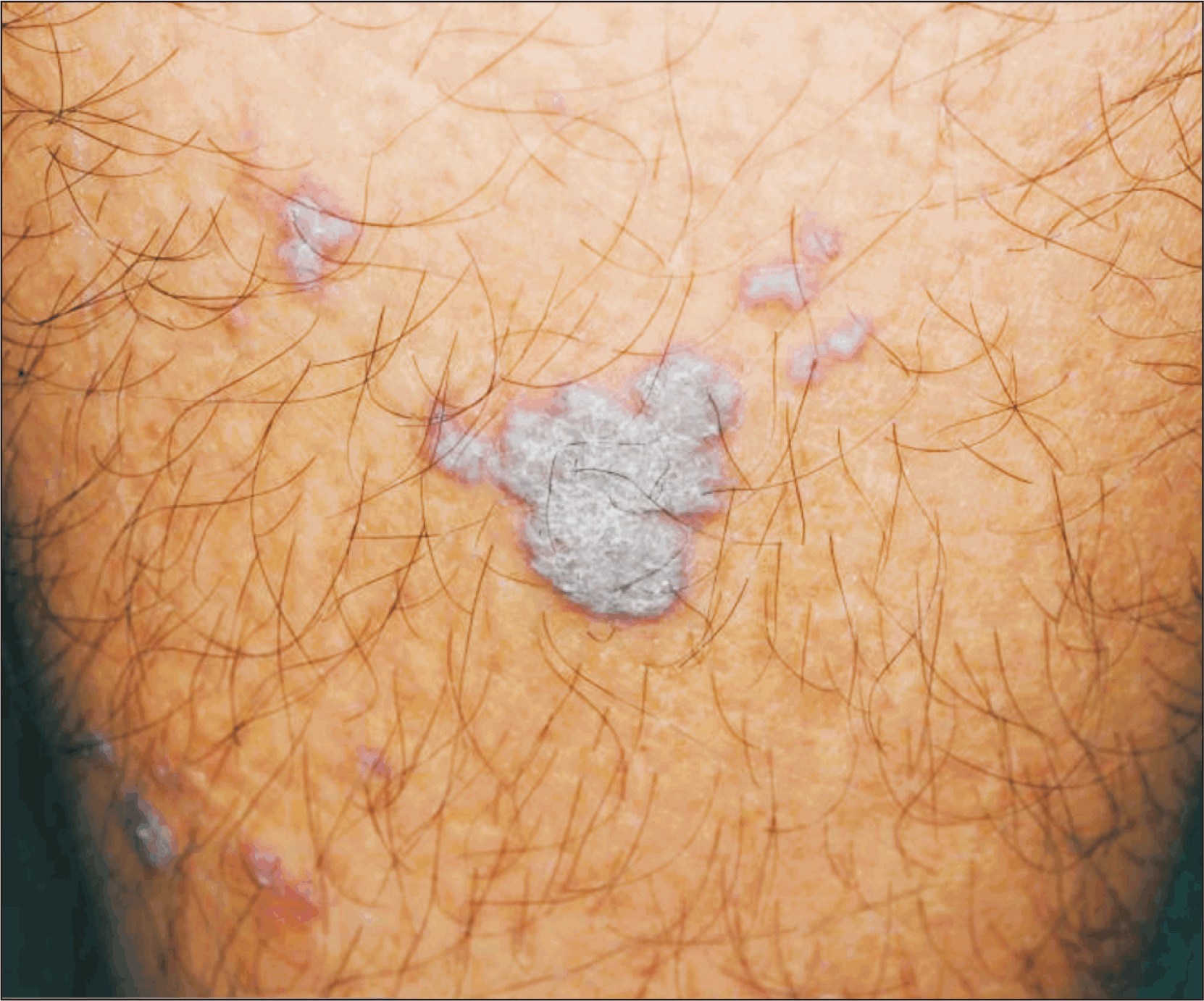
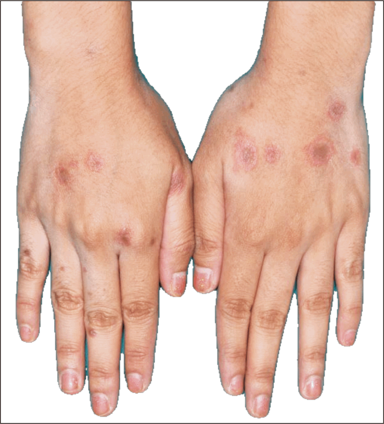
Lichen planus is a chronic inflammatory skin lesion found to present in association with LE [40]. An association known as LE-lichen planus overlap syndrome is rare and accounts for approximately 50 cases known to date [124-127]. The diagnosis is based on a combination of clinical, histopathological, and immunopathological features [128]. Although there is some controversy, true overlap syndrome is suggested to have both diseases in the same lesion [128]. Bluish-red plaques with scaly centers primarily on extremities and verrucous papulonodular lesions on hands and arms are the two most common presentations (Figure 7) [127]. Important histologic findings for lichen planus (LP) include the presence of band-like lymphocytic infiltration along the derma-epidermal junction and vacuolar degeneration in the basal layers [129,130]. Immunofluorescent microscopy may show clusters of cytoid bodies in the papillary dermis in contrast to linear arrangements in LE. Basement membrane clefts and linear fibrinogen bands also determine LP [127]. Lichen planus is caused by cell-mediated immune response [130]. Due to similar etiology, it is explained to coexist with LE [131]. Physicians need to raise suspicion for LP-LE overlap syndrome if the symptoms last for over a year or not improved with treatment [124]. Conversion to SLE is higher in overlap syndromes at 5%~10% of cases [132].

Papulonodular mucinosis

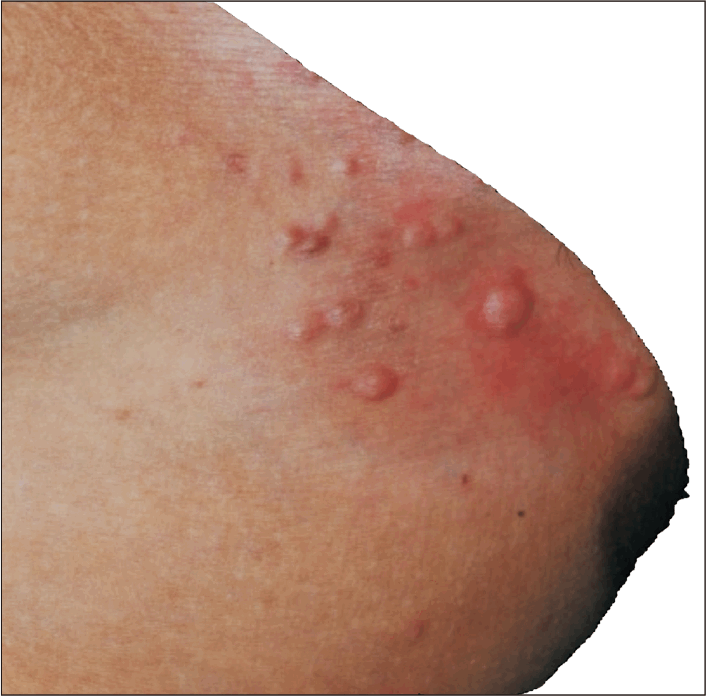
Papulonodular mucinosis (PNM), a subtype of cutaneous mucinosis, is presented as multiple asymptomatic raised flesh-colored papules and nodules located mainly on the trunk and upper body (Figure 8) [133]. The deposition of mucin and acid glycosaminoglycans in the dermis and epidermal change leads to a distinct dermatological presentation [134]. The etiology remains unclear, but some studies extrapolated that the elevated serum immunoglobulins, autoantibodies, and cytokines play a significant role in developing PNM by autoantibody-driven synthesis of glycosaminoglycans by cutaneous fibroblasts [135]. PNM is a rare non-specific cutaneous lesion in SLE with a 1%~2% prevalence. Interestingly, it is more prevalent in males with the male-to-female ratio of 18:13, in which sex-related factors, including androgenic hormone, might play a role in the pathogenesis of the PNM [136,137].
Not enough study available to determine the temporal relationship between PNM and SLE; in some cases, PNM was presented as an initial SLE presentation or wax and wane during the disease activity [138,139]. Approximately 80% of PNM cases were reported to be associated with joint and kidney involvement in SLE [138]. With the rarity of PNM, the studies regarding PNM and SLE were mainly case reports.

Acanthosis nigricans

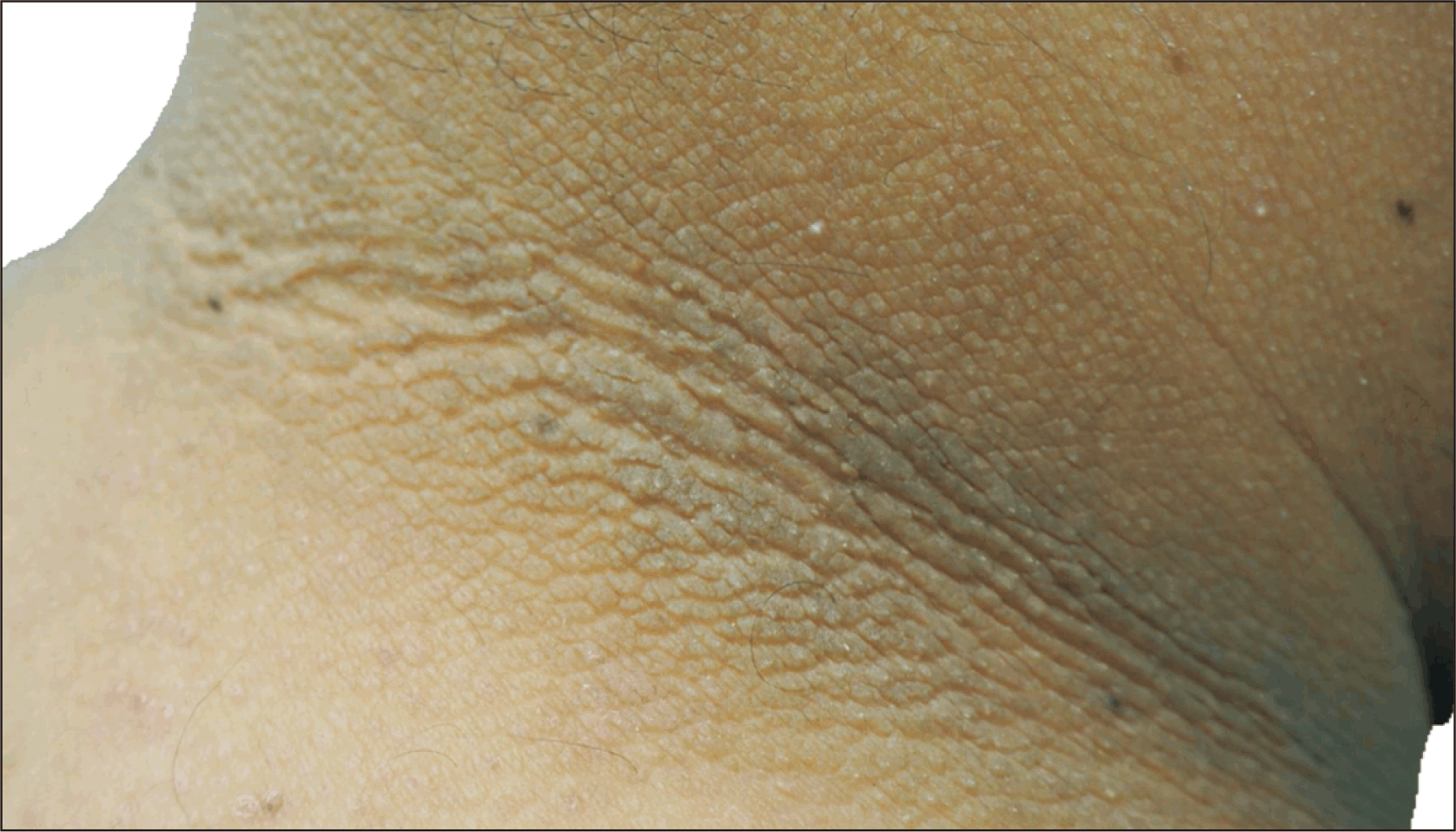
Acanthosis nigricans (AN) commonly presents as skin hyperpigmentation and thickening, mainly on the neck, axilla, and mucosal areas (Figure 9). Notably, AN was reported to be associated with metabolic disease and is well-known as a clinical marker of some malignancies. However, AN is also reported to be associated with many autoimmune disorders, including SLE [140]. Although it remains unclear, type B insulin resistance (TBIR) was believed to be the potential cause of AN in SLE, which is mostly positive for anti-insulin receptor antibodies (AIRA) [141]. SLE was found to be the most frequent autoimmune disease associated with TBIR syndrome in 35% of all cases [142]. SLE with AIRA was reported to be associated with high SLE disease activity and poor prognosis but mainly reported in case reports and not yet determined the correlation in larger cohorts or its mechanism [143].

Rheumatoid nodules

The incidence of subcutaneous nodules in SLE is 5%~7% [144]. Pathogenesis remains elusive; solid evidence other than case reports and case series are required to conclude whether rheumatoid nodules can occur in SLE alone or only in RA overlapping SLE. The nodules histologically resemble those of rheumatoid nodules (palisading granulomas) seen in rheumatoid arthritis (RA) [145]; however, the incidence of histologically proven rheumatoid granulomas in SLE is unknown [146]. The nodules are commonly present on the olecranon and dorsal aspect of the interphalangeal joints. The joint destruction in these patients was similar to RA, which may suggest a superimpose RA or lupus-related arthropathy [147]. The majority of the patients with subcutaneous nodules have persistent joint inflammation/Jaccoud arthritis [148,149]. HCQ has been effective in controlling the number, size, and development of the nodules [145].

Calcinosis cutis

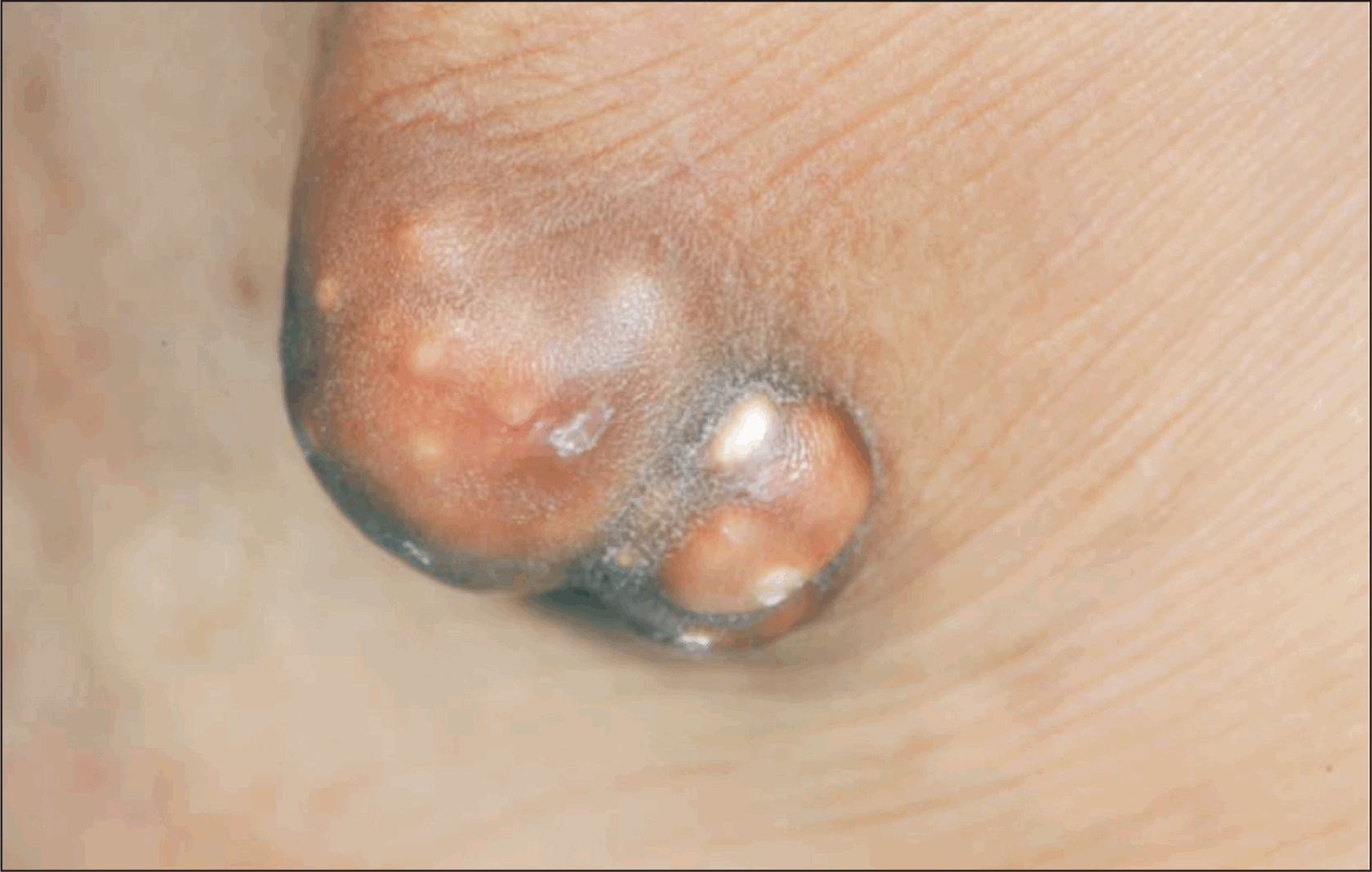
Calcinosis cutis (CC) is the deposition of calcium in previously damaged tissues regardless of calcium metabolism (Figure 10). It is less common in SLE (up to 17%) [150,151] and rarer with specific cutaneous LE compared to dermatomyositis and SSc [150]. Fewer than 40 case reports of dystrophic CC in SLE have been reports [152,153], and fewer than 5 cases of CC in pre-existing discoid LE [153,154].
CC is classified into dystrophic, metastatic, idiopathic, iatrogenic, and calciphylaxis dystrophic type is most commonly associated with SLE, typically occurs in women, in a localized pattern (circumscripta) and long-standing disease [151,154,155]. The calcification in SLE tends to be on the periauricular area skin or subcutaneous tissue underneath cutaneous lupus lesions, and buttocks [156,157]. However, some cases developed CC before the onset of SLE without any abnormal calcium metabolism [158].
CC universalis, on the other hand, has been reported in approximately 10 cases [151,152,159-162]. The mechanism of CC in SLE is not well understood but was presumed to be caused by local tissue damage [163]. It is believed that interleukin (IL)-6, IL-1, and tumor necrosis factor-alpha (TNF-α) may play a role in the formation of calcinosis [162]. Six out of seven cases had active lupus or lupus flare during the calcinosis process [160]. Patients are usually asymptomatic and may not require treatment.
The longstanding recalcitrant nature of dystrophic CC can occur even when SLE is under control [164]. However, it is a relatively non-serious SLE complication [158]. A review by Jeremic et al. [165] had concluded that CC associated with SLE patients have higher levels of anti-dsDNA antibodies, low complement, and anti-Ro antibodies [157,166-168].
Although all retrieved studies in this review were case reports and case series, the systemic inflammation or cutaneous inflammation in SLE can precipitate CC undoubtedly.
Spontaneous resolution is rare. No single effective treatment for CC. Medical treatment such as steroid injections, etidronate disodium, aluminum hydroxide, low-dose warfarin, and diltiazem have been reported effective [155,169,170]. Cyclosporine, tacrolimus, TNF-α inhibitors, and IVIg are possible treatments in difficult to control autoimmune diseases with a reported reduction in CC [171]. Surgical removal, considered as a last resort, is indicated when CC causes pain, infection, ulceration, functional impairment or disfigurement [158,170,172].

Leg ulcers

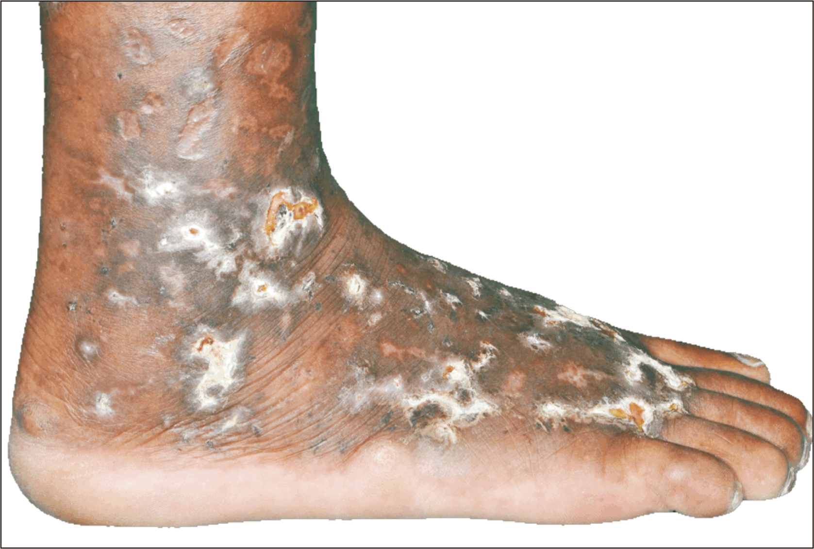
Leg ulcers are found to be nonspecific findings in SLE [40]. They vary in size, ranging from 1~10 cm in diameter and number, usually multiple. The most common site is the malleolus though they can be found on the feet, pretibial area, shins, or elsewhere [173-178]. Leg ulcers in SLE can be secondary to many causes and the pathological findings vary accordingly. The causes include vasculitis, vasculopathy, peripheral arterial disease, and aPL that can develop leg ulcers [178-180]. Another rare cause would be PG, a ND [179]. The prevalence of leg ulcers in LE patients ranges from 5.6%~10.0%, mostly associated with APS or LCV [115,173,177]. The presence of leg ulcers may reflect higher disease activity. A study showed low serum complements and high antinuclear anti-antibody (ANA) titers, which usually correlate with high disease activity, were associated with the enlargement of ulcers [174]. Leg ulcers secondary to PG in the setting of SLE can be related to disease activity PG [176,181].

Rowell syndrome

Rowell syndrome (RS) classification remains a subject of debate, with some considering it a distinct syndrome and others categorizing it as a subset of SCLE [182]. RS is an uncommon condition, with an estimated prevalence of 2.0% to 2.7% among LE patients [38,183]. Since its initial description, fewer than 100 cases have been reported [184]. The syndrome predominantly affects females, with a female-to-male ratio of approximately 8:1. The median age of onset is 32 years; however, cases in both pediatric and elderly populations have been documented [85].
It is a rare clinical entity characterized by the coexistence of LE and erythema multiforme (EM)-like lesions, target lesions, which may be typical or atypical, displaying concentric color variation (Figure 11) [40]. RS presents with painful, pruritic, annular, erythematous plaques with blistering features resembling EM. The lesions primarily affect the arms and legs; involvement of the face, neck, chest, and oral mucosa is less common. These lesions may persist for a duration ranging from several days to over a month [185]. It is distinguished from EM by specific immunologic markers, including a speckled ANA pattern, anti-Ro/SSA or anti-La/SSB antibodies, and positive RF [2,186]. The speckled ANA pattern is the most consistent immunologic feature of RS, observed in approximately 88% of cases, while RF is the least prevalent, present in only 41%. The immunologic abnormalities of RS are also observed in SCLE [182,183].
Histopathologically, RS exhibits vacuolar interface dermatitis, sharing features with both EM and LE. Necrotic keratinocytes, a hallmark of EM, are present alongside peri-adnexal lymphocytic inflammation and parakeratosis. Peri-adnexal CD123-positive plasmacytoid dendritic cells further support its classification within the LE spectrum. Direct immunofluorescence studies of RS and EM reveal similar C3 and IgM deposits in the dermal capillaries, akin to findings observed in SLE [185-187].
The precise pathogenesis of RS remains unclear. However, it is hypothesized to involve a type IV immune response, particularly in patients with anti-Ro antibodies. In the absence of classical exogenous antigens, anti-Ro antibodies may trigger an EM-like type IV immune response and antibody-dependent cellular cytotoxicity, leading to a lupus-like diathesis [188,189].
RS has been observed in association with various forms of LE, including SLE, DLE, and SCLE. The presence of EM-like lesions in lupus patients indicates a severe form of ACLE and SCLE, systemic disease involvement such as SLE exacerbation or overlap syndromes. It may serve as a disease activity marker representing high lupus disease activity and increased risk of vasculitis [190,191].
RS generally follows a benign clinical course, with most patients achieving remission within one year. Recurrence is uncommon, as reported in follow-up studies. Compared to other severe cutaneous manifestations of LE, the prognosis of RS is favorable [186]. It is important to note that EM major with severe mucocutaneous involvement in lupus patients may progress to Stevens-Johnson syndrome or toxic epidermal necrolysis, requiring aggressive management [190,192,193].

Non-scarring alopecia

Non-scarring alopecia is a common dermatologic manifestation of SLE. Non-scaring alopecia, presented as the first sign of disease in up to 20% of cases in certain studies, is a component in the 2019 EULAR (European Alliance of Associations for Rheumatology)/ACR criteria for SLE [194]. It presents as diffuse hair thinning or localized hair loss without evidence of scarring or permanent follicular destruction. Non-scarring alopecia in lupus is not merely a cosmetic concern but may indicate underlying disease activity. It occurs during the evolutionary stage or SLE acute exacerbations [195].
The pathogenesis of non-scarring alopecia in SLE is not fully elucidated but is assumed to be immune-mediated damage to hair follicles and systemic inflammation. High levels of proinflammatory cytokines may negatively affect the hair growth cycle. Type I interferons are well known to play a role in SLE and CLE, as they inhibit the anagen phase and induce premature hair shedding, whereby hairs are broken due to hair follicle growth retardation [196,197].
Non-scarring alopecia in LE typically manifests as diffuse hair thinning, increased hair fragility, or patchy hair loss, primarily affecting the scalp. Despite the hair and scalp appearing normal, microscopic inflammation and hair loss are observed. Most frequently, hair loss occurs at the onset of the illness. When the disease is under control, the hair should re-grow. Alopecia in LE has been sub-classified as LE-specific and LE-nonspecific according to histopathological findings [198].
Among LE nonspecific skin lesions, nonscarring alopecia was the common manifestation. Some research found the most frequently reported LE-nonspecific skin lesions were diffuse alopecia [34]. Diffuse non-scarring alopecia is characterized by diffuse thinning and fragility of hair, often correlating with disease activity. This form is typically reversible with appropriate management of SLE [198]. Multiple patches all over the scalp account for 48% of the cases, the posterior scalp for 20%, the temporal scalp for 16%, and the central scalp for 12% [199]. Excessive trichorrhexis, dry and thinning, brittle, short hair shafts, that vary from 5~25 mm in length, are characteristics of lupus hair and may be a form of telogen effluvium (TE) [85,198] Alopecia may occur before, during, or after the diagnosis of SLE and is often associated with severe disease and may mimic alopecia areata (AA) [35,36,198].
A dermoscopic image of “lupus hair” shows thinning and hypopigmentation of the hair shafts, telangiectasia, red dots around the hair follicles, white dots, honeycomb pigment macules, and polymorphic interfollicular vessels [85,198]. Histopathological findings of patches of partial or total hair loss in SLE are peribulbar mononuclear cell infiltrates around anagen bulbs [200].
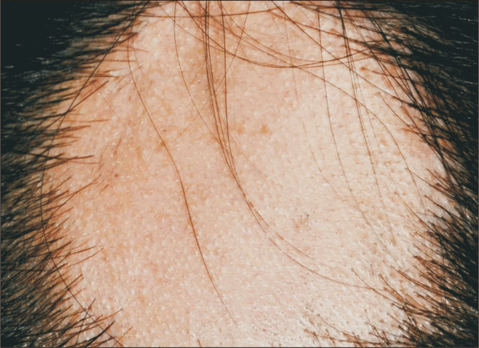
Other types of non-scarring alopecia in non-specific LE, which can co-exist in SLE patients, are AA (Figure 12) and TE. There was a high incidence of AA in SLE patients. AA in SLE represented either a coincidental finding or had been confused with non-scarring patch alopecia of SLE. A significant positive association has been observed between AA and SLE [201,202].
TE can occur in the context of SLE as a transient phenomenon, occurring during increased LE activity [85]. Anagen effluvium, if observed in SLE, is likely secondary to specific triggering events, cytotoxic agents (e.g., cyclophosphamide), or antimetabolites (e.g., MTX). Disease flares can cause the rapidly dividing matrical cells of the anagen follicle to shut down temporarily [203]. With all these affirming evidences, the association of non-scarring alopecia and SLE activity is not incongruous.

Research gaps

A lot of non-specific cutaneous LE still lack firm evidence of its association with SLE disease activities and its occurrence with autoantibodies or internal organ involvement. We have summarized the clinical significance of each non-specific cutaneous LE from all literature reviewed, which includes prevalence, organ/systemic involvement, antibodies and clinical correlation with SLE disease activity/severity in Table 2. Due to the fairly uncommon incidence of some cutaneous manifestations, for instance, papulonodular mucinosis, literature regarding its association with SLE was made difficult. These missing data may be valuable as a clue to SLE diagnosis or even serve as SLE prognostic factors. Reports on a few cutaneous manifestations like CC, thrombophlebitis, or erythromelalgia were majorly case reports or case series and were outdated with no recent reports, unlike RP or vasculitis which have been reported and known to be associated with SLE with retrospective cohort studies. Further research is still needed to explore the relationship between many non-specific LE and SLE disease activity or severity. Additional focus on targeted immunomodulating therapies may offer novel treatment avenues for non-specific cutaneous LE to improve quality of life and decrease morbidity.

CONCLUSION

Rheumatologists and dermatologists need to recognize these conditions as they may represent the first manifestation of SLE or the sole manifestation of SLE. Many known cutaneous manifestations portend different prognoses, disease activity of SLE, or other organ involvement. However, some non-specific cutaneous LE may co-exist but indicate no clinical significance and are debilitating or result in disfigurement. The treatment of each cutaneous lesion must also be accompanied by specific treatment for SLE or other cutaneous LE. Early recognition and prompt treatment of each cutaneous manifestation and screening for other systemic involvement to improve outcome and quality of life by controlling disease activity and avoiding further organ damage.
ACKNOWLEDGMENTS

None.

Notes

FUNDING

None.

CONFLICT OF INTEREST

No potential conflict of interest relevant to this article was reported.

AUTHOR CONTRIBUTIONS

All co-authors take full responsibility for the integrity and accuracy of all aspects of the work. Nattanicha Chaisrimaneepan: substantial contribution to conception, drafting, editing, resource and final approval. Thunyaporn Khoruamklang: substantial contribution to interpretation, and drafting, final approval. Tulaton Sodsri: substantial to drafting, final approval. Natnicha Jakramonpreeya: substantial contribution to drafting, final approval.

REFERENCES

1. Wenzel J. 2019; Cutaneous lupus erythematosus: new insights into pathogenesis and therapeutic strategies. Nat Rev Rheumatol. 15:519–32. DOI: 10.1038/s41584-019-0272-0. PMID: 31399711.
2. Vale ECSD, Garcia LC. 2023; Cutaneous lupus erythematosus: a review of etiopathogenic, clinical, diagnostic and therapeutic aspects. An Bras Dermatol. 98:355–72. DOI: 10.1016/j.abd.2022.09.005. PMID: 36868923. PMCID: PMC10173173.
3. Ribero S, Sciascia S, Borradori L, Lipsker D. 2017; The cutaneous spectrum of lupus erythematosus. Clin Rev Allergy Immunol. 53:291–305. DOI: 10.1007/s12016-017-8627-2. PMID: 28752372.
4. Gheita TA, Abaza NM, Sayed S, El-Azkalany GS, Fishawy HS, Eissa AH. 2018; Cutaneous vasculitis in systemic lupus erythematosus patients: potential key players and implications. Lupus. 27:738–43. DOI: 10.1177/0961203317739134. PMID: 29087263.
5. Zhizho Y, Bo L, Jianso L, Fenlia G, Xun W, Zhihu Y. 2008; Clinical characteristics of cutaneous vasculitis secondary to systemic lupus erythematosus. Pediatr Rheumatol. 6:P239. DOI: 10.1186/1546-0096-6-S1-P239. PMCID: PMC3334044.
6. Chen KR, Carlson JA. 2008; Clinical approach to cutaneous vasculitis. Am J Clin Dermatol. 9:71–92. DOI: 10.2165/00128071-200809020-00001. PMID: 18284262.
7. Kuhn A, Sticherling M, Bonsmann G. 2007; Clinical manifestations of cutaneous lupus erythematosus. J Dtsch Dermatol Ges. 5:1124–37. DOI: 10.1111/j.1610-0387.2007.06554.x. PMID: 18042093.
8. Fraticelli P, Benfaremo D, Gabrielli A. 2021; Diagnosis and management of leukocytoclastic vasculitis. Intern Emerg Med. 16:831–41. DOI: 10.1007/s11739-021-02688-x. PMID: 33713282. PMCID: PMC8195763.
9. Ramos-Casals M, Nardi N, Lagrutta M, Brito-Zerón P, Bové A, Delgado G, et al. 2006; Vasculitis in systemic lupus erythematosus: prevalence and clinical characteristics in 670 patients. Medicine (Baltimore). 85:95–104. DOI: 10.1097/01.md.0000216817.35937.70. PMID: 16609348.
10. Chottawornsak N, Rodsaward P, Suwannachote S, Rachayon M, Rattananupong T, Deekajorndech T, et al. 2018; Skin signs in juvenile- and adult-onset systemic lupus erythematosus: clues to different systemic involvement. Lupus. 27:2069–75. DOI: 10.1177/0961203318805851. PMID: 30336755.
11. Kallas R, Goldman D, Petri MA. 2020; Cutaneous vasculitis in SLE. Lupus Sci Med. 7:e000411. DOI: 10.1136/lupus-2020-000411. PMID: 32963114. PMCID: PMC7509964.
12. Chen KR, Toyohara A, Suzuki A, Miyakawa S. 2002; Clinical and histopathological spectrum of cutaneous vasculitis in rheumatoid arthritis. Br J Dermatol. 147:905–13. DOI: 10.1046/j.1365-2133.2002.04933.x. PMID: 12410699.
13. Burling F, Ng J, Thein H, Ly J, Marshall MR, Gow P. 2007; Ethnic, clinical and immunological factors in systemic lupus erythematosus and the development of lupus nephritis: results from a multi-ethnic New Zealand cohort. Lupus. 16:830–7. DOI: 10.1177/0961203307080225. PMID: 17895308.
14. Kawakami T, Soma Y, Mizoguchi M. 2005; Initial cutaneous manifestations associated with histopathological leukocytoclastic vasculitis in two patients with antiphospholipid antibody syndrome. J Dermatol. 32:1032–7. DOI: 10.1111/j.1346-8138.2005.tb00896.x. PMID: 16471472.
15. Shinjo SK, Bonfá E. 2011; Cutaneous vasculitis in systemic lupus erythematosus: association with anti-ribosomal P protein antibody and Raynaud phenomenon. Clin Rheumatol. 30:173–7. DOI: 10.1007/s10067-010-1432-4. PMID: 20349257.
16. Fukuda MV, Lo SC, de Almeida CS, Shinjo SK. 2009; Anti-Ro antibody and cutaneous vasculitis in systemic lupus erythematosus. Clin Rheumatol. 28:301–4. DOI: 10.1007/s10067-008-1043-5. PMID: 19031093.
17. Bombardier C, Gladman DD, Urowitz MB, Caron D, Chang CH. 1992; Derivation of the SLEDAI. A disease activity index for lupus patients. The Committee on Prognosis Studies in SLE. Arthritis Rheum. 35:630–40. DOI: 10.1002/art.1780350606. PMID: 1599520.
18. Vitali C, Bencivelli W, Isenberg DA, Smolen JS, Snaith ML, Sciuto M, et al. 1992; Disease activity in systemic lupus erythematosus: report of the Consensus Study Group of the European Workshop for Rheumatology Research. II. Identification of the variables indicative of disease activity and their use in the development of an activity score. The European Consensus Study Group for Disease Activity in SLE. Clin Exp Rheumatol. 10:541–7.
19. Isenberg DA, Rahman A, Allen E, Farewell V, Akil M, Bruce IN, et al. 2005; BILAG 2004. Development and initial validation of an updated version of the British Isles Lupus Assessment Group's disease activity index for patients with systemic lupus erythematosus. Rheumatology (Oxford). 44:902–6. DOI: 10.1093/rheumatology/keh624. PMID: 15814577.
20. Toubi E, Kessel A, Bamberger E, Golan TD. 2004; Systemic lupus erythematosus vasculitis: a current therapeutic overview. Curr Treat Options Cardiovasc Med. 6:87–97. DOI: 10.1007/s11936-004-0036-1. PMID: 15066237.
21. Wisnieski JJ. 2000; Urticarial vasculitis. Curr Opin Rheumatol. 12:24–31. DOI: 10.1097/00002281-200001000-00005. PMID: 10647951.
22. Venzor J, Lee WL, Huston DP. 2002; Urticarial vasculitis. Clin Rev Allergy Immunol. 23:201–16. DOI: 10.1385/CRIAI:23:2:201. PMID: 12221865.
23. Siegert CE, Kazatchkine MD, Sjöholm A, Würzner R, Loos M, Daha MR. 1999; Autoantibodies against C1q: view on clinical relevance and pathogenic role. Clin Exp Immunol. 116:4–8. DOI: 10.1046/j.1365-2249.1999.00867.x. PMID: 10209498. PMCID: PMC1905233.
24. Davis MD, Daoud MS, Kirby B, Gibson LE, Rogers RS 3rd. 1998; Clinicopathologic correlation of hypocomplementemic and normocomplementemic urticarial vasculitis. J Am Acad Dermatol. 38:899–905. DOI: 10.1016/S0190-9622(24)00193-2. PMID: 9631995.
25. Jachiet M, Flageul B, Deroux A, Le Quellec A, Maurier F, Cordoliani F, et al. French Vasculitis Study Group. 2015; The clinical spectrum and therapeutic management of hypocomplementemic urticarial vasculitis: data from a French nationwide study of fifty-seven patients. Arthritis Rheumatol. 67:527–34. DOI: 10.1002/art.38956. PMID: 25385679.
26. Breillat P, Jachiet M, Ditchi Y, Lenormand C, Costedoat-Chalumeau N, et al. for EMSED study group (Etude des maladies systémiques en dermatologie) and the Swiss SLE Cohort Study (SSCS). 2023; Cutaneous vasculitis occurring in the setting of systemic lupus erythematosus: a multicentre cohort study. Rheumatology (Oxford). 62:2189–96. DOI: 10.1093/rheumatology/keac566. PMID: 36190335.
27. Yasue T. 1986; Livedoid vasculitis and central nervous system involvement in systemic lupus erythematosus. Arch Dermatol. 122:66–70. DOI: 10.1001/archderm.1986.01660130070028. PMID: 3942409.
28. Sopeña B, Pérez-Rodríguez MT, Rivera A, Ortiz-Rey JA, Lamas J, Freire-Dapena MC. 2010; Livedoid vasculopathy and recurrent thrombosis in a patient with lupus: seronegative antiphospholipid syndrome? Lupus. 19:1340–3. DOI: 10.1177/0961203310373783. PMID: 20659971.
29. Khenifer S, Thomas L, Balme B, Dalle S. 2010; Livedoid vasculopathy: thrombotic or inflammatory disease? Clin Exp Dermatol. 35:693–8. DOI: 10.1111/j.1365-2230.2009.03732.x. PMID: 19874344.
30. Pyrpasopoulou A, Chatzimichailidou S, Aslanidis S. 2012; Vascular disease in systemic lupus erythematosus. Autoimmune Dis. 2012:876456. DOI: 10.1155/2012/876456. PMID: 22957213. PMCID: PMC3432322.
31. Hidayati A, Earlia N, Liana MR. 2022; Successful treatment of refractory livedoid vasculopathy in a patient with systemic lupus erythematosus. Int J Res Dermatol. 9:36–9. DOI: 10.18203/issn.2455-4529.IntJResDermatol20223343.
32. Yoshioka K, Tateishi C, Kato H, Chen KR. 2018; Systemic lupus erythematosus with refractory ulcerated livedoid vasculopathy: successful treatment with intravenous immunoglobulin and warfarin. Clin Case Rep. 6:2045–7. DOI: 10.1002/ccr3.1803. PMID: 30455888. PMCID: PMC6230605.
33. Bilgic A, Ozcobanoglu S, Bozca BC, Alpsoy E. 2021; Livedoid vasculopathy: a multidisciplinary clinical approach to diagnosis and management. Int J Womens Dermatol. 7:588–99. DOI: 10.1016/j.ijwd.2021.08.013. PMID: 35024414. PMCID: PMC8721056.
34. Biazar C, Sigges J, Patsinakidis N, Ruland V, Amler S, Bonsmann G, et al. EUSCLE co-authors. 2013; Cutaneous lupus erythematosus: first multicenter database analysis of 1002 patients from the European Society of Cutaneous Lupus Erythematosus (EUSCLE). Autoimmun Rev. 12:444–54. DOI: 10.1016/j.autrev.2012.08.019. PMID: 23000206.
35. Uva L, Miguel D, Pinheiro C, Freitas JP, Marques Gomes M, Filipe P. 2012; Cutaneous manifestations of systemic lupus erythematosus. Autoimmune Dis. 2012:834291. DOI: 10.1155/2012/834291. PMID: 22888407. PMCID: PMC3410306.
36. Fijałkowska A, Kądziela M, Żebrowska A. 2024; The spectrum of cutaneous manifestations in lupus erythematosus: a comprehensive review. J Clin Med. 13:2419. DOI: 10.3390/jcm13082419. PMID: 38673692. PMCID: PMC11050979.
37. Cardinali C, Caproni M, Bernacchi E, Amato L, Fabbri P. 2000; The spectrum of cutaneous manifestations in lupus erythematosus--the Italian experience. Lupus. 9:417–23. DOI: 10.1191/096120300678828569. PMID: 10981645.
38. Mowla MR, Barua DP, Zaman S, Chowdhury MIH, Ara S, Reich A. 2022; Clinicopathological characteristics of cutaneous lupus erythematosus patients in Bangladesh. Postepy Dermatol Alergol. 39:782–7. DOI: 10.5114/ada.2021.110254. PMID: 36090729. PMCID: PMC9454348.
39. Obermoser G, Sontheimer RD, Zelger B. 2010; Overview of common, rare and atypical manifestations of cutaneous lupus erythematosus and histopathological correlates. Lupus. 19:1050–70. DOI: 10.1177/0961203310370048. PMID: 20693199.
40. Stull C, Sprow G, Werth VP. 2023; Cutaneous involvement in systemic lupus erythematosus: a review for the rheumatologist. J Rheumatol. 50:27–35. DOI: 10.3899/jrheum.220089. PMID: 36109075. PMCID: PMC10152495.
41. Verdelli A, Coi A, Marzano AV, Antiga E, Cozzani E, Quaglino P, et al. 2019; Autoantibody profile and clinical patterns in 619 Italian patients with cutaneous lupus erythematosus. J Eur Acad Dermatol Venereol. 33:742–52. DOI: 10.1111/jdv.15147. PMID: 29924416.
42. Zhou W, Wu H, Zhao M, Lu Q. 2020; New insights into the progression from cutaneous lupus to systemic lupus erythematosus. Expert Rev Clin Immunol. 16:829–37. DOI: 10.1080/1744666X.2020.1805316. PMID: 32746644.
43. Block JA, Sequeira W. 2001; Raynaud's phenomenon. Lancet. 357:2042–8. DOI: 10.1016/S0140-6736(00)05118-7. PMID: 11438158.
44. Pavlov-Dolijanovic S, Damjanov NS, Vujasinovic Stupar NZ, Marcetic DR, Sefik-Bukilica MN, Petrovic RR. 2013; Is there a difference in systemic lupus erythematosus with and without Raynaud's phenomenon? Rheumatol Int. 33:859–65. DOI: 10.1007/s00296-012-2449-6. PMID: 22618491.
45. Baba Ö, Kısaoğlu H, Kalyoncu M. 2024; Associations of Raynaud's phenomenon in juvenile onset systemic lupus erythematosus. Van Med J. 31:172–8. DOI: 10.5505/vmj.2024.66375.
46. Nakano M, Ohuchi Y, Hasegawa H, Kuroda T, Ito S, Gejyo F. 2000; Clinical significance of anticentromere antibodies in patients with systemic lupus erythematosus. J Rheumatol. 27:1403–7.
47. Butkiewicz F, Kaszuba M, Brzeziński M, Iżbicki J, Kubiś M, Lopiński H, et al. 2014; Associations between the incidence of antiphosphatidylserine and antiphosphatidylethanolamine antibodies and clinical manifestations of systemic lupus erythematosus. Pol Arch Med Wewn. 124:573–8. DOI: 10.20452/pamw.2489. PMID: 25220366.
48. Fischer K, Przepiera-Będzak H, Brzosko I, Sawicki M, Walecka A, Brzosko M. 2022; Anti-phosphatidylethanolamine and anti-phosphatidylserine antibodies-association with renal involvement, atherosclerosis, cardiovascular manifestations, Raynaud phenomenon and disease activity in Polish patients with systemic lupus erythematosus. Biomolecules. 12:1328. DOI: 10.3390/biom12101328. PMID: 36291537. PMCID: PMC9599205.
49. Caccavo D, Del Porto F, Garzia P, Mitterhofer AP, Galluzzo S, Rigon A, et al. 2003; Raynaud's phenomenon and antiphospholipid antibodies in systemic lupus erythematosus: is there an association? Ann Rheum Dis. 62:1003–5. DOI: 10.1136/ard.62.10.1003. PMID: 12972482. PMCID: PMC1754341.
50. Vayssairat M, Abuaf N, Baudot N, Deschamps A, Gaitz JP. 1998; Abnormal IgG cardiolipin antibody titers in patients with Raynaud's phenomenon and/or related disorders: prevalence and clinical significance. J Am Acad Dermatol. 38:555–8. DOI: 10.1016/S0190-9622(98)70116-1. PMID: 9555793.
51. To CH, Petri M. 2005; Is antibody clustering predictive of clinical subsets and damage in systemic lupus erythematosus? Arthritis Rheum. 52:4003–10. DOI: 10.1002/art.21414. PMID: 16320348.
52. Dimant J, Ginzler E, Schlesinger M, Sterba G, Diamond H, Kaplan D, et al. 1979; The clinical significance of Raynaud's phenomenon in systemic lupus erythematosus. Arthritis Rheum. 22:815–9. DOI: 10.1002/art.1780220802. PMID: 465096.
53. Kim JS, Kim D, Joo YB, Won S, Lee J, Shin J, et al. 2018; Factors associated with development and mortality of pulmonary hypertension in systemic lupus erythematosus patients. Lupus. 27:1769–77. DOI: 10.1177/0961203318788163. PMID: 30028258.
54. Andraos R, Ahmad A, Eriksson P, Dahlström Ö, Wirestam L, Dahle C, et al. 2022; Autoantibodies associated with systemic sclerosis in three autoimmune diseases imprinted by type I interferon gene dysregulation: a comparison across SLE, primary Sjögren's syndrome and systemic sclerosis. Lupus Sci Med. 9:e000732. DOI: 10.1136/lupus-2022-000732. PMID: 36581379. PMCID: PMC9806068.
55. Shen JY, Chen SL, Wu YX, Tao RQ, Gu YY, Bao CD, et al. 1999; Pulmonary hypertension in systemic lupus erythematosus. Rheumatol Int. 18:147–51. DOI: 10.1007/s002960050074. PMID: 10220835.
56. Schreiber BE, Connolly MJ, Coghlan JG. 2013; Pulmonary hypertension in systemic lupus erythematosus. Best Pract Res Clin Rheumatol. 27:425–34. DOI: 10.1016/j.berh.2013.07.011. PMID: 24238697.
57. Chen CH, Chen HA, Wang HP, Liao HT, Chou CT, Huang DF. 2006; Pulmonary arterial hypertension in autoimmune diseases: an analysis of 19 cases from a medical center in northern Taiwan. J Microbiol Immunol Infect. 39:162–8.
58. Artim-Esen B, Çene E, Şahinkaya Y, Ertan S, Pehlivan Ö, Kamali S, et al. 2014; Cluster analysis of autoantibodies in 852 patients with systemic lupus erythematosus from a single center. J Rheumatol. 41:1304–10. DOI: 10.3899/jrheum.130984. PMID: 24833757.
59. Kasparian A, Floros A, Gialafos E, Kanakis M, Tassiopoulos S, Kafasi N, et al. 2007; Raynaud's phenomenon is correlated with elevated systolic pulmonary arterial pressure in patients with systemic lupus erythematosus. Lupus. 16:505–8. DOI: 10.1177/0961203307080629. PMID: 17670849.
60. Heimovski FE, Simioni JA, Skare TL. 2015; Systemic lupus erythematosus and Raynaud's phenomenon. An Bras Dermatol. 90:837–40. DOI: 10.1590/abd1806-4841.20153881. PMID: 26734864. PMCID: PMC4689071.
61. Bhatt SP, Handa R, Gulati GS, Sharma S, Pandey RM, Aggarwal P, et al. 2007; Peripheral vascular disease in systemic lupus erythematosus. Lupus. 16:720–3. DOI: 10.1177/0961203307081123. PMID: 17728365.
62. Barbacki A, Rached-d'Astous N, Pineau CA, Vinet E, Grenier LP, Kalache F, et al. 2022; Clinical significance of Raynaud phenomenon in systemic lupus erythematosus. J Clin Rheumatol. 28:e488–90. DOI: 10.1097/RHU.0000000000001773. PMID: 35192594.
63. Sakamoto K, Houya I, Inoue K, Tanaka M, Suzuki T, Sakamoto Y, et al. 1999; An imbalance in plasma prostanoids in patients with Raynaud's phenomenon and pulmonary vasospasm. Eur Respir J. 13:137–44. DOI: 10.1183/09031936.99.13113799. PMID: 10836338.
64. Min HK, Lee JH, Jung SM, Lee J, Kang KY, Kwok SK, et al. 2015; Pulmonary hypertension in systemic lupus erythematosus: an independent predictor of patient survival. Korean J Intern Med. 30:232–41. DOI: 10.3904/kjim.2015.30.2.232. PMID: 25750566. PMCID: PMC4351331.
65. Fries JF, Holman HR. 1975; Systemic lupus erythematosus: a clinical analysis. Major Probl Intern Med. 6:v–199.
66. Lavras Costallat LT, Valente Coimbra AM. 1995; Raynaud's phenomenon in systemic lupus erythematosus. Rev Rhum Engl Ed. 62:349–53.
67. Costallat LT, Coimbra AM. 1994; Systemic lupus erythematosus: clinical and laboratory aspects related to age at disease onset. Clin Exp Rheumatol. 12:603–7.
68. Iqbal S, Sher MR, Good RA, Cawkwell GD. 1999; Diversity in presenting manifestations of systemic lupus erythematosus in children. J Pediatr. 135:500–5. DOI: 10.1016/S0022-3476(99)70174-5. PMID: 10518085.
69. Lian F, Chen D, Wang Y, Ye Y, Wang X, Zhan Z, et al. 2012; Clinical features and independent predictors of pulmonary arterial hypertension in systemic lupus erythematosus. Rheumatol Int. 32:1727–31. DOI: 10.1007/s00296-011-1880-4. PMID: 21437686.
70. Tarachandani D, Ingle V, Singhal K, Panda S. 2023; Rare case of systemic lupus erythematosus presenting as erythromelalgia. J Datta Meghe Inst Med Sci Univ. 18:772–5. DOI: 10.4103/jdmimsu.jdmimsu_264_23.
71. Alarcon-Segovia D, Babb RR, Fairbairn JF 2nd. 1963; Systemic lupus erythematosus with erythermalgia. Arch Intern Med. 112:688–92. DOI: 10.1001/archinte.1963.03860050075006. PMID: 14064989.
72. Badeloe S, Henquet CJ, Nieuwhof CM, Frank J. 2007; Secondary erythromelalgia involving the ears probably preceding lupus erythematosus. Int J Dermatol. 46(Suppl 3):6–8. DOI: 10.1111/j.1365-4632.2007.03500.x. PMID: 17973878.
73. Kraus A. 1990; Erythromelalgia in a patient with systemic lupus erythematosus treated with clonazepam. J Rheumatol. 17:120.
74. Alarcon-Segovia D, Diaz-Jouanen E. 1973; Case report: erythermalgia in systemic lupus erythematosus. Am J Med Sci. 266:149–51. DOI: 10.1097/00000441-197308000-00011. PMID: 4742370.
75. Cailleux N, Levesque H, Courtois H. 1996; [Erythermalgia and systemic lupus erythematosus]. J Mal Vasc. 21:88–91. French.
76. Tham SW, Giles M. 2018; Current pain management strategies for patients with erythromelalgia: a critical review. J Pain Res. 11:1689–98. DOI: 10.2147/JPR.S154462. PMID: 30214279. PMCID: PMC6121769.
77. Vinay K, Sawatkar G, Dogra S, Saikia UN. 2017; Systemic lupus erythematosus with Degos disease: role of dermatoscopy in diagnosis. Int J Dermatol. 56:770–2. DOI: 10.1111/ijd.13629. PMID: 28444743.
78. Ball E, Newburger A, Ackerman AB. 2003; Degos' disease: a distinctive pattern of disease, chiefly of lupus erythematosus, and not a specific disease per se. Am J Dermatopathol. 25:308–20. DOI: 10.1097/00000372-200308000-00005. PMID: 12876488.
79. Doutre MS, Beylot C, Bioulac P, Busquet M, Conte M. 1987; Skin lesion resembling malignant atrophic papulosis in lupus erythematosus. Dermatologica. 175:45–6. DOI: 10.1159/000248781. PMID: 3609417.
80. Stavorn T, Chanprapaph K. 2019; Degos-like lesions in association with connective tissue diseases: a report of three cases and literature review. Clin Cosmet Investig Dermatol. 12:815–22. DOI: 10.2147/CCID.S230081. PMID: 31807047. PMCID: PMC6848981.
81. Riyaz N, Saleem R, Shafeeq R. 2011; Degos disease-like presentation in systemic lupus erythematosus. Indian J Dermatol Venereol Leprol. 77:219–21. DOI: 10.4103/0378-6323.77477. PMID: 21393965.
82. Dubin HV, Stawiski MA. 1974; Systemic lupus erythematosus resembling malignant atrophic papulosis. Arch Intern Med. 134:321–3. DOI: 10.1001/archinte.1974.00320200131019.
83. Mutizwa MM, Tang MB, Ng SK. 2010; Prominent Degos-like skin lesions in a patient with chronic cutaneous lupus erythematosus. Dermatol Online J. 16:5. DOI: 10.5070/D36SC6K7WX. PMID: 20673533.
84. Jang MS, Park JB, Yang MH, Jang JY, Kim JH, Lee KH, et al. 2017; Degos-like lesions associated with systemic lupus erythematosus. Ann Dermatol. 29:215–8. DOI: 10.5021/ad.2017.29.2.215. PMID: 28392651. PMCID: PMC5383749.
85. Rothfield N, Sontheimer RD, Bernstein M. 2006; Lupus erythematosus: systemic and cutaneous manifestations. Clin Dermatol. 24:348–62. DOI: 10.1016/j.clindermatol.2006.07.014. PMID: 16966017.
86. Singal A, Arora R. 2015; Nail as a window of systemic diseases. Indian Dermatol Online J. 6:67–74. DOI: 10.4103/2229-5178.153002. PMID: 25821724. PMCID: PMC4375768.
87. Cutolo M, Melsens K, Wijnant S, Ingegnoli F, Thevissen K, De Keyser F, et al. 2018; Nailfold capillaroscopy in systemic lupus erythematosus: a systematic review and critical appraisal. Autoimmun Rev. 17:344–52. DOI: 10.1016/j.autrev.2017.11.025. PMID: 29427827.
88. Ansari A, Larson PH, Bates HD. 1985; Cardiovascular manifestations of systemic lupus erythematosus: current perspective. Prog Cardiovasc Dis. 27:421–34. DOI: 10.1016/0033-0620(85)90003-9. PMID: 2860699.
89. Pilz P, Wallnöfer H, Klein J. 1980; [Thrombophlebitis of internal cerebral veins in a case of systemic lupus erythematosus (author's transl)]. Arch Psychiatr Nervenkr (1970). 228:31–43. German. DOI: 10.1007/BF00365741. PMID: 7387348.
90. Shalhout SZ, Nahas MR, Drews RE, Miller DM. 2020; Generalized acquired cutis laxa associated with monoclonal gammopathy of dermatological significance. Case Rep Dermatol Med. 2020:7480607. DOI: 10.1155/2020/7480607. PMID: 32099688. PMCID: PMC7037480.
91. Nabatanzi A, Da S, Male M, Chen S, Huang C. 2020; Type II acquired cutis laxa associated with recurrent urticarial vasculitis: brief report. Allergy Asthma Clin Immunol. 16:1. DOI: 10.1186/s13223-019-0401-y. PMID: 31911804. PMCID: PMC6942328.
92. Golisch KB, Gottesman SP, Ferrer P, Culpepper KS. 2018; Cutis laxa acquisita after urticarial vasculitis in SLE patients. Am J Dermatopathol. 40:433–7. DOI: 10.1097/DAD.0000000000001084. PMID: 29329127.
93. Lewis PG, Hood AF, Barnett NK, Holbrook KA. 1990; Postinflammatory elastolysis and cutis laxa. A case report. J Am Acad Dermatol. 22:40–8. DOI: 10.1016/0190-9622(90)70005-3. PMID: 2298964.
94. Schwartz E, Cruickshank FA, Lebwohl MG. 1988; Elastase-like protease and elastolytic activities expressed in cultured dermal fibroblasts derived from lesional skin of patients with pseudoxanthoma elasticum, actinic elastosis, and cutis laxa. Clin Chim Acta. 176:219–24. DOI: 10.1016/0009-8981(88)90211-2. PMID: 3180471.
95. Venencie PY, Winkelmann RK, Moore BA. 1984; Anetoderma. Clinical findings, associations, and long-term follow-up evaluations. Arch Dermatol. 120:1032–9. DOI: 10.1001/archderm.1984.01650440062021. PMID: 6465909.
96. Montilla C, Alarcón-Segovia D. 2000; Anetoderma in systemic lupus erythematosus: relationship to antiphospholipid antibodies. Lupus. 9:545–7. DOI: 10.1177/096120330000900712. PMID: 11035422.
97. Gibson GE, Su WP, Pittelkow MR. 1997; Antiphospholipid syndrome and the skin. J Am Acad Dermatol. 36:970–82. DOI: 10.1016/S0190-9622(97)80283-6. PMID: 9204065.
98. Jeong NJ, Park SB, Im M, Seo YJ, Lee JH, Lee Y. 2014; Eruptive anetoderma in a patient with systemic lupus erythematosus. Ann Dermatol. 26:621–3. DOI: 10.5021/ad.2014.26.5.621. PMID: 25324656. PMCID: PMC4198591.
99. Stephansson EA, Niemi KM. 1995; Antiphospholipid antibodies and anetoderma: are they associated? Dermatology. 191:204–9. DOI: 10.1159/000246547. PMID: 8534938.
100. Alvarez-Cuesta CC, Raya-Aguado C, Fernández-Rippe ML, Sánchez TS, Pérez-Oliva N. 2001; Anetoderma in a systemic lupus erythematosus patient with anti-PCNA and antiphospholipid antibodies. Dermatology. 203:348–50. DOI: 10.1159/000051789. PMID: 11752829.
101. Hasbún Acuña P, Cullen Aravena R, Queirolo González A, Corredoira Salum Y. 2018; Primary anetoderma: a cutaneous predictor of autoimmunity. Reumatol Clin (Engl Ed). 14:242–3. DOI: 10.1016/j.reuma.2017.01.009. PMID: 28363348.
102. Haider M, Alenazi M, Almutawa A, Alfadley A. 2012; Lupus erythematosus-associated primary and secondary anetoderma. J Cutan Med Surg. 16:64–7. DOI: 10.1177/120347541201600113. PMID: 22417999.
103. Owen CE, Malone JC, Callen JP. 2008; Sweet-like dermatosis in 2 patients with clinical features of dermatomyositis and underlying autoimmune disease. Arch Dermatol. 144:1486–90. DOI: 10.1001/archderm.144.11.1486. PMID: 19015424.
104. Cárdenas AGS, Lima MS, Mendez-Flores S. 2021; Systemic lupus erythematosus-associated neutrophilic dermatosis presenting with an erythema gyratum repens-like appearance. Int J Dermatol. 60:857–9. DOI: 10.1111/ijd.15520. PMID: 33792025.
105. Larson AR, Granter SR. 2014; Systemic lupus erythematosus-associated neutrophilic dermatosis--an underrecognized neutrophilic dermatosis in patients with systemic lupus erythematosus. Hum Pathol. 45:598–605. DOI: 10.1016/j.humpath.2013.10.029. PMID: 24457080.
106. Gusdorf L, Bessis D, Lipsker D. 2014; Lupus erythematosus and neutrophilic urticarial dermatosis: a retrospective study of 7 patients. Medicine (Baltimore). 93:e351. DOI: 10.1097/MD.0000000000000351. PMID: 25546692. PMCID: PMC4602611.
107. Brinster NK, Nunley J, Pariser R, Horvath B. 2012; Nonbullous neutrophilic lupus erythematosus: a newly recognized variant of cutaneous lupus erythematosus. J Am Acad Dermatol. 66:92–7. DOI: 10.1016/j.jaad.2009.05.019. PMID: 22050914.
108. Gleason BC, Zembowicz A, Granter SR. 2006; Non-bullous neutrophilic dermatosis: an uncommon dermatologic manifestation in patients with lupus erythematosus. J Cutan Pathol. 33:721–5. DOI: 10.1111/j.1600-0560.2006.00559.x. PMID: 17083690.
109. Quatrano NA, Criscito MC, Femia AN, Brinster NK. 2016; Systemic lupus erythematosus-associated neutrophilic dermatosis with palmoplantar involvement. JAAD Case Rep. 2:329–33. DOI: 10.1016/j.jdcr.2016.07.005. PMID: 27570816. PMCID: PMC4992001.
110. Lee WJ, Kang HJ, Shin HJ, Won CH, Chang SE, Choi JH, et al. 2018; Neutrophilic urticarial dermatosis and Sweet-like neutrophilic dermatosis: under-recognized neutrophilic dermatoses in lupus erythematosus. Lupus. 27:628–36. DOI: 10.1177/0961203317736145. PMID: 29058993.
111. Pavlidakey P, Mills O, Bradley S, Casey J, Morgan MB. 2012; Neutrophilic dermatosis revisited: an initial presentation of lupus? J Am Acad Dermatol. 67:e29–35. DOI: 10.1016/j.jaad.2011.05.048. PMID: 21907449.
112. Das A, Burmeister R, Chhaya R, Eisenga B, Kumar A. 2020; Sweet syndrome in a patient with systemic lupus erythematosus. J Clin Rheumatol. 26:e85–6. DOI: 10.1097/RHU.0000000000000904. PMID: 32453302.
113. Hou TY, Chang DM, Gao HW, Chen CH, Chen HC, Lai JH. 2005; Sweet's syndrome as an initial presentation in systemic lupus erythematosus: a case report and review of the literature. Lupus. 14:399–402. DOI: 10.1191/0961203305lu2083cr. PMID: 15934441.
114. Lebrun D, Robbins A, Hentzien M, Toquet S, Plee J, Durlach A, et al. 2018; Two case reports of pyoderma gangrenosum and systemic lupus erythematosus: a rare but nonfortuitous association? Medicine (Baltimore). 97:e11933. DOI: 10.1097/MD.0000000000011933. PMID: 30142811. PMCID: PMC6112963.
115. Kole AK, Ghosh A. 2009; Cutaneous manifestations of systemic lupus erythematosus in a tertiary referral center. Indian J Dermatol. 54:132–6. DOI: 10.4103/0019-5154.53189. PMID: 20101308. PMCID: PMC2807152.
116. Vassileva S. 2004; Bullous systemic lupus erythematosus. Clin Dermatol. 22:129–38. DOI: 10.1016/j.clindermatol.2003.12.020. PMID: 15234014.
117. Contestable JJ, Edhegard KD, Meyerle JH. 2014; Bullous systemic lupus erythematosus: a review and update to diagnosis and treatment. Am J Clin Dermatol. 15:517–24. DOI: 10.1007/s40257-014-0098-0. PMID: 25358414.
118. Sebaratnam DF, Murrell DF. 2011; Bullous systemic lupus erythematosus. Dermatol Clin. 29:649–53. DOI: 10.1016/j.det.2011.06.002. PMID: 21925012.
119. Koch K, Tikly M. 2019; Spectrum of cutaneous lupus erythematosus in South Africans with systemic lupus erythematosus. Lupus. 28:1021–6. DOI: 10.1177/0961203319856091. PMID: 31215844.
120. Martin Calderon L, Pope JE. 2022; Precursors to systemic sclerosis and systemic lupus erythematosus: from undifferentiated connective tissue disease to the development of identifiable connective tissue diseases. Front Immunol. 13:869172. DOI: 10.3389/fimmu.2022.869172. PMID: 35603174. PMCID: PMC9118990.
121. Scherlinger M, Guillotin V, Truchetet ME, Contin-Bordes C, Sisirak V, Duffau P, et al. 2018; Systemic lupus erythematosus and systemic sclerosis: all roads lead to platelets. Autoimmun Rev. 17:625–35. DOI: 10.1016/j.autrev.2018.01.012. PMID: 29635077.
122. Asano Y. 2020; The pathogenesis of systemic sclerosis: an understanding based on a common pathologic cascade across multiple organs and additional organ-specific pathologies. J Clin Med. 9:2687. DOI: 10.3390/jcm9092687. PMID: 32825112. PMCID: PMC7565034.
123. Zecević RD, Vojvodić D, Ristić B, Pavlović MD, Stefanović D, Karadaglić D. 2001; Skin lesions--an indicator of disease activity in systemic lupus erythematosus? Lupus. 10:364–7. DOI: 10.1191/096120301675962535. PMID: 11403268.
124. Schmitz S, Vatanchi M, Alapati U. 2018; Seven-year itch: a perplexing case of lichen planus-lupus erythematosus overlap syndrome. Dermatol Online J. 24:13030/qt40g268t4. DOI: 10.5070/D3249041418. PMID: 30677835.
125. Lospinoso DJ, Fernelius C, Edhegard KD, Finger DR, Arora NS. 2013; Lupus erythematosus/lichen planus overlap syndrome: successful treatment with acitretin. Lupus. 22:851–4. DOI: 10.1177/0961203313492243. PMID: 23761099.
126. Patil P, Nayak C, Tambe S, Das D. 2016; Lupus erythematosus-lichen planus overlap syndrome in an HIV-infected individual. Int J STD AIDS. 27:1117–22. DOI: 10.1177/0956462415618109. PMID: 26582481.
127. Inalöz HS, Chowdhury MM, Motley RJ. 2001; Lupus erythematosus/lichen planus overlap syndrome with scarring alopecia. J Eur Acad Dermatol Venereol. 15:171–4. DOI: 10.1046/j.1468-3083.2001.00223.x. PMID: 11495530.
128. Demirci GT, Altunay IK, Sarıkaya S, Sakiz D. 2011; Lupus erythematosus and lichen planus overlap syndrome:a case report with a rapid response to topical corticosteroid therapy. Dermatol Reports. 3:e48. DOI: 10.4081/dr.2011.e48. PMID: 25386300. PMCID: PMC4211510.
129. Farhi D, Dupin N. 2010; Pathophysiology, etiologic factors, and clinical management of oral lichen planus, part I: facts and controversies. Clin Dermatol. 28:100–8. DOI: 10.1016/j.clindermatol.2009.03.004. PMID: 20082959.
130. Boch K, Langan EA, Kridin K, Zillikens D, Ludwig RJ, Bieber K. 2021; Lichen planus. Front Med (Lausanne). 8:737813. DOI: 10.3389/fmed.2021.737813. PMID: 34790675. PMCID: PMC8591129.
131. Kiyani A, Shahroz N. 2018; Synchronous presentation of systemic lupus erythematosus and oral reticular lichen planus. JAAD Case Rep. 4:135–7. DOI: 10.1016/j.jdcr.2017.08.008. PMID: 29387764. PMCID: PMC5789516.
132. Jablonska S, Blaszczyk M. 2001; Lupus erythematosus. What's new? J Eur Acad Dermatol Venereol. 15:103–5. DOI: 10.1046/j.1468-3083.2001.00109.x. PMID: 11495512.
133. Gold SC. 1954; An unusual papular eruption associated with lupus erythematosus. Br J Dermatol. 66:429–33. DOI: 10.1111/j.1365-2133.1954.tb12574.x. PMID: 13230393.
134. Sonntag M, Lehmann P, Megahed M, Ruzicka T, Kuhn A. 2003; Papulonodular mucinosis associated with subacute cutaneous lupus erythematosus. Dermatology. 206:326–9. DOI: 10.1159/000069945. PMID: 12771474.
135. Pandya AG, Sontheimer RD, Cockerell CJ, Takashima A, Piepkorn M. 1995; Papulonodular mucinosis associated with systemic lupus erythematosus: possible mechanisms of increased glycosaminoglycan accumulation. J Am Acad Dermatol. 32:199–205. DOI: 10.1016/0190-9622(95)90126-4. PMID: 7829703.
136. Dallo C, Lee K, Cragun WC, Lee M. 2020; A case of papulonodular mucinosis in a patient with systemic lupus erythematosus. Am J Dermatopathol. 42:280–2. DOI: 10.1097/DAD.0000000000001518. PMID: 31567136.
137. Kanda N, Tsuchida T, Watanabe T, Tamaki K. 1997; Cutaneous lupus mucinosis: a review of our cases and the possible pathogenesis. J Cutan Pathol. 24:553–8. DOI: 10.1111/j.1600-0560.1997.tb01459.x. PMID: 9404852.
138. Rongioletti F, Parodi A, Rebora A. 1990; Papular and nodular mucinosis as a sign of lupus erythematosus. Dermatologica. 180:221–3. DOI: 10.1159/000248034. PMID: 2192926.
139. Gammon WR, Caro I, Long JC, Wheeler CE Jr. 1978; Secondary cutaneous mucinosis with systemic lupus erythematosus. A case presentation and review of the literature. Arch Dermatol. 114:432–5. DOI: 10.1001/archderm.1978.01640150068020. PMID: 415667.
140. Rosenstein ED, Advani S, Reitz RE, Kramer N. 2001; The prevalence of insulin receptor antibodies in patients with systemic lupus erythematosus and related conditions. J Clin Rheumatol. 7:371–3. DOI: 10.1097/00124743-200112000-00004. PMID: 17039177.
141. Kondo Y, Umegaki N, Terao M, Murota H, Kimura T, Katayama I. 2012; A case of generalized acanthosis nigricans with positive lupus erythematosus-related autoantibodies and antimicrosomal antibody: autoimmune acanthosis nigricans? Case Rep Dermatol. 4:85–91. DOI: 10.1159/000337751. PMID: 22649336. PMCID: PMC3362225.
142. Martins LM, Fernandes VO, Carvalho MMD, Gadelha DD, Queiroz PC, Montenegro Junior RM. 2020; Type B insulin resistance syndrome: a systematic review. Arch Endocrinol Metab. 64:337–48. DOI: 10.20945/2359-3997000000257. PMID: 32813762. PMCID: PMC10522085.
143. Huang Q, Yan Y, Zhao H, Zuo L. 2017; A systemic lupus erythematosus patient presenting as type B insulin resistance complicated with cryoglobulinemia. Lupus. 26:95–7. DOI: 10.1177/0961203316655209. PMID: 27416845.
144. Hahn BH, Yardley JH, Stevens MB. 1970; "Rheumatoid" nodules in systemic lupus erythematosus. Ann Intern Med. 72:49–58. DOI: 10.7326/0003-4819-72-1-49. PMID: 5308132.
145. Schofield JK, Cerio R, Grice K. 1992; Systemic lupus erythematosus presenting with 'rheumatoid nodules'. Clin Exp Dermatol. 17:53–5. DOI: 10.1111/j.1365-2230.1992.tb02536.x. PMID: 1424263.
146. Dubois EL, Friou GJ, Chandor S. 1972; Rheumatoid nodules and rheumatoid granulomas in systemic lupus erythematosus. JAMA. 220:515–8. DOI: 10.1001/jama.1972.03200040035007. PMID: 5067126.
147. Fernández A, Quintana G, Rondón F, Restrepo JF, Sánchez A, Matteson EL, et al. 2006; Lupus arthropathy: a case series of patients with rhupus. Clin Rheumatol. 25:164–7. DOI: 10.1007/s10067-005-1149-y. PMID: 15980935.
148. Bautista A, Restrepo JF, Matteson EL, Iglesias A. 2007; Rheumatoid nodulosis in a patient with lupus erythematosus: case report and review. Curr Rheumatol Rev. 3:37–40. DOI: 10.2174/157339707779815803.
149. Moore CP, Willkens RF. 1977; The subcutaneous nodule: its significance in the diagnosis of rheumatic disease. Semin Arthritis Rheum. 7:63–79. DOI: 10.1016/S0049-0172(77)80005-X. PMID: 302987.
150. Balin SJ, Wetter DA, Andersen LK, Davis MD. 2012; Calcinosis cutis occurring in association with autoimmune connective tissue disease: the Mayo Clinic experience with 78 patients, 1996-2009. Arch Dermatol. 148:455–62. DOI: 10.1001/archdermatol.2011.2052. PMID: 22184719.
151. Huang HL, Wu WT, Ou TT. 2014; Extensive calcinosis cutis universalis in a patient with systemic lupus erythematosus: 10-year treatment experience. Kaohsiung J Med Sci. 30:639–40. DOI: 10.1016/j.kjms.2014.03.009. PMID: 25476103. PMCID: PMC11916690.
152. Tristano AG, Villarroel JL, Rodríguez MA, Millan A. 2006; Calcinosis cutis universalis in a patient with systemic lupus erythematosus. Clin Rheumatol. 25:70–4. DOI: 10.1007/s10067-005-1134-5. PMID: 15902514.
153. Kabir DI, Malkinson FD. 1969; Lupus erythematosus and calcinosis cutis. Arch Dermatol. 100:17–22. DOI: 10.1001/archderm.1969.01610250023005.
154. Marzano AV, Kolesnikova LV, Gasparini G, Alessi E. 1999; Dystrophic calcinosis cutis in subacute lupus. Dermatology. 198:90–2. DOI: 10.1159/000018074. PMID: 10026412.
155. Cousins MA, Jones DB, Whyte MP, Monafo WW. 1997; Surgical management of calcinosis cutis universalis in systemic lupus erythematosus. Arthritis Rheum. 40:570–2. DOI: 10.1002/art.1780400325. PMID: 9082946.
156. Bhatia S, Silverberg NB, Don PC, Weinberg J. 2001; Extensive calcinosis cutis in association with systemic lupus erythematosus. Acta Dermato-Venereologica. 81:446–7. DOI: 10.1080/000155501317208516. PMID: 11859960.
157. Achebe I, Mbachi C, Asotibe JC, Paintsil I. 2020; Dystrophic calcinosis cutis in systemic lupus erythematosus. Cureus. 12:e8727. DOI: 10.7759/cureus.8727.
158. Rothe MJ, Grant-Kels JM, Rothfield NF. 1990; Extensive calcinosis cutis with systemic lupus erythematosus. Arch Dermatol. 126:1060–3. DOI: 10.1001/archderm.1990.01670320084015. PMID: 2383031.
159. Freire de Carvalho J. 2021; Calcinosis cutis universalis in an inactive systemic lupus erythematosus patient. Isr Med Assoc J. 23:674–5.
160. Dönmez O, Durmaz O. 2010; Calcinosis cutis universalis with pediatric systemic lupus erythematosus. Pediatr Nephrol. 25:1375–6. DOI: 10.1007/s00467-010-1441-1. PMID: 20130918.
161. Khudadah M, Jawad A, Pyne D. 2020; Calcinosis cutis universalis in a patient with systemic lupus erythematosus: a case report. Lupus. 29:1630–2. DOI: 10.1177/0961203320942946. PMID: 32693683.
162. Carocha AP, Torturella DM, Barreto GR, Estrella RR, Rochael MC. 2010; Calcinosis cutis universalis associated with systemic lupus erythematosus: an exuberant case. An Bras Dermatol. 85:883–7. DOI: 10.1590/S0365-05962010000600016. PMID: 21308314.
163. Carette S, Urowitz MB. 1983; Systemic lupus erythematosus and diffuse soft tissue calcifications. Int J Dermatol. 22:416–8. DOI: 10.1111/j.1365-4362.1983.tb02161.x. PMID: 6629605.
164. Eastham AB, Velez NF, Chesebro AL, Townsend HB, Vleugels RA. 2013; Diffuse dystrophic calcinosis cutis of the scalp in a patient with scalp discoid lupus erythematosus and systemic lupus erythematosus. JAMA Dermatol. 149:246–8. DOI: 10.1001/jamadermatol.2013.1420. PMID: 23426499.
165. Jeremic IR, Simeunovic B, Dolijanovic SP, Gacic EM. 2023; Calcinosis cutis in a patient with systemic lupus erythematosus: case-based review. Egypt Rheumatol. 45:333–7. DOI: 10.1016/j.ejr.2023.09.003.
166. Lopez AT, Grossman ME. 2017; Facial calcinosis cutis in a patient with systemic lupus erythematosus: a case report of tissue injury owing to photosensitivity as the cause of dystrophic calcification. JAAD Case Rep. 3:460–3. DOI: 10.1016/j.jdcr.2017.06.018. PMID: 28936472. PMCID: PMC5597880.
167. Pérez Pampín E, Franco JC, Rodríguez JB, Varela AM. 2010; [Calcinosis cutis in a patient with systemic lupus erythematosus]. Reumatol Clin. 6:180–1. Spanish. DOI: 10.1016/S2173-5743(10)70039-3.
168. Tiao J, Gaffney R, Fedeles F. 2022; A retrospective study of calcinosis cutis in patients with systemic lupus erythematosus. J Drugs Dermatol. 21:1137.
169. Matsuoka Y, Miyajima S, Okada N. 1998; A case of calcinosis universalis successfully treated with low-dose warfarin. J Dermatol. 25:716–20. DOI: 10.1111/j.1346-8138.1998.tb02490.x. PMID: 9863284.
170. Gutierrez A Jr, Wetter DA. 2012; Calcinosis cutis in autoimmune connective tissue diseases. Dermatol Ther. 25:195–206. DOI: 10.1111/j.1529-8019.2012.01492.x. PMID: 22741938.
171. Boulman N, Slobodin G, Rozenbaum M, Rosner I. 2005; Calcinosis in rheumatic diseases. Semin Arthritis Rheum. 34:805–12. DOI: 10.1016/j.semarthrit.2005.01.016. PMID: 15942915.
172. Sousa FC, Figueiras M, Parente AR, Santos S, Miranda M, Teixeira M, et al. 2021; Dystrophic calcinosis cutis associated with systemic lupus erythematosus: a case report. Pan Afr Med J. 40:258. DOI: 10.11604/pamj.2021.40.258.28215. PMID: 35251452. PMCID: PMC8856974.
173. Tuffanelli DL, Dubois EL. 1964; Cutaneous manifestations of systemic lupus erythematosus. Arch Dermatol. 90:377–86. DOI: 10.1001/archderm.1964.01600040005001. PMID: 14184481.
174. Kirsner AB, Diller JG, Sheon RP. 1971; Systemic lupus erythematosus with cutaneous ulceration. Correlation of immunologic factors with therapy and clinical activity. JAMA. 217:821–3. DOI: 10.1001/jama.1971.03190060059017. PMID: 5314965.
175. Lederhandler M, Valins W, Zoghbi Z, Grossman ME. 2016; Leg ulcers in systemic lupus erythematosus associated with underlying dystrophic calcinosis and bone infarcts in the absence of antiphospholipid antibodies. JAAD Case Rep. 2:164–7. DOI: 10.1016/j.jdcr.2016.02.009. PMID: 27222878. PMCID: PMC4864114.
176. Reddy V, Dziadzio M, Hamdulay S, Boyce S, Prasad N, Keat A. 2007; Lupus and leg ulcers--a diagnostic quandary. Clin Rheumatol. 26:1173–5. DOI: 10.1007/s10067-006-0306-2. PMID: 16633706.
177. Dass TA. 2010; What is your diagnosis? Leg ulcers in SLE. Indian J Rheumatol. 5:46–8. DOI: 10.1177/0973369820100108.
178. Kulkarni S, Biradar S. 2023; An interesting case of peripheral arterial disease as presentation in systemic lupus erythematosus. APIK J Int Med. 11:56–9. DOI: 10.4103/ajim.ajim_63_21.
179. Werth VP. 2005; Clinical manifestations of cutaneous lupus erythematosus. Autoimmun Rev. 4:296–302. DOI: 10.1016/j.autrev.2005.01.003. PMID: 15990077.
180. Shanmugam VK, Steen VD, Cupps TR. 2008; Lower extremity ulcers in connective tissue disease. Isr Med Assoc J. 10:534–6.
181. González-Moreno J, Ruíz-Ruigomez M, Callejas Rubio JL, Ríos Fernández R, Ortego Centeno N. 2015; Pyoderma gangrenosum and systemic lupus erythematosus: a report of five cases and review of the literature. Lupus. 24:130–7. DOI: 10.1177/0961203314550227. PMID: 25199808.
182. Bhat RY, Varma C, Bhatt S, Balachandran C. 2014; Rowell syndrome. Indian Dermatol Online J. 5:S33–5. DOI: 10.4103/2229-5178.144526. PMID: 25506561. PMCID: PMC4252948.
183. Grönhagen CM, Gunnarsson I, Svenungsson E, Nyberg F. 2010; Cutaneous manifestations and serological findings in 260 patients with systemic lupus erythematosus. Lupus. 19:1187–94. DOI: 10.1177/0961203310367656. PMID: 20501526.
184. Duarte AF, Mota A, Pereira M, Baudrier T, Azevedo F. 2008; Rowell syndrome - case report and review of the literature. Dermatol Online J. 14:15. DOI: 10.5070/D32SV231H1.
185. Sethy M, Padhan P, Abirami C, Maikap D. 2021; Rowell's syndrome: a case report and literature overview. Indian Dermatol Online J. 12:608–10. DOI: 10.4103/idoj.IDOJ_554_20. PMID: 34430473. PMCID: PMC8354387.
186. Aldousari HFA, Aldaihani D, Al-Mutairi NBSN. 2023; Rowell syndrome. Postepy Dermatol Alergol. 40:577–80. DOI: 10.5114/ada.2023.130514. PMID: 37692268. PMCID: PMC10485764.
187. Imtiaz R, Hornback C, Eslam M, Crowson AN, Levin J. 2021; Targetoid lesions in a patient with systemic lupus erythematosus. Dermatol Online J. 27:13030/qt1m50r4xk. DOI: 10.5070/D3272052390. PMID: 33818986.
188. Lee D, Waseh S, Motaparthi K, Hsu S. 2023; Rowell syndrome in a middle-aged woman: a case report. Cureus. 15:e39631. DOI: 10.7759/cureus.39631.
189. Kim SS, Magro C, Granstein RD, Bass A, Erkan D. 2013; Systemic lupus erythematosus associated with Rowell's syndrome: a clinical pathology conference held by the division of rheumatology at hospital for special surgery. HSS J. 9:289–92. DOI: 10.1007/s11420-013-9356-6. PMID: 24426883. PMCID: PMC3772164.
190. Aydogan K, Karadogan S, Balaban Adim S, Tunali S. 2005; Lupus erythematosus associated with erythema multiforme: report of two cases and review of the literature. J Eur Acad Dermatol Venereol. 19:621–7. DOI: 10.1111/j.1468-3083.2005.01233.x. PMID: 16164723.
191. Chandra A, Saha SK, Ray AK, Karmakar P. 2020; Rowell's syndrome: a rare but distinct entity in rheumatology. BMJ Case Rep. 13:e235173.
192. Torchia D, Romanelli P, Kerdel FA. 2012; Erythema multiforme and Stevens-Johnson syndrome/toxic epidermal necrolysis associated with lupus erythematosus. J Am Acad Dermatol. 67:417–21. DOI: 10.1016/j.jaad.2011.10.012. PMID: 22101216.
193. Antiga E, Caproni M, Bonciani D, Bonciolini V, Fabbri P. 2012; The last word on the so-called 'Rowell's syndrome'? Lupus. 21:577–85. DOI: 10.1177/0961203311430513. PMID: 22170759.
194. Aringer M, Costenbader K, Daikh D, Brinks R, Mosca M, Ramsey-Goldman R, et al. 2019; 2019 European League Against Rheumatism/American College of Rheumatology classification criteria for systemic lupus erythematosus. Arthritis Rheumatol. 71:1400–12. DOI: 10.1002/art.40930. PMID: 31385462. PMCID: PMC6827566.
195. Trüeb RM. 2004; Hair and nail involvement in lupus erythematosus. Clin Dermatol. 22:139–47. DOI: 10.1016/j.clindermatol.2003.12.021. PMID: 15234015.
196. Rodríguez-Tamez G, Herz-Ruelas ME, Gómez-Flores M, Ocampo-Candiani J, Chavez-Alvarez S. 2023; Hair disorders in autoimmune diseases. Skin Appendage Disord. 9:84–93. DOI: 10.1159/000527933. PMID: 36937160. PMCID: PMC10015649.
197. Chanprapaph K, Udompanich S, Visessiri Y, Ngamjanyaporn P, Suchonwanit P. 2019; Nonscarring alopecia in systemic lupus erythematosus: a cross-sectional study with trichoscopic, histopathologic, and immunopathologic analyses. J Am Acad Dermatol. 81:1319–29. DOI: 10.1016/j.jaad.2019.05.053. PMID: 31150712.
198. Concha JSS, Werth VP. 2018; Alopecias in lupus erythematosus. Lupus Sci Med. 5:e000291. DOI: 10.1136/lupus-2018-000291. PMID: 30397497. PMCID: PMC6203010.
199. Argobi Y, Smith GP. 2021; Characterization and clinical significance of non-scarring alopecia in systemic lupus erythematosus. J Drugs Dermatol. 20:584.
200. Yun SJ, Lee JW, Yoon HJ, Lee SS, Kim SY, Lee JB, et al. 2007; Cross-sectional study of hair loss patterns in 122 Korean systemic lupus erythematosus patients: a frequent finding of non-scarring patch alopecia. J Dermatol. 34:451–5. DOI: 10.1111/j.1346-8138.2007.00309.x. PMID: 17584322.
201. Kridin K, Shalom G, Comaneshter D, Cohen AD. 2020; Is there an association between alopecia areata and systemic lupus erythematosus? A population-based study. Immunol Res. 68:1–6. DOI: 10.1007/s12026-020-09115-x. PMID: 31970630.
202. Ueda Y, Mizuta T, Murata S, Yokogawa N. 2023; Alopecia areata-like patchy non-scarring alopecia as an initial presentation of systemic lupus erythematosus. Rheumatol Adv Pract. 7:rkad004. DOI: 10.1093/rap/rkad004. PMID: 36726733. PMCID: PMC9872446.
203. Anaba EL, Dawodu OO, Cole-Adeife O, Olaosebikan H. 2023; Comparative histopathological analyses of non-scarring alopecia in systemic lupus erythematosus patients in Lagos Nigeria. Niger J Clin Pract. 26:324–30. DOI: 10.4103/njcp.njcp_308_22. PMID: 37056107.

Figure 1
Palpable purpura.
jrd-33-3-169-f1.tif
Figure 2
Urticarial vasculitis.
jrd-33-3-169-f2.tif
Figure 3
Livedoid vasculopathy.
jrd-33-3-169-f3.tif
Figure 4
Livedo reticularis.
jrd-33-3-169-f4.tif
Figure 5
Anetoderma.
jrd-33-3-169-f5.tif
Figure 6
Sweet syndrome.
jrd-33-3-169-f6.tif
Figure 7
Lichen planus.
jrd-33-3-169-f7.tif
Figure 8
Papulonodular mucinosis.
jrd-33-3-169-f8.tif
Figure 9
Acanthosis nigricans.
jrd-33-3-169-f9.tif
Figure 10
Calcinosis cutis.
jrd-33-3-169-f10.tif
Figure 11
Erythema multiforme.
jrd-33-3-169-f11.tif
Figure 12
Alopecia areata.
jrd-33-3-169-f12.tif
Table 1
Classification of non-specific cutaneous LE
Vascular/thrombotic pathology Neutrophilic cutaneous LE/neutrophilic dermatosis of LE Others
Palpable purpura/leukocytoclastic vasculitis LE-nonspecific bullous lesion Non-scarring alopecia
Polyarteritis nodosa-like Neutrophilic urticarial dermatosis Telogen effluvium
Livedoid vasculopathy/secondary atrophie blanche Sweet’s syndrome Alopecia areata
Degos disease-like lesions Pyoderma gangrenosum Sclerodactyly
Thrombophlebitis Amicrobial pustulosis of skin folds Rheumatoid nodule
Cutis laxa/anetoderma Calcinosis cutis
Livedo reticularis/livedo racemosa Urticaria
Raynaud phenomenon Papulonodular mucinosis
Periungual telangiectasia Acanthosis nigricans
Eryhthromelalgia Leg ulcers
Urticarial vasculitis Lichen planus
Erythema multiforme

LE: lupus erythematosus.

Table 2
Summary of non-specific cutaneous LE with clinical significance
Non-specific cutaneous LE Prevalence in SLE Systemic/non-cutaneous organ involvement Antibodies/immunology Association with disease activity/severity
Vascular/thrombotic pathology
Cutaneous vasculitis 30%~70% Musculoskeletal, LN Hypocomplementemia, anti-Smith, anti-RNP, anti-Ro Increased
Urticarial vasculitis 7%~8% (HUV) N/A Hypocomplementemia, anti-dsDNA, RF N/A
Livedoid vasculopathy/secondary atrophie blanche ~17% CNS, LN N/A N/A
Livedo reticularis 11%~26% Arterial/venous thrombotic risk aCL and LA Increased
Degos disease-like lesions Rare CNS, LN N/A N/A
Thrombophlebitis 3%~5% N/A N/A N/A
Raynaud phenomenon 10%~45% PH, peripheral neuropathy aPS, aCL, RF, dsDNA (jSLE) Variable
Periungual telangiectasia 2%~15% N/A Anti-U1RNP, anti-dsDNA N/A
Cutis laxa/anetoderma Rare N/A aPL N/A
Eryhthromelalgia Rare N/A N/A N/A
Neutrophilic cutaneous LE/neutrophilic dermatosis of LE
LE-nonspecific bullous lesion ~5% N/A N/A Flares, uncontrolled, relapsing-remitting
NUD Rare N/A Anti-Ro/SSA, anti-La/SSB Variable
Others
Non-scarring alopecia 20% N/A N/A Increased, flare
Sclerodactyly 0%~3% N/A N/A N/A
Rheumatoid nodule 5%~7% Arthritis N/A N/A
Calcinosis cutis Rare N/A Anti-dsDNA, low complement, anti-Ro N/A
Papulonodular mucinosis 1%~2% Joint, LN N/A N/A
Leg ulcers 5.6%~10.0% APS Low complements, ANA N/A
Lichen planus Rare N/A N/A N/A
Rowell syndrome/EM 2.0%~2.7% N/A ANA, anti-Ro/SSA, anti-La/SSB, RF Increased

LE: lupus erythematosus, SLE: systemic lupus erythematosus, LN: lupus nephritis, RNP: ribonucleoprotein, HUV: hypo-complementemic urticarial vasculitis, N/A: not applicable, dsDNA: double-stranded DNA, RF: rheumatoid factor, CNS: central nervous system, aCL: anticardiolipin, LA: lupus anticoagulant, PH: pulmonary hypertension, aPS: anti-phosphatidylserine, jSLE: juvenile SLE, U1RNP: U1 ribonucleoprotein, aPL: anti-phospholipid antibodies, NUD: neutrophilic urticarial dermatosis, APS: antiphospholipid syndrome, ANA: antinuclear anti-antibody, EM: erythema multiforme.

TOOLS
Similar articles