

Journal List > J Rheum Dis > v.33(3) > 1516096074

Hwang, Lee, and Hong: Large vessel vasculitis reframed: integrating conceptual shifts and evidence into practice for Takayasu arteritis and giant cell arteritis

Abstract

Large vessel vasculitis, encompassing giant cell arteritis and Takayasu arteritis, represents a spectrum of granulomatous aortitides with substantially similar clinical presentation, pathophysiology, and therapeutic response. Recent advances have redefined epidemiological trends, expanded the role of multimodal imaging, and clarified the genetic and immunological pathways that shape disease heterogeneity. This review integrates contemporary data on clinical phenotypes, vascular distribution, pathogenesis, and treatment strategies, with attention to unmet clinical needs in Korean practice. Imaging has assumed a central role in diagnosis and monitoring, supported by updated classification criteria and novel activity scores. Although glucocorticoids remain the therapeutic cornerstone, accumulating evidence indicates the use of targeted biologics, and emerging small molecules. However, the optimal sequencing and long-term outcomes remain unelucidated. Persistent gaps include a lack of validated composite outcome measures, limited availability of reliable biomarkers, and uneven implementation of imaging standards. Coordinated multicenter research and improved access to advanced therapies can support the next steps in patient care.

INTRODUCTION

Large vessel vasculitis (LVV) is a group of inflammatory disorders that primarily affect the aorta and its major branches. It has long been categorized into two distinct entities, Takayasu arteritis (TAK) and giant cell arteritis (GCA) [1]. The differentiation is generally based on the age at onset, with TAK typically occurring before 40 years of age and GCA after 50 years. This paradigm is now being reconsidered. Advances in immunology and the routine use of noninvasive vascular imaging have revealed a substantial overlap and discrepancies in clinical presentation, pathophysiology, and therapeutic response, particularly with the increased recognition of large vessel GCA (LV-GCA).
The historical context highlights the limitations of past classifications. TAK was first described in 1908 by the Japanese ophthalmologist Mikito Takayasu, who reported distinctive retinal vascular changes in a young female with progressive visual loss [2]. The condition was subsequently referred to as “pulseless disease” and “aortic arch syndrome,” before being officially named “Takayasu’s arteritis” in 1962 [3]. GCA was long regarded as synonymous with “temporal arteritis,” a term tracing back to Bayard T. Horton’s 1931 report describing arteritis in biopsied temporal arteries from a 50-year-old female [4]. The condition was also called cranial arteritis, Horton’s disease, and senile arteritis [5]. The 2012 revised International Chapel Hill Consensus Conference Nomenclature discouraged using the term “temporal arteritis” because temporal artery involvement is frequently absent [1].
This review synthesizes contemporary evidence to refine the current understanding of LVV, including highlighting diagnostic tools and evolving therapeutic strategies. It builds on the 2022 American College of Rheumatology/European Alliance of Associations for Rheumatology (ACR/EULAR) classification criteria [6,7] and other recent international guidelines [8,9], underlining the shift toward an imaging-centric approach to LVV. It offers an updated, clinically relevant synthesis for TAK and GCA that clarifies shared features and key distinctions in this new era.

MAIN SUBJECTS

Epidemiology

Although first described in Japan, TAK is rare worldwide [10]. Its overall incidence is 1.29 per million person-years, which is higher in Asia than in Europe (1.94 vs. 1.07 per million person-years) [11]. The prevalence peaks exceed 40 per million in Japan and reaches 33 per million in Türkiye, whereas most European regions report a prevalence of 4 to 25 per million [10]. Data from the United States revealed a rising prevalence from 2.6 to 8.4 per million from 1985 to 2021 [12,13], whereas those from Korea revealed a rising prevalence from 2.82 to 3.52 per 100,000 from 2012 to 2018 [14,15]. By contrast, GCA is the most common type of primary vasculitis, though estimates vary according to case definitions. The pooled incidence among adults aged ≥50 years is 10.0 to 15.5 per 100,000, which is highest in Scandinavia and lowest in Asia (21.57 vs. 0.34 per 100,000). European countries show a latitude gradient, with higher rates noted in north regions and lower in south regions [11]. Korean GCA data are limited to two retrospective multicenter cohorts [16,17].
TAK typically presents before 40 of age, peaking at 20 to 30 years, though late-onset TAK (>40 years) accounts for 13% to 43% of cases, reflecting diagnostic delay and distinct phenotypes, potentially complicating its distinction from GCA [18]. GCA occurs almost exclusively in individuals aged ≥50 years, with an incidence peaking at 70 to 79 years, and its burden is likely to rise with population aging [19]. Female predominance is modest at approximately 2 to 4:1, contrasting with the male predominance observed in granulomatosis with polyangiitis, microscopic polyangiitis, and polyarteritis nodosa [10,19]. Evidence for seasonality and other environmental influences in GCA remains inconsistent [10]. In TAK, the proposed associations with Mycobacterium tuberculosis are limited by epidemiological confounding, and confirmatory studies are required [20].

Evolving diagnostic landscapes from clinical assessment to imaging centered approaches

1) Clinical phenotypes

The clinical manifestations of TAK and GCA are attributable to vascular inflammation, downstream ischemia, or a combination of these. Vascular abnormalities may also be detected upon examination or imaging in asymptomatic individuals [21]. Presentations are often insidious and nonspecific, with constitutional symptoms, such as anorexia, fatigue, lethargy, arthralgia, myalgia, low-grade fever, night sweats, and weight loss. Such presentations can delay timely diagnosis and, therefore, warrant prompt evaluation to exclude mimics, including infection, non-infectious inflammatory disease, and malignancy.
TAK follows a chronic, waxing-and-waning course [22]. The early pre-stenotic phase features inflammation with nonspecific constitutional symptoms and vessel wall tenderness, such as carotidynia. With disease progression, the late pulseless phase manifests as stenosis, occlusion, or aneurysmal dilation of the aorta and its major branches, producing ischemic manifestations. Inter-arm blood pressure discrepancies, pulse deficits, bruits, and limb claudication are highly indicative of TAK [18]. Valvular disease is the most common cardiac manifestation, and aortic root dilatation with regurgitation occurs in approximately 5% to 55% of cases. Coronary artery lesions develop in 5.9% to 58.2% of cases [23]. Pulmonary artery (PA) involvement occurs in 6.9% to 86% of cases, potentially leading to pulmonary hypertension, whereas isolated PA disease and nonvascular pulmonary involvement are rare [24]. Extravascular features include inflammatory bowel disease, spondyloarthritis, recurrent oral ulcers, and cutaneous lesions such as erythema nodosum and pyoderma gangrenosum [25].
TAK phenotype varies according to the age at onset and sex. Pediatric-onset TAK (onset before 16 to 18 years) exhibits less female preponderance, more prominent constitutional features such as fever and weight loss, and preferential abdominal aortic involvement (Hata type IV), and it often requires more intensive immunosuppression [26]. Adult-onset TAK (18 to 40 years) classically presents with pulse deficit and upper limb claudication (Hata type I). Late-onset disease (onset after 40 years of age) is predominant in males and has more coronary involvement, though findings may be confounded by age-related comorbidities [18]. Sex-related differences in the anatomical distribution are evident. Females more often have supradiaphragmatic disease, whereas males more commonly have abdominal vessel involvement [27].
In GCA, disease onset may be abrupt over days, and high fever up to 39°C to 40°C can be the presenting feature in approximately 15% of patients [28]. The most common presenting symptom in GCA is new-onset headache, reported in 71% to 85% of patients, varying in location, quality, and course [29]. Jaw and tongue claudication is highly suggestive of GCA; however, scalp tenderness is nonspecific. Visual loss is the most serious manifestation, occurring in up to 20% of patients [30]. Ocular involvement is discussed in the next section on “Key differences and overlaps in vascular beds”, including a focused comparison with TAK. Stroke occurs in 1.5% to 7% of patients, caused by stenosis of the vertebral or carotid arteries. Although historically regarded as cranial-limited GCA (C-GCA), GCA also involves large extracranial arteries (LV-GCA). Each clinical phenotype is discussed in the section on “Cranial and large-vessel involvement in GCA”.
Polymyalgia rheumatica (PMR) occurs in 40% to 60% of patients with GCA at disease onset or during relapse. Although GCA is notably rare in East Asian population, it is still worth briefly outlining the epidemiology of its closely related condition, PMR, in Korea. PMR appears to be rare, with a nationwide claims-based analysis reporting an annual incidence of 2.06 per 100,000 individuals aged ≥50 years and a prevalence of 8.21 per 100,000 in 2012, while the age and sex distribution resembles Western cohorts [31]. In more recent Korean multicenter retrospective series, PMR were present in 14 of 27 (51.9%) and 6 of 18 (33.3%) patients with GCA, and a single-center study reported PMR in 7 of 9 (77.8%) GCA cases, indicating that PMR is observed in a substantial proportion of Korean patients with GCA [16,17,32].

2) Key differences and overlaps in vascular beds

Patterns of aortic and branch involvement have been extensively compared between TAK and GCA, and distinct predilections have been recognized, supported by patient-level unsupervised clusters validated across international cohorts and multimodal imaging that separate inflammation-driven from damage-driven patterns [33]. In aortitis, TAK more often affects the abdominal aorta and its branches, including the renal and mesenteric arteries, and it frequently presents with bilateral carotid and subclavian or isolated left subclavian patterns at the patient level. In contrast, GCA more often demonstrates bilateral axillary and subclavian involvement and a diffuse aortic arch inflammatory pattern with relatively little concurrent structural damage. In general, TAK tends to involve more proximal portions of major arterial branches, whereas GCA more often affects the distal segments, including the axillary arteries beyond the subclavian segment and extradural branches such as the temporal and ophthalmic arteries. Vessel wall inflammation drives remodeling, resulting in stenosis or aneurysmal dilatation in a substantial proportion of patients; however, the patterns differ between the two conditions [34]. Upper extremity claudication is a frequent finding in both conditions, though it is reported more often in GCA imaging series. Similarly, although aortic stenosis and occlusion are the predominant features of TAK, aneurysmal changes in the thoracic aorta are more frequently observed in GCA. Nevertheless, large vessel complications are more frequent in TAK than in GCA, including new arterial occlusion, stroke or transient ischemic attack, myocardial infarction, and new or worsening aneurysm. Moreover, the overall cardiovascular disease burden is higher in TAK, particularly during disease progression [35].
Ocular ischemia in both diseases can cause severe bilateral vision loss and transient monocular vision loss (amaurosis fugax); however, episcleritis or scleritis occurs infrequently [36]. In GCA, arteritic anterior ischemic optic neuropathy (AAION) is the hallmark manifestation, caused by inflammation of the short posterior ciliary arteries supplying the optic nerve head [30]. GCA may also cause occlusion of the ophthalmic artery or its distal branches, such as the central retinal, cilioretinal, or posterior ciliary artery, and diplopia is common. In TAK, ocular symptoms typically result from hypoperfusion of the proximal great vessels. Therefore, the severity of visual impairment depends on the degree of available blood supply. This classic phenotype is known as Takayasu retinopathy, with early stages characterized by venous distension and microaneurysm formation. Later stages can progress to arteriovenous anastomoses and eventually to total cataract, neovascular glaucoma, vitreous hemorrhage, and retinal detachment [37]. Other ocular complications including optic neuropathy, retinal artery occlusion, hypertensive retinopathy, and retinal vasculitis occur, but they are far less common than Takayasu retinopathy [38].

3) Central role of imaging in diagnosis

Temporal artery biopsy (TAB) has long been considered the gold standard for diagnosing GCA; however, it has no role in the diagnosis of TAK. Its use is becoming less common across centers, as the widespread adoption of noninvasive, advanced vascular imaging modalities has begun to provide critical diagnostic insights [39]. This shift has also deepened our understanding of the clinical heterogeneity within LVV, making the distinction between extracranial GCA and TAK increasingly challenging. In parallel, the ACR and EULAR endorsed the Diagnostic and Classification Criteria for Vasculitis project to revise the classification criteria for primary vasculitis [40]. Contemporary guidance now favors imaging-first pathways, advocating temporal and axillary ultrasonography for suspected GCA, and magnetic resonance imaging (MRI) as the preferred initial test for suspected TAK [9]. The ACR’s preference for TAB over ultrasonography is related to the limited technical expertise with ultrasonography in some settings [8].
For TAK, the 1990 ACR classification criteria were derived from a small North American cohort (n=63) [41], whereas the 2022 ACR/EULAR criteria were developed using a larger, international LVV dataset (n=462 for TAK, n=450 for comparators) [6]. Crucially, the revised framework makes imaging foundational and introduces two absolute entry criteria: (1) age at diagnosis ≤60 years, raised from a previous threshold of ≤40 years and (2) imaging evidence of vasculitis (Table 1). Additional items capture specific clinical and imaging phenotypes and contribute to weighted scores. These changes increased the sensitivity from 93.8% to 84% while maintaining high specificity (99.2%) [6]. For GCA, the 1990 ACR criteria focused on cranial features [42], whereas the 2022 ACR/EULAR criteria adopted a weighted scoring system that incorporates advanced imaging and clinical features to capture LV-GCA in addition to TAB positivity (Table 2) [7]. The new criteria are more sensitive and specific than the previous criteria (87% vs. 80.3% for sensitivity; 94.8% vs. 92.5% for specificity), with particular gains in sensitivity for LV-GCA (55.7% vs. 37.1%) [7]. Although classification criteria themselves are not diagnostic tools, their improved performance when applied to validated patient cohorts highlights the central role of imaging in the diagnosis of LVV.
In bedside practice, the initial imaging choice depends on the patient’s pretest probability, local availability, and operator expertise in the relevant modalities. Although imaging should not delay treatment initiation [9], the diagnostic yield of several imaging modalities is affected by glucocorticoids (GCs). Thus, ultrasonography and MRI should be performed within the first 7 days after GCs initiation and positron emission tomography (PET) within 3 days [43].

(1) Ultrasonography

The 2023 EULAR imaging update highlights the role of imaging in LVV in clinical practice, particularly ultrasonography, for suspected GCA [9]. Ultrasonography is safe, inexpensive, and widely available; however, it requires modern high-quality equipment and is highly operator-dependent [43]. The noncompressible halo sign on ultrasonography reflects intima-media thickening, which indicates arterial wall inflammation and is characteristic of GCA. Ultrasonography demonstrates higher sensitivity than TAB for clinically diagnosing GCA, supporting its use in contemporary diagnostic strategies [44]. Diagnostic performance improves with experience, with sensitivity increasing by 17% after ten scans, and targeted training enhances inter-reader reliability even among non-experts [45]. Few studies have evaluated ultrasonography for TAK, partly because the descending thoracic aorta is difficult to visualize sonographically [43]. Nevertheless, ultrasonography can be used for assessing carotid wall involvement and new arterial stenosis in TAK.

(2) MRI and magnetic resonance angiography (MRA)

High-resolution MRI and MRA are valuable for evaluating cranial and extracranial arteries [43]. Their key advantages include the absence of radiation and superior assessment of vessel wall edema or inflammation and luminal abnormalities. The main limitations of this technique include its high cost, long scan time, and limited accessibility for patients with metal implants, pacemakers, and claustrophobia. Moreover, radiologists with dedicated expertise in cranial arterial imaging are scarce. Despite these challenges, high-resolution MRI offers diagnostic accuracy comparable to that of ultrasonography in suspected C-GCA when ultrasonography is not feasible [46].

(3) Computed tomography (CT) including CT angiography (CTA)

CT is faster and more widely available than MRI. However, it is less sensitive and specific for diagnosing LV-GCA because MRI provides superior vessel wall evaluation [47]. Moreover, the EULAR does not recommend CT for examining cranial arteries [9]. Nevertheless, CTA may serve as a useful initial diagnostic tool for suspected TAK.

(4) [18F]-fluorodeoxyglucose PET (FDG-PET)

FDG-PET is a functional imaging modality that sensitively detects vascular FDG uptake along the arterial territory. It also helps differentiate LVV from mimics, such as occult malignancies. FDG-PET can be used to evaluate cranial and extracranial arteries. The pooled sensitivities and specificities are 90% and 98% for GCA and 87% and 73% for TAK, respectively [48]. Limitations include high cost, limited availability, prolonged acquisition time, and challenges in distinguishing FDG uptake from atherosclerosis and accounting for background activity in the brain and heart. PET/CT is commonly used, combining structural and functional imaging. More recently, PET/MRI has emerged as a promising approach for reducing radiation exposure while providing superior evaluation [49].

4) Cranial and large-vessel involvement in GCA

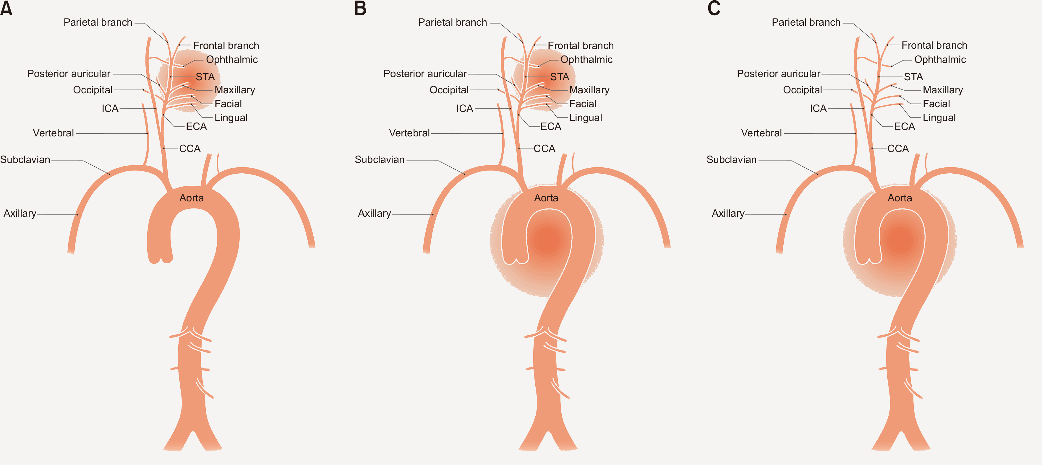
GCA is classified into two clinical phenotypes: C-GCA and LV-GCA. However, mixed cranial and large-vessel disease is observed in more than half of patients (Figure 1) [50]. The characteristics of each phenotype are summarized in Table 3 [51-53]. C-GCA affects older patients (mean age in the mid-70s) with a short diagnostic delay, while LV-GCA presents in slightly younger individuals (late 60s to early 70s) with a longer delay [54]. C-GCA presents with new-onset headache, visual symptoms, and jaw or tongue claudication, whereas LV-GCA more often manifests with as extracranial bruits, limb claudication, and inter-arm blood pressure discrepancy [55]. LV-GCA is more often associated with systemic inflammation with fever, and weight loss, while C-GCA shows milder systemic symptoms despite severe cranial ischemic events. While relapse rates are comparable, LV-GCA has a higher risk of aortic dilatation and aneurysm formation, requiring systematic vascular imaging and long-term surveillance of the aorta. The association of PMR with C-GCA versus LV-GCA varies according to cohort and case definition [53]. Despite this variability, large vessel involvement in patients with PMR was first recognized decades ago [56]. In this context, the concept of GCA-PMR spectrum disease has been proposed as a unifying framework that encompasses PMR and GCA on the basis of shared clinical features and common pathophysiology [57].

Pathology, pathogenesis, and therapeutic perspectives

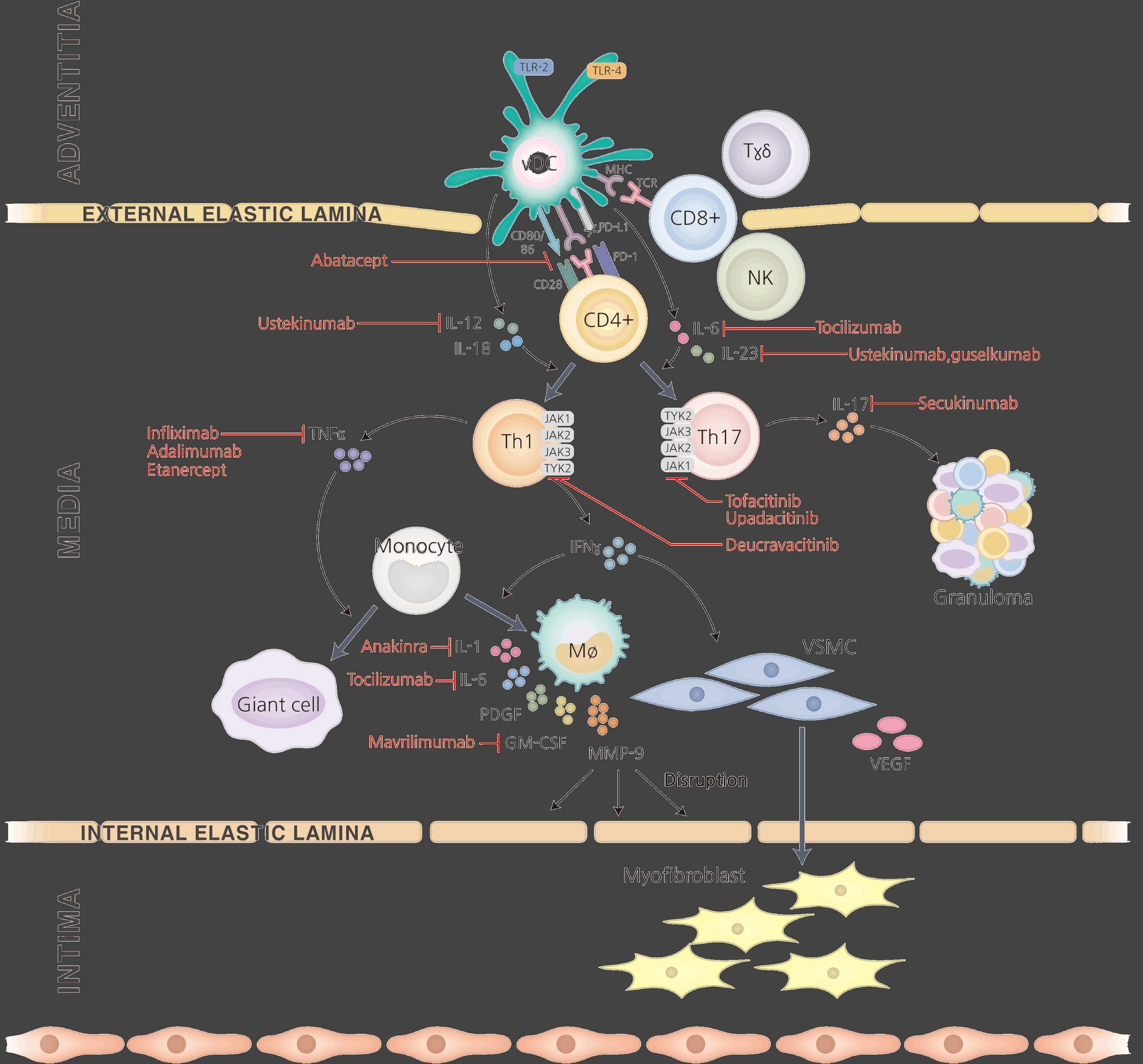
Recent advancements in the treatment of LVV have been substantially propelled by a deeper understanding of the underlying pathology and pathogenesis of this disease (Figure 2).

1) Sustained relevance of TAB

Despite the shift toward imaging, TAB remains a crucial diagnostic tool for cranial GCA because of its excellent specificity. This is particularly relevant when imaging findings are inconclusive due to factors such as atherosclerosis or healed arteritis [58].
While TAB’s sensitivity can be influenced by patient selection and specimen handling, some reports have indicated that overreliance on a positive TAB result could lead to unnecessarily prolonged steroid use [59]. The sensitivity can be enhanced by obtaining longer specimens and examining a greater number of sections. Nevertheless, both imaging and TAB are operator-dependent, and their results can be influenced by the timing of GCs treatment. A Cochrane meta-analysis was unable to conclude whether imaging could replace TAB owing to heterogeneous data and inconsistent diagnostic thresholds [60].
A sequential strategy that employs high-sensitivity imaging for initial screening, such as ultrasonography in GCA fast-tract clinics, and reserves a high-specificity tool, such as TAB, for ambiguous cases presents a logical and economically efficient approach. This finding is supported by the TABUL study, which demonstrated the cost-effectiveness of imaging-led algorithms while still acknowledging TAB’s essential role in resolving equivocal findings [44]. Ultimately, TAB and imaging are not mutually exclusive, but rather complementary tools, each holding a distinct and valuable role in the diagnostic pathway for GCA.

2) Histopathology

Our understanding of the histopathologic features of LVV, particularly GCA, is largely derived from TAB specimens, whereas obtaining sufficient tissue for TAK remains highly limited. Although GCA and TAK target the aortic wall, they have different histologic characteristics. In both diseases, immune infiltrates, granulomas, and multinucleated giant cells primarily involve the media and extend toward the intima and are infrequently observed in the adventitia [61]. Granulomas and giant cells are not invariably present, and their absence does not exclude diagnosis [62]. When present, granulomas in TAK tend to be more compact and well-formed [63]. Despite a remarkable heterogeneity of GCA specimens, four histological patterns have been described: (1) a transmural inflammatory pattern, (2) inflammation confined to the adventitia, (3) small vessel vasculitis distal to the adventitia, and (4) vasa vasoritis [64]. Loss of smooth muscle cells in the media with resultant aneurysmal remodeling can occur in both diseases. By contrast, stenosis of the distal aortic branches is often severe in TAK, whereas such lesions are unusual in GCA. The cellular composition of the inflammatory infiltrate is also different, as GCA lesions are dominated by CD4+ T cells, whereas TAK lesions show prominent CD8+ T cell and natural killer (NK) cell components. Tissue compartment involvement further distinguishes the two entities, as GCA more commonly involves the intima and inner media, whereas TAK often exhibits marked adventitial expansion with accumulation of collagen-rich, newly formed tissue that appears blue with Masson’s trichrome staining [61]. In later phases, adventitial and periadventitial fibrosis are typical features of TAK and are generally absent in GCA [61].

3) Pathogenesis

Explanations of LVV pathogenesis have largely been derived from studies on GCA because access to TAK arterial lesions is typically limited to surgical specimens [65]. Although TAK and GCA share many histopathological features, important differences in clinical phenotype persist. Emerging evidence on the genetic architecture and immunological phenotypes is beginning to map areas of overlap and divergence when extrapolating GCA-derived mechanisms to TAK.

(1) Genetic background

Marked geo-ethnic variability in disease incidence supports a substantial genetic contribution and etiological differences between GCA and TAK [11]. GCA shows a long-recognized association with human leukocyte antigen (HLA) class II, particularly HLA-DRB1*04. This finding was confirmed and refined by a large multicenter genome-wide association study (GWAS) involving more than 2,000 individuals of European ancestry [66]. By contrast, TAK is consistently linked to HLA class I, with the strongest signal for HLA-B*52:01 across cohorts [67]. These class-specific patterns parallel the cellular milieu in tissues, with CD4+ T cells predominating in GCA lesions and CD8+ T cells predominating in TAK. The non-HLA risk variants further distinguish between the two diseases. In GCA, associations have been reported for the genes tumor necrosis factor (TNF) and its receptors [68], intracellular adhesion molecule 1 (ICAM1) [69], vascular endothelial growth factor (VEGF) [70], toll-like receptor 4 (TLR4) [71], protein tyrosine phosphatase non-receptor 22 (PTPN22) [72], plasminogen (PLG) and an isoform of the α-subunit of collagen prolyl 4-hydroxylase (P4HA2) [66]. In TAK, a multi-ancestral GWAS revealed susceptible loci in SVEP1, CFL2, VPS8, and chr13q21 and confirmed earlier signals in IL12B, PTK2B, and chr21q22 [67]. Despite these differences, both conditions are susceptibility to IL12B, which encodes the p40 subunit common to interleukin (IL)-12 and IL-23, driving the differentiation between Th1 and Th17 cells, respectively [73].

(2) Vascular inflammation and remodeling

The healthy arterial wall is an immune-privileged site, largely devoid of resident leukocytes, except for a sparse population of vascular dendritic cells (vDCs) at the adventitia-media interface [74]. Loss of tolerance leads to immune activation of vDCs via TLR2/4 by an unknown trigger [75]. Upon activation, vDCs upregulate the major histocompatibility complex class II (MHC II) and costimulatory molecules CD83, CD80, and CD86, leading to the recruitment and priming of T cells [76]. Concurrently, low expression of programmed cell death-ligand 1 (PD-L1) on vDCs fails to restrain PD-1+ effector T cells [77], permitting a massive influx of T cells and their polarization toward the Th1 and Th17 lineages [78]. This cascade is partially mediated by the Janus kinase-signal transducer and activator of transcription (JAK-STAT) signaling [79]. Th1 cells release interferon-gamma (IFN-γ), which promotes the differentiation and activation of monocyte-derived macrophages, and TNF-α, which contributes multinucleated giant cell formation [80]. Activated macrophages overexpress MMP-9, disrupting the elastic laminae and creating conduits into the intima [81]. They also release a range of effector molecules, including IL-6, IL-12, IL-23, IL-1β, VEGF, and platelet-derived growth factor (PDGF) [20]. VEGF induces Jagged1 expression on adventitial microvascular endothelial cells of the vasa vasorum, which engages Notch1 on T cells and amplifies T cell recruitment, thereby promoting vascular remodeling [82]. The skewing of macrophage phenotypes leads to the release of granulocyte macrophage colony-stimulating factor (GM-CSF) and macrophage colony-stimulating factor (M-CSF), which further contribute to tissue destruction and intimal hyperplasia [83]. Mammalian target of rapamycin complex 1 (mTORC1) further drives pro-inflammatory T cell polarization in GCA and TAK [84]. In TAK, the expansion of Th17.1 and PD-1+ Th17 subsets is prominent and relatively glucocorticoid-refractory [85]. In contrast, the IL-6/Th17 axis in GCA is typically glucocorticoid-responsive, whereas the persistence of glucocorticoid-resistant Th1 cells often leads to chronic diseases [86]. Vascular inflammation drives maladaptive vascular remodeling, mediated primarily by endothelial cells and vascular smooth muscle cells (VSMCs) [79]. This process is characterized by medial destruction and concomitant development of a neointima composed of myofibroblasts and extracellular matrix (ECM), leading to intimal hyperplasia and arterial occlusion [87]. Although both GCA and TAK manifest as panarteritis, TAK is distinguished by more pronounced vascular fibrosis and a stronger cytotoxic signature contributed by perforin-secreting CD8+ T cells and NK cells [61]. Arterial tertial lymphoid organs are observed in both GCA and TAK; however, the role of B cells in GCA remains unclear, whereas multiple functions are recognized in TAK [20,76]. Advanced fibrosis in TAK is driven by transforming growth factor-β (TGF-β) produced by M2-polarized macrophages and PD-1+ Th17 cells, with mast-cell mediators acting in concert, and activated fibroblasts then perpetuating fibrosis by depositing ECM and releasing additional TGF-β [88].
Aging likely contributes to a high incidence of GCA in two aspects [89]: first, immunosenescence, characterized by the contraction of naïve and regulatory T cell pools, dysregulated cytokine networks, and diminished responsiveness to immune signals and second, age-related arterial remodeling, including endothelial dysfunction, elastin fragmentation, and adventitial neovascularization, which lowers the threshold for vascular immune activation.

4) Therapeutic strategies and monitoring

(1) GCs and non-GC immunosuppressants

Although GCs remain the cornerstone of LVV treatment, systematic studies on the optimal dose, tapering schedule, and treatment duration are lacking. Both the 2018 EULAR and 2021 ACR/Vasculitis Foundation (VF) guidelines recommend high-dose GC induction followed by tapering; however, the recommended pace differs slightly between GCA and TAK [8,90]. Even with a high risk of relapse with GC monotherapy in both diseases, it may still be considered for selected patients with GCA; however, the treatment is generally not advised for TAK because of its chronic and relapsing nature.
In GCA, methotrexate (MTX) has yielded modest benefits in randomized controlled trials (RCTs). Other agents such as leflunomide (LEF), mycophenolate mofetil (MMF), dapsone, and cyclophosphamide have only been evaluated in small, low-quality studies [20]. However, for TAK, both the EULAR and ACR/VF guidelines recommend initiating non-GC immunosuppressants with GCs as a first-line therapy. While evidence for MTX, LEF, MMF, and azathioprine is limited to small, low quality studies, cyclophosphamide is typically avoided because of its adverse effects and significant concern regarding fertility, especially considering the younger age of patients with TAK [91].

(2) Targeted therapy

Tocilizumab, an IL-6 receptor-blocking monoclonal antibody, is the best established treatment for GCA. Initially recommended by the EULAR in refractory or relapsing disease, the 2021 ACR/VF guideline expanded its role to first-line therapy in combination with GCs. Although the pivotal Giant-Cell Arteritis Actemra (GiACTA) trial confirmed its steroid-sparing effect, uncertainties remain regarding the duration of therapy, patient selection for long-term use, and impact on aortic complications [92]. Contrary to expectations, TNF inhibitors, including infliximab, etanercept, and adalimumab, have failed in multiple trials. Abatacept, a fusion protein comprising cytotoxic T-lymphocyte protein 4 (CTLA-4) and the Fc region of IgG1, demonstrated higher relapse-free survival than placebo in a phase II trial [93]. Baricitinib, a JAK1/JAK2 inhibitor, showed favorable outcomes in a small proof-of-concept phase II study [94]. Upadacitinib, a JAK1 inhibitor, was recently seen to achieve the primary endpoint in the phase III SELECT-GCA trial by reducing relapse and cumulative GC exposure [95]. Mavrilimumab, an anti-GM-CSF receptor antibody, was superior to placebo in a phase II study [96]. In TAK, tocilizumab failed to meet its primary endpoint in an RCT but demonstrated benefits in an observational cohort study. TNF inhibitors are extensively used and associated with improved clinical and radiographic stability, despite limited evidence from RCTs [20]. Without head-to-head trials, the ACR/VF guideline for TAK prioritizes TNF inhibition over tocilizumab, whereas the EULAR guidelines allow either agent in relapsing or refractory disease [8,90]. Abatacept did not demonstrate efficacy in a phase II trial for TAK. Trials of other targeted therapies for GCA and TAK are shown in Table 4.

(3) Special considerations

Regarding revascularization, the EULAR and ACR/VF guidelines advise a conservative, “medical-first” approach for GCA and TAK, reserving endovascular or surgical procedures for selected patients [8,90]. In cases of new or worsening ischemia without a life-threatening compromise, escalating or optimizing immunosuppression is preferred over immediate intervention. Elective revascularization should be deferred during active disease and performed in stable remission. If the procedure is not delayed, even in patients with active disease, high-dose GCs should be given pre-procedurally to control vascular inflammation.
Females with TAK have higher rates of adverse pregnancy outcomes than the general population, with 16% having a miscarriage, 37% having hypertension, and 14% having preeclampsia [97]. Preconception counseling, careful blood pressure control, and stringent maintenance of remission are essential elements of care for females considering pregnancy.
LVV increases cardiovascular risk through chronic low-grade inflammation and cumulative GC exposure. This risk is amplified by advanced age in GCA and by prolonged exposure over a longer life expectancy in TAK. Nevertheless, routine antiplatelet therapy is not recommended for either disease [8,90]. Imminent critical ischemia, such as threatened cerebral perfusion or coronary involvement, warrants the use of aspirin. Statins should not be used because of their supposed immunosuppressive effects in GCA. Lipid-lowering therapy should adhere to the standard cardiovascular risk-based indications.

(4) Monitoring disease activity and treatment response

Despite strong recommendations in the guidelines, monitoring disease activity and treatment response in LVV remains challenging owing to the nonspecific nature of the symptoms and the absence of a single, validated composite measure. Nevertheless, imaging is becoming increasingly critical, particularly because of its ability to detect inflammatory changes in the vessel walls. Several imaging-based scoring systems have been proposed, including the Southend Score [98] and the Outcome Measures in Rheumatology (OMERACT) Giant Cell Arteritis Ultrasound Score (OGUS) [99] using ultrasonography, MR Vasculitis Activity Score (MRVAS) using MRI [100], PET Vascular Activity Score (PETVAS) using PET/CT [101], and the Vascular Activity using MR/PET (VAMP) [102], although their use in clinical practice remains limited except for PETVAS. Historically, the monitoring of inflammation relied on classical markers such as C-reactive protein (CRP) and erythrocyte sedimentation rate (ESR) [90]. However, their use is challenging in patients treated with IL-6 inhibitors. Candidate biomarkers, such as S100A8/S100A9 proteins (calprotectin), pentraxin-3, VEGF, and angiopoietins offer potential alternative [103-106].

Gaps, controversies, and research agenda in Korea

Considerable advances have been clarified the similarities and differences in LVV (Table 5). However, critical gaps and controversies remain in the Korean literature. From an epidemiological standpoint, robust domestic data are scarce for GCA and are outdated for TAK, limiting accurate estimates of disease burden and impeding cross-ethnic comparisons. In parallel, harmonized genetic characterization of both diseases is needed to clarify the geo-ethnic differences and shared versus distinct pathways. Given Korea’s rapid transition to a super-aged society, the prevalence of GCA is likely to increase, either coexisting with PMR or emerging subsequently, raising public health concerns. Moreover, the traditional age-based dichotomy between GCA and TAK warrants reexamination in Korean cohorts, with a focus on the spectrum and overlap of large vessel involvement. Although the 2023 EULAR imaging update provides clear technical and operational standards, a uniform implementation across Korean centers is not yet realistic. Only a limited number of centers routinely perform TAB, and the rapid integration of vascular ultrasonography into clinical practice remains difficult, partly because of the perceived low pre-test probability in Korean patients. Targeted competency-based training in vascular ultrasonography for rheumatologists may be strategically important to avoid the under-recognition of GCA in older adults presenting with visual symptoms and new-onset headaches. Finally, the field is constrained by the absence of a standardized and validated composite outcome measure for LVV, limited availability of novel serum biomarkers, and insufficient integration of advanced imaging tools to quantify vascular wall disease activity and treatment response. Addressing these deficits will require coordinated, multi-center efforts, and alignment with international standards. Nonetheless, two priorities are particularly pressing in Korea: first, to ascertain population-level incidence and prevalence with disease characterization from contemporary, high-quality data; and second, to improve access to high-cost biological therapies for the small but clinically severe subset of patients who would benefit most.

CONCLUSION

LVV care is advancing through improved imaging, deepened immunological insights, and expanding therapeutic options. Further progress depends on validated outcomes, practical biomarkers, and uniform imaging practices. Multidisciplinary, patient-centered care and coordinated work across centers can translate current evidence into routine practice in Korea and lay the groundwork for future trials.
ACKNOWLEDGMENTS

None.

Notes

FUNDING

None.

CONFLICT OF INTEREST

No potential conflict of interest relevant to this article was reported.

AUTHOR CONTRIBUTIONS

JH, YAL, and SJH contributed to the study conception, design, data collection, and the interpretation of data. JH drafted the manuscript, and all authors contributed to revising the manuscript. All authors reviewed and approved the final version of the manuscript.

REFERENCES

1. Jennette JC, Falk RJ, Bacon PA, Basu N, Cid MC, Ferrario F, et al. 2013; 2012 revised International Chapel Hill Consensus Conference Nomenclature of Vasculitides. Arthritis Rheum. 65:1–11. DOI: 10.1002/art.37715. PMID: 23045170.
2. Takayasu M. 1908; A case with curious change in the central retinal vessel. J Jpn Ophthalmol Soc. 12:554–5.
3. Sugiyama K, Ijiri S, Tagawa S, Shimizu K. 2009; Takayasu disease on the centenary of its discovery. Jpn J Ophthalmol. 53:81–91. DOI: 10.1007/s10384-009-0650-2. PMID: 19333690.
4. Horton BT, Magath TB, Brown GE. 1934; Arteritis of the temporal vessels: a previously undescribed form. Arch Intern Med (Chic). 53:400–9. DOI: 10.1001/archinte.1934.00160090077007.
5. Hunder GG. 2006; The early history of giant cell arteritis and polymyalgia rheumatica: first descriptions to 1970. Mayo Clin Proc. 81:1071–83. DOI: 10.4065/81.8.1071. PMID: 16901030.
6. Grayson PC, Ponte C, Suppiah R, Robson JC, Gribbons KB, Judge A, et al. 2022; 2022 American College of Rheumatology/EULAR classification criteria for Takayasu arteritis. Ann Rheum Dis. 81:1654–60. DOI: 10.1136/ard-2022-223482. PMID: 36351705.
7. Ponte C, Grayson PC, Robson JC, Suppiah R, Gribbons KB, Judge A, et al. 2022; 2022 American College of Rheumatology/EULAR classification criteria for giant cell arteritis. Ann Rheum Dis. 81:1647–53. DOI: 10.1136/ard-2022-223480. PMID: 36351706.
8. Maz M, Chung SA, Abril A, Langford CA, Gorelik M, Guyatt G, et al. 2021; 2021 American College of Rheumatology/Vasculitis Foundation guideline for the management of giant cell arteritis and Takayasu arteritis. Arthritis Rheumatol. 73:1349–65. DOI: 10.1002/art.41774. PMID: 34235884. PMCID: PMC12344528.
9. Dejaco C, Ramiro S, Bond M, Bosch P, Ponte C, Mackie SL, et al. 2024; EULAR recommendations for the use of imaging in large vessel vasculitis in clinical practice: 2023 update. Ann Rheum Dis. 83:741–51. DOI: 10.1136/ard-2023-224543. PMID: 37550004.
10. Watts RA, Hatemi G, Burns JC, Mohammad AJ. 2022; Global epidemiology of vasculitis. Nat Rev Rheumatol. 18:22–34. DOI: 10.1038/s41584-021-00718-8. PMID: 34853411. PMCID: PMC8633913.
11. Sun Y, Kong X, Dai X, Jiang L. 2024; Epidemiology of large vasculitis in Shanghai, China: a 10-year multicenter hospital-based study and systematic review. Int J Rheum Dis. 27:e15360. DOI: 10.1111/1756-185X.15360. PMID: 39402757.
12. Sanchez-Alvarez C, Crowson CS, Koster MJ, Warrington KJ. 2021; Prevalence of Takayasu arteritis: a population-based study. J Rheumatol. 48:952. DOI: 10.3899/jrheum.201463. PMID: 33589549. PMCID: PMC8456169.
13. Hall S, Barr W, Lie JT, Stanson AW, Kazmier FJ, Hunder GG. 1985; Takayasu arteritis. A study of 32 North American patients. Medicine (Baltimore). 64:89–99. DOI: 10.1097/00005792-198503000-00002. PMID: 2858047.
14. Park SJ, Kim HJ, Park H, Hann HJ, Kim KH, Han S, et al. 2017; Incidence, prevalence, mortality and causes of death in Takayasu arteritis in Korea - a nationwide, population-based study. Int J Cardiol. 235:100–4. DOI: 10.1016/j.ijcard.2017.02.086. PMID: 28283361.
15. Ahn SS, Han M, Park YB, Jung I, Lee SW. 2022; Incidence, prevalence and risk of stroke in patients with Takayasu arteritis: a nationwide population-based study in South Korea. Stroke Vasc Neurol. 7:149–57. DOI: 10.1136/svn-2020-000809. PMID: 34880114. PMCID: PMC9067261.
16. Lee JI, Park JW, Jung Y, Shin K, Choi SR, Kang EH, et al. 2024; Clinical characteristics and courses of Korean patients with giant cell arteritis: a multi-center retrospective study. J Rheum Dis. 31:160–70. DOI: 10.4078/jrd.2024.0007. PMID: 38957359. PMCID: PMC11215252.
17. Kang MK, Hong Y, Kim YH, Park HK, Kim SK, Sohn JH, et al. 2024; Diagnosis, treatment, and follow-up of giant-cell arteritis: a retrospective multicenter study. J Clin Neurol. 20:306–14. DOI: 10.3988/jcn.2023.0169. PMID: 38627226. PMCID: PMC11076186.
18. Kermani TA. 2019; Takayasu arteritis and giant cell arteritis: are they a spectrum of the same disease? Int J Rheum Dis. 22 Suppl 1:41–8. DOI: 10.1111/1756-185X.13288. PMID: 29624864.
19. Nordborg E, Nordborg C. 2003; Giant cell arteritis: epidemiological clues to its pathogenesis and an update on its treatment. Rheumatology (Oxford). 42:413–21. DOI: 10.1093/rheumatology/keg116. PMID: 12626790.
20. Pugh D, Karabayas M, Basu N, Cid MC, Goel R, Goodyear CS, et al. 2022; Large-vessel vasculitis. Nat Rev Dis Primers. 7:93. DOI: 10.1038/s41572-021-00327-5. PMID: 34992251. PMCID: PMC9115766.
21. Michailidou D, Rosenblum JS, Rimland CA, Marko J, Ahlman MA, Grayson PC. 2020; Clinical symptoms and associated vascular imaging findings in Takayasu's arteritis compared to giant cell arteritis. Ann Rheum Dis. 79:262–7. DOI: 10.1136/annrheumdis-2019-216145. PMID: 31649025. PMCID: PMC12207754.
22. Zaldivar Villon MLF, de la Rocha JAL, Espinoza LR. 2019; Takayasu arteritis: recent developments. Curr Rheumatol Rep. 21:45. DOI: 10.1007/s11926-019-0848-3. PMID: 31321560.
23. Dzhus M, Mostbauer H. 2023; Coronary artery lesions in Takayasu arteritis. Reumatologia. 61:460–72. DOI: 10.5114/reum/176483. PMID: 38322104. PMCID: PMC10839913.
24. Liao H, Zhang N, Pan L, Du J, Liu J, Zheng Y. 2023; Predictors for pulmonary artery involvement in Takayasu arteritis and its cluster analysis. Arthritis Res Ther. 25:9. DOI: 10.1186/s13075-022-02987-4. PMID: 36639641. PMCID: PMC9840297.
25. Kwon OC, Lee SW, Park YB, Oh JS, Lee SH, Hong S, et al. 2018; Extravascular manifestations of Takayasu arteritis: focusing on the features shared with spondyloarthritis. Arthritis Res Ther. 20:142. DOI: 10.1186/s13075-018-1643-7. PMID: 29996949. PMCID: PMC6042334.
26. Misra DP, Rathore U, Kopp CR, Patro P, Agarwal V, Sharma A. 2022; Presentation and clinical course of pediatric-onset versus adult-onset Takayasu arteritis-a systematic review and meta-analysis. Clin Rheumatol. 41:3601–13. DOI: 10.1007/s10067-022-06318-5. PMID: 35927524.
27. Tomelleri A, Campochiaro C, Sartorelli S, Cavalli G, De Luca G, Baldissera E, et al. 2019; Gender differences in clinical presentation and vascular pattern in patients with Takayasu arteritis. Scand J Rheumatol. 48:482–90. DOI: 10.1080/03009742.2019.1581838. PMID: 31064248.
28. Calamia KT, Hunder GG. 1981; Giant cell arteritis (temporal arteritis) presenting as fever of undetermined origin. Arthritis Rheum. 24:1414–8. DOI: 10.1002/art.1780241113. PMID: 7317119.
29. Soulages A, Sibon I, Vallat JM, Ellie E, Bourdain F, Duval F, et al. 2022; Neurologic manifestations of giant cell arteritis. J Neurol. 269:3430–42. DOI: 10.1007/s00415-022-10991-6. PMID: 35124749.
30. Soriano A, Muratore F, Pipitone N, Boiardi L, Cimino L, Salvarani C. 2017; Visual loss and other cranial ischaemic complications in giant cell arteritis. Nat Rev Rheumatol. 13:476–84. DOI: 10.1038/nrrheum.2017.98. PMID: 28680132.
31. Kim IY, Seo GH, Lee S, Jeong H, Kim H, Lee J, et al. 2014; Epidemiology of polymyalgia rheumatica in Korea. J Rheum Dis. 21:297–302. DOI: 10.4078/jrd.2014.21.6.297.
32. Ha JW, Song JJ, Park YB, Lee SW. 2025; Validation of the 2022 ACR/EULAR classification criteria for giant cell arteritis in Korean patients with giant cell arteritis. Mod Rheumatol. 35:524–8. DOI: 10.1093/mr/roae110. PMID: 39786768.
33. Gribbons KB, Ponte C, Carette S, Craven A, Cuthbertson D, Hoffman GS, et al. 2020; Patterns of arterial disease in Takayasu arteritis and giant cell arteritis. Arthritis Care Res (Hoboken). 72:1615–24. DOI: 10.1002/acr.24055. PMID: 31444857. PMCID: PMC7035996.
34. Kermani TA, Crowson CS, Muratore F, Schmidt J, Matteson EL, Warrington KJ. 2015; Extra-cranial giant cell arteritis and Takayasu arteritis: how similar are they? Semin Arthritis Rheum. 44:724–8. DOI: 10.1016/j.semarthrit.2015.01.005. PMID: 25712813.
35. Comarmond C, Biard L, Lambert M, Mekinian A, Ferfar Y, Kahn JE, et al. 2017; Long-term outcomes and prognostic factors of complications in Takayasu arteritis: a multicenter study of 318 patients. Circulation. 136:1114–22. DOI: 10.1161/CIRCULATIONAHA.116.027094. PMID: 28701469.
36. Carreño E, Olivas-Vergara OM. 2023; Systemic vasculitis and its association with the eye. Ophthalmologica. 246:174–80. DOI: 10.1159/000531395. PMID: 37331330.
37. Chun YS, Park SJ, Park IK, Chung H, Lee J. 2001; The clinical and ocular manifestations of Takayasu arteritis. Retina. 21:132–40. DOI: 10.1097/00006982-200104000-00006. PMID: 11321139.
38. Szydełko-Paśko U, Przeździecka-Dołyk J, Nowak Ł, Małyszczak A, Misiuk-Hojło M. 2023; Ocular manifestations of Takayasu's arteritis-a case-based systematic review and meta-analysis. J Clin Med. 12:3745. DOI: 10.3390/jcm12113745. PMID: 37297942. PMCID: PMC10253919.
39. Mackie SL, Dejaco C, Appenzeller S, Camellino D, Duftner C, Gonzalez-Chiappe S, et al. 2020; British Society for Rheumatology guideline on diagnosis and treatment of giant cell arteritis. Rheumatology (Oxford). 59:e1–23. DOI: 10.1093/rheumatology/kez672. PMID: 31970405.
40. Craven A, Robson J, Ponte C, Grayson PC, Suppiah R, Judge A, et al. 2013; ACR/EULAR-endorsed study to develop Diagnostic and Classification Criteria for Vasculitis (DCVAS). Clin Exp Nephrol. 17:619–21. DOI: 10.1007/s10157-013-0854-0. PMID: 23996327.
41. Arend WP, Michel BA, Bloch DA, Hunder GG, Calabrese LH, Edworthy SM, et al. 1990; The American College of Rheumatology 1990 criteria for the classification of Takayasu arteritis. Arthritis Rheum. 33:1129–34. DOI: 10.1002/art.1780330811. PMID: 1975175.
42. Hunder GG, Bloch DA, Michel BA, Stevens MB, Arend WP, Calabrese LH, et al. 1990; The American College of Rheumatology 1990 criteria for the classification of giant cell arteritis. Arthritis Rheum. 33:1122–8. DOI: 10.1002/art.1780330810. PMID: 2202311. PMCID: PMC3627791.
43. Schmidt WA, Schäfer VS. 2024; Diagnosing vasculitis with ultrasound: findings and pitfalls. Ther Adv Musculoskelet Dis. 16:1759720X241251742. DOI: 10.1177/1759720X241251742. PMID: 38846756. PMCID: PMC11155338.
44. Luqmani R, Lee E, Singh S, Gillett M, Schmidt WA, Bradburn M, et al. 2016; The role of ultrasound compared to biopsy of temporal arteries in the diagnosis and treatment of giant cell arteritis (TABUL): a diagnostic accuracy and cost-effectiveness study. Health Technol Assess. 20:1–238. DOI: 10.3310/hta20900. PMID: 27925577. PMCID: PMC5165283.
45. Duftner C, Redlinger N, Bruyn GA, Chadachan V, Chrysidis S, Correyero M, et al. 2025; Reliability of the OMERACT giant cell arteritis ultrasonography score (OGUS): results of a patient-based exercise involving experts and non-experts in vascular ultrasonography. RMD Open. 11:e004667. DOI: 10.1136/rmdopen-2024-004667. PMID: 39753294. PMCID: PMC11749318.
46. Duftner C, Dejaco C, Sepriano A, Falzon L, Schmidt WA, Ramiro S. 2018; Imaging in diagnosis, outcome prediction and monitoring of large vessel vasculitis: a systematic literature review and meta-analysis informing the EULAR recommendations. RMD Open. 4:e000612. DOI: 10.1136/rmdopen-2017-000612. PMID: 29531788. PMCID: PMC5845406.
47. Lariviere D, Benali K, Coustet B, Pasi N, Hyafil F, Klein I, et al. 2016; Positron emission tomography and computed tomography angiography for the diagnosis of giant cell arteritis: a real-life prospective study. Medicine (Baltimore). 95:e4146. DOI: 10.1097/MD.0000000000004146. PMID: 27472684. PMCID: PMC5265821.
48. Soussan M, Nicolas P, Schramm C, Katsahian S, Pop G, Fain O, et al. 2015; Management of large-vessel vasculitis with FDG-PET: a systematic literature review and meta-analysis. Medicine (Baltimore). 94:e622. DOI: 10.1097/MD.0000000000000622. PMID: 25860208. PMCID: PMC4554050.
49. Martin O, Schaarschmidt BM, Kirchner J, Suntharalingam S, Grueneisen J, Demircioglu A, et al. 2020; PET/MRI versus PET/CT for whole-body staging: results from a single-center observational study on 1,003 sequential examinations. J Nucl Med. 61:1131–6. DOI: 10.2967/jnumed.119.233940. PMID: 31806777.
50. van der Geest KSM, Sandovici M, Bley TA, Stone JR, Slart RHJA, Brouwer E. 2024; Large vessel giant cell arteritis. Lancet Rheumatol. 6:e397–408. DOI: 10.1016/S2665-9913(23)00300-4. PMID: 38574745. PMCID: PMC13022965.
51. Saadoun D, Vautier M, Cacoub P. 2021; Medium- and large-vessel vasculitis. Circulation. 143:267–82. DOI: 10.1161/CIRCULATIONAHA.120.046657. PMID: 33464968.
52. Koster MJ, Matteson EL, Warrington KJ. 2018; Large-vessel giant cell arteritis: diagnosis, monitoring and management. Rheumatology (Oxford). 57:ii32–42. DOI: 10.1093/rheumatology/kex424. PMID: 29982778.
53. González-Gay MÁ, Heras-Recuero E, Blázquez-Sánchez T, Caraballo-Salazar C, Rengifo-García F, Castañeda S, et al. 2024; Broadening the clinical spectrum of giant cell arteritis: from the classic cranial to the predominantly extracranial pattern of the disease. Expert Rev Clin Immunol. 20:1089–100. DOI: 10.1080/1744666X.2024.2356741. PMID: 38757894.
54. Monti S, Milanesi A, Klersy C, Tomelleri A, Dagna L, Campochiaro C, et al. 2023; Age at diagnosis influences the clinical phenotype, treatment strategies and outcomes in patients with giant cell arteritis: results from the observational GCAGE study on a large cohort of 1004 patients. Ann Rheum Dis. 82:1098–106. DOI: 10.1136/ard-2023-223895. PMID: 37188498.
55. Estrada P, Narváez J, Moya P, Roig-Vilaseca D, Camacho O, Navarro V, et al. 2024; Clinical phenotypes of giant cell arteritis: insights into complications and survival outcomes. Eur J Rheumatol. 11:33–8. DOI: 10.5152/eurjrheum.2024.23065. PMID: 39576031. PMCID: PMC11448687.
56. HAMRIN B, JONSSON N, LANDBERG T. 1965; Involvement of large vessels in polymyalgia arteritica. Lancet. 1:1193–6. DOI: 10.1016/S0140-6736(65)92723-6. PMID: 14288559.
57. Dejaco C, Duftner C, Buttgereit F, Matteson EL, Dasgupta B. 2017; The spectrum of giant cell arteritis and polymyalgia rheumatica: revisiting the concept of the disease. Rheumatology (Oxford). 56:506–15. DOI: 10.1093/rheumatology/kew273. PMID: 27481272. PMCID: PMC12199278.
58. Stamatis P, Turesson C, Mohammad AJ. 2024; Temporal artery biopsy in giant cell arteritis: clinical perspectives and histological patterns. Front Med (Lausanne). 11:1453462. DOI: 10.3389/fmed.2024.1453462. PMID: 39386746. PMCID: PMC11461189.
59. Ponich B, Lafreniere AS, Hartley R, Temple-Oberle CF. 2022; Temporal artery biopsy debate: positive TAB result prolongs steroid use in giant cell arteritis. Plast Reconstr Surg Glob Open. 10:e4652. DOI: 10.1097/GOX.0000000000004652. PMID: 36438469. PMCID: PMC9681624.
60. Pouncey AL, Yeldham G, Magan T, Lucenteforte E, Jaffer U, Virgili G. 2024; Halo sign on temporal artery ultrasound versus temporal artery biopsy for giant cell arteritis. Cochrane Database Syst Rev. 2:CD013199. DOI: 10.1002/14651858.CD013199.pub2. PMID: 38323659. PMCID: PMC10848297.
61. Watanabe R, Berry GJ, Liang DH, Goronzy JJ, Weyand CM. 2020; Pathogenesis of giant cell arteritis and Takayasu arteritis-similarities and differences. Curr Rheumatol Rep. 22:68. DOI: 10.1007/s11926-020-00948-x. PMID: 32845392. PMCID: PMC9112376.
62. Maleszewski JJ, Younge BR, Fritzlen JT, Hunder GG, Goronzy JJ, Warrington KJ, et al. 2017; Clinical and pathological evolution of giant cell arteritis: a prospective study of follow-up temporal artery biopsies in 40 treated patients. Mod Pathol. 30:788–96. DOI: 10.1038/modpathol.2017.10. PMID: 28256573. PMCID: PMC5650068.
63. Stone JR, Bruneval P, Angelini A, Bartoloni G, Basso C, Batoroeva L, et al. 2015; Consensus statement on surgical pathology of the aorta from the Society for Cardiovascular Pathology and the Association for European Cardiovascular Pathology: I. Inflammatory diseases. Cardiovasc Pathol. 24:267–78. DOI: 10.1016/j.carpath.2015.05.001. PMID: 26051917.
64. Cavazza A, Muratore F, Boiardi L, Restuccia G, Pipitone N, Pazzola G, et al. 2014; Inflamed temporal artery: histologic findings in 354 biopsies, with clinical correlations. Am J Surg Pathol. 38:1360–70. DOI: 10.1097/PAS.0000000000000244. PMID: 25216320.
65. Tombetti E, Mason JC. 2019; Takayasu arteritis: advanced understanding is leading to new horizons. Rheumatology (Oxford). 58:206–19. DOI: 10.1093/rheumatology/key040. PMID: 29635396.
66. Carmona FD, Vaglio A, Mackie SL, Hernández-Rodríguez J, Monach PA, Castañeda S, et al. 2017; A genome-wide association study identifies risk alleles in plasminogen and P4HA2 associated with giant cell arteritis. Am J Hum Genet. 100:64–74. DOI: 10.1016/j.ajhg.2016.11.013. PMID: 28041642. PMCID: PMC5223025.
67. Ortiz-Fernández L, Saruhan-Direskeneli G, Alibaz-Oner F, Kaymaz-Tahra S, Coit P, Kong X, et al. 2021; Identification of susceptibility loci for Takayasu arteritis through a large multi-ancestral genome-wide association study. Am J Hum Genet. 108:84–99. DOI: 10.1016/j.ajhg.2020.11.014. PMID: 33308445. PMCID: PMC7820633.
68. Mattey DL, Hajeer AH, Dababneh A, Thomson W, González-Gay MA, García-Porrúa C, et al. 2000; Association of giant cell arteritis and polymyalgia rheumatica with different tumor necrosis factor microsatellite polymorphisms. Arthritis Rheum. 43:1749–55. DOI: 10.1002/1529-0131(200008)43:8<1749::AID-ANR11>3.0.CO;2-K. PMID: 10943865.
69. Salvarani C, Casali B, Boiardi L, Ranzi A, Macchioni P, Nicoli D, et al. 2000; Intercellular adhesion molecule 1 gene polymorphisms in polymyalgia rheumatica/giant cell arteritis: association with disease risk and severity. J Rheumatol. 27:1215–21.
70. Rueda B, Lopez-Nevot MA, Lopez-Diaz MJ, Garcia-Porrua C, Martín J, Gonzalez-Gay MA. 2005; A functional variant of vascular endothelial growth factor is associated with severe ischemic complications in giant cell arteritis. J Rheumatol. 32:1737–41.
71. Palomino-Morales R, Torres O, Vazquez-Rodriguez TR, Morado IC, Castañeda S, Callejas-Rubio JL, et al. 2009; Association between toll-like receptor 4 gene polymorphism and biopsy-proven giant cell arteritis. J Rheumatol. 36:1501–6. DOI: 10.3899/jrheum.081286. PMID: 19531762.
72. Serrano A, Márquez A, Mackie SL, Carmona FD, Solans R, Miranda-Filloy JA, et al. 2013; Identification of the PTPN22 functional variant R620W as susceptibility genetic factor for giant cell arteritis. Ann Rheum Dis. 72:1882–6. DOI: 10.1136/annrheumdis-2013-203641. PMID: 23946333. PMCID: PMC4053592.
73. Carmona FD, Coit P, Saruhan-Direskeneli G, Hernández-Rodríguez J, Cid MC, Solans R, et al. 2017; Analysis of the common genetic component of large-vessel vasculitides through a meta-Immunochip strategy. Sci Rep. 7:43953. DOI: 10.1038/srep43953. PMID: 28277489. PMCID: PMC5344032.
74. Weyand CM, Goronzy JJ. 2003; Medium- and large-vessel vasculitis. N Engl J Med. 349:160–9. DOI: 10.1056/NEJMra022694. PMID: 12853590.
75. Ma-Krupa W, Kwan M, Goronzy JJ, Weyand CM. 2005; Toll-like receptors in giant cell arteritis. Clin Immunol. 115:38–46. DOI: 10.1016/j.clim.2005.02.009. PMID: 15870019.
76. Greigert H, Genet C, Ramon A, Bonnotte B, Samson M. 2022; New insights into the pathogenesis of giant cell arteritis: mechanisms involved in maintaining vascular inflammation. J Clin Med. 11:2905. DOI: 10.3390/jcm11102905. PMID: 35629030. PMCID: PMC9143803.
77. Zhang H, Watanabe R, Berry GJ, Vaglio A, Liao YJ, Warrington KJ, et al. 2017; Immunoinhibitory checkpoint deficiency in medium and large vessel vasculitis. Proc Natl Acad Sci U S A. 114:E970–9. DOI: 10.1073/pnas.1616848114. PMID: 28115719. PMCID: PMC5307483.
78. Weyand CM, Younge BR, Goronzy JJ. 2011; IFN-γ and IL-17: the two faces of T-cell pathology in giant cell arteritis. Curr Opin Rheumatol. 23:43–9. DOI: 10.1097/BOR.0b013e32833ee946. PMID: 20827207. PMCID: PMC3081721.
79. Ciccia F, Macaluso F, Mauro D, Nicoletti GF, Croci S, Salvarani C. 2021; New insights into the pathogenesis of giant cell arteritis: are they relevant for precision medicine? Lancet Rheumatol. 3:e874–85. DOI: 10.1016/S2665-9913(21)00253-8. PMID: 38287633.
80. Szekeres D, Al Othman B. 2022; Current developments in the diagnosis and treatment of giant cell arteritis. Front Med (Lausanne). 9:1066503. DOI: 10.3389/fmed.2022.1066503. PMID: 36582285. PMCID: PMC9792614.
81. Watanabe R, Maeda T, Zhang H, Berry GJ, Zeisbrich M, Brockett R, et al. 2018; MMP (matrix metalloprotease)-9-producing monocytes enable T cells to invade the vessel wall and cause vasculitis. Circ Res. 123:700–15. DOI: 10.1161/CIRCRESAHA.118.313206. PMID: 29970365. PMCID: PMC6202245.
82. Wen Z, Shen Y, Berry G, Shahram F, Li Y, Watanabe R, et al. 2017; The microvascular niche instructs T cells in large vessel vasculitis via the VEGF-Jagged1-Notch pathway. Sci Transl Med. 9:eaal3322. DOI: 10.1126/scitranslmed.aal3322. PMID: 28724574. PMCID: PMC5708299.
83. Jiemy WF, van Sleen Y, van der Geest KS, Ten Berge HA, Abdulahad WH, Sandovici M, et al. 2020; Distinct macrophage phenotypes skewed by local granulocyte macrophage colony-stimulating factor (GM-CSF) and macrophage colony-stimulating factor (M-CSF) are associated with tissue destruction and intimal hyperplasia in giant cell arteritis. Clin Transl Immunology. 9:e1164. DOI: 10.1002/cti2.1164. PMID: 32884747. PMCID: PMC7453134.
84. Maciejewski-Duval A, Comarmond C, Leroyer A, Zaidan M, Le Joncour A, Desbois AC, et al. 2018; MTOR pathway activation in large vessel vasculitis. J Autoimmun. 94:99–109. DOI: 10.1016/j.jaut.2018.07.013. PMID: 30061014.
85. Singh K, Rathore U, Rai MK, Behera MR, Jain N, Ora M, et al. 2022; Novel Th17 lymphocyte populations, Th17.1 and PD1+Th17, are increased in Takayasu arteritis, and both Th17 and Th17.1 sub-populations associate with active disease. J Inflamm Res. 15:1521–41. DOI: 10.2147/JIR.S355881. PMID: 35256852. PMCID: PMC8898181.
86. Deng J, Younge BR, Olshen RA, Goronzy JJ, Weyand CM. 2010; Th17 and Th1 T-cell responses in giant cell arteritis. Circulation. 121:906–15. DOI: 10.1161/CIRCULATIONAHA.109.872903. PMID: 20142449. PMCID: PMC2837465.
87. Parreau S, Vedrenne N, Regent A, Richard L, Sindou P, Mouthon L, et al. 2021; An immunohistochemical analysis of fibroblasts in giant cell arteritis. Ann Diagn Pathol. 52:151728. DOI: 10.1016/j.anndiagpath.2021.151728. PMID: 33798926.
88. Misra DP, Singh K, Sharma A, Agarwal V. 2023; Arterial wall fibrosis in Takayasu arteritis and its potential for therapeutic modulation. Front Immunol. 14:1174249. DOI: 10.3389/fimmu.2023.1174249. PMID: 37256147. PMCID: PMC10225504.
89. Weyand CM, Goronzy JJ. 2023; Immunology of giant cell arteritis. Circ Res. 132:238–50. DOI: 10.1161/CIRCRESAHA.122.322128. PMID: 36656970. PMCID: PMC9870348.
90. Hellmich B, Agueda A, Monti S, Buttgereit F, de Boysson H, Brouwer E, et al. 2020; 2018 Update of the EULAR recommendations for the management of large vessel vasculitis. Ann Rheum Dis. 79:19–30. DOI: 10.1136/annrheumdis-2019-215672. PMID: 31270110.
91. Kaymakci MS, Warrington KJ, Kermani TA. 2024; New therapeutic approaches to large-vessel vasculitis. Annu Rev Med. 75:427–42. DOI: 10.1146/annurev-med-060622-100940. PMID: 37683286.
92. Stone JH, Tuckwell K, Dimonaco S, Klearman M, Aringer M, Blockmans D, et al. 2017; Trial of tocilizumab in giant-cell arteritis. N Engl J Med. 377:317–28. DOI: 10.1056/NEJMoa1613849. PMID: 28745999.
93. Langford CA, Cuthbertson D, Ytterberg SR, Khalidi N, Monach PA, Carette S, et al. 2017; A randomized, double-blind trial of abatacept (CTLA-4Ig) for the treatment of giant cell arteritis. Arthritis Rheumatol. 69:837–45. DOI: 10.1002/art.40044. PMID: 28133925. PMCID: PMC5378642.
94. Koster MJ, Crowson CS, Giblon RE, Jaquith JM, Duarte-García A, Matteson EL, et al. 2022; Baricitinib for relapsing giant cell arteritis: a prospective open-label 52-week pilot study. Ann Rheum Dis. 81:861–7. DOI: 10.1136/annrheumdis-2021-221961. PMID: 35190385. PMCID: PMC9592156.
95. Blockmans D, Penn SK, Setty AR, Schmidt WA, Rubbert-Roth A, Hauge EM, et al. 2025; A phase 3 trial of upadacitinib for giant-cell arteritis. N Engl J Med. 392:2013–24. DOI: 10.1056/NEJMoa2413449. PMID: 40174237.
96. Cid MC, Unizony SH, Blockmans D, Brouwer E, Dagna L, Dasgupta B, et al. 2022; Efficacy and safety of mavrilimumab in giant cell arteritis: a phase 2, randomised, double-blind, placebo-controlled trial. Ann Rheum Dis. 81:653–61. DOI: 10.1136/annrheumdis-2021-221865. PMID: 35264321. PMCID: PMC8995812.
97. Partalidou S, Mamopoulos A, Dimopoulou D, Dimitroulas T. 2023; Pregnancy outcomes in Takayasu arteritis patients: a systematic review and meta-analysis. Sci Rep. 13:546. DOI: 10.1038/s41598-023-27379-9. PMID: 36631504. PMCID: PMC9834209.
98. van der Geest KSM, Borg F, Kayani A, Paap D, Gondo P, Schmidt W, et al. 2020; Novel ultrasonographic Halo Score for giant cell arteritis: assessment of diagnostic accuracy and association with ocular ischaemia. Ann Rheum Dis. 79:393–9. DOI: 10.1136/annrheumdis-2019-216343. PMID: 31900304. PMCID: PMC7034352.
99. Dejaco C, Ponte C, Monti S, Rozza D, Scirè CA, Terslev L, et al. 2023; The provisional OMERACT ultrasonography score for giant cell arteritis. Ann Rheum Dis. 82:556–64. DOI: 10.1136/ard-2022-223367. PMID: 36600183.
100. Froehlich M, Guggenberger KV, Vogt M, Mihatsch PW, Dalla Torre G, Werner RA, et al. 2024; MRVAS-introducing a standardized magnetic resonance scoring system for assessing the extent of inflammatory burden in giant cell arteritis. Rheumatology (Oxford). 63:2781–90. DOI: 10.1093/rheumatology/keae056. PMID: 38305463.
101. Grayson PC, Alehashemi S, Bagheri AA, Civelek AC, Cupps TR, Kaplan MJ, et al. 2018; 18 F-fluorodeoxyglucose-positron emission tomography as an imaging biomarker in a prospective, longitudinal cohort of patients with large vessel vasculitis. Arthritis Rheumatol. 70:439–49. DOI: 10.1002/art.40379. PMID: 29145713. PMCID: PMC5882488.
102. Pugh D, Patel D, Macnaught G, Czopek A, Bruce L, Donachie J, et al. 2024; 18F-FDG-PET/MR imaging to monitor disease activity in large vessel vasculitis. Nat Commun. 15:7314. DOI: 10.1038/s41467-024-51613-1. PMID: 39183340. PMCID: PMC11345444.
103. Dagna L, Salvo F, Tiraboschi M, Bozzolo EP, Franchini S, Doglioni C, et al. 2011; Pentraxin-3 as a marker of disease activity in Takayasu arteritis. Ann Intern Med. 155:425–33. DOI: 10.7326/0003-4819-155-7-201110040-00005. PMID: 21969341.
104. Baldini M, Maugeri N, Ramirez GA, Giacomassi C, Castiglioni A, Prieto-González S, et al. 2012; Selective up-regulation of the soluble pattern-recognition receptor pentraxin 3 and of vascular endothelial growth factor in giant cell arteritis: relevance for recent optic nerve ischemia. Arthritis Rheum. 64:854–65. DOI: 10.1002/art.33411. PMID: 21989653.
105. Springer JM, Monach P, Cuthbertson D, Carette S, Khalidi NA, McAlear CA, et al. 2018; Serum S100 proteins as a marker of disease activity in large vessel vasculitis. J Clin Rheumatol. 24:393–5. DOI: 10.1097/RHU.0000000000000729. PMID: 29470262. PMCID: PMC6105726.
106. van Sleen Y, Sandovici M, Abdulahad WH, Bijzet J, van der Geest KSM, Boots AMH, et al. 2019; Markers of angiogenesis and macrophage products for predicting disease course and monitoring vascular inflammation in giant cell arteritis. Rheumatology (Oxford). 58:1383–92. DOI: 10.1093/rheumatology/kez034. PMID: 30805622. PMCID: PMC6649803.

Figure 1
Schematic distribution of arterial involvement in GCA. (A) Cranial-limited GCA. Predominant disease in the extracranial branches of the carotid system, including the superficial temporal artery and its frontal/parietal branches, maxillary, facial, lingual, occipital, and ophthalmic arteries. (B) Mixed cranial and large-vessel GCA. Concurrent involvement of cranial arteries and the aorta with its major branches. (C) Large-vessel-limited GCA. Disease is most commonly confined to the thoracic aorta, abdominal aorta, brachiocephalic trunk, subclavian, axillary, carotid, vertebral, iliofemoral arteries. Shaded areas denote the involvement of typical vascular territories. Pooled estimates from prospective studies suggest ~33% cranial-limited GCA, ~12% large-vessel-limited GCA, and ~55% mixed cranial and large-vessel GCA. GCA: giant cell arteritis, STA: superficial temporal artery, ICA: internal carotid artery, ECA: external carotid artery, CCA: common carotid artery. Schematic concept adapted from the article of van der Geest et al. (Lancet Rheumatol 2024;6:e397-408) [50].
jrd-33-3-152-f1.tif
Figure 2
Immunopathogenesis and therapeutic targets in large vessel vasculitis. At the adventitia–media interface, vDCs sense danger signals via TLR2 and TLR4. Upon activation, vDCs upregulate MHC II and engage TCR on CD4+ T cells; CD80/86 on vDCs deliver costimulatory signals through CD28 on CD4+ T cells, while low PD-L1 fails to restrain PD-1+ effector T cells. CD4+ T cells polarize toward Th1 and Th17, with downstream signaling integrated by JAK1/2/3 and TYK2. Th1 cells produce IFN-γ and TNF-α, promoting activation of monocytes and macrophages. Th17 cells produce IL-17, contributing to giant-cell formation. Activated macrophages overexpress MMP-9, disrupting the elastic lamina and enabling intimal infiltration, and they release IL-1, IL-6, PDGF, GM-CSF, and VEGF that drive myofibroblast activation and VSMC proliferation, leading to intimal hyperplasia. CD8+ T cells, NK cells, and Tγδ cells are additionally implicated in Takayasu arteritis. Red T-bar denotes blockade by targeted therapies. Abatacept blocks CD28–CD80/CD86 interactions. Tocilizumab inhibits IL-6 signaling, while ustekinumab, guselkumab, and secukinumab target IL-12/23 or IL-17 pathways. TNF-α inhibitors (infliximab, adalimumab, etanercept) suppress TNF-driven inflammation. Anakinra blocks IL-1, and mavrilimumab antagonizes GM-CSF signaling. JAK inhibitors such as tofacitinib and upadacitinib interfere with downstream JAK-STAT pathways while deucravacitinib blocks TYK2. These interventions collectively modulate the inflammatory cascade and vascular injury that characterize LVV. TLR: toll-like receptor, vDC: vascular dendritic cell, MHC: major histocompatibility complex, TCR: T cell receptor, PD-L1: programmed cell death-ligand 1, PD-1: programmed cell death 1, CD4+: CD4+ T cell, CD8+: CD8+ T cell, Tγδ: gamma delta T cell, NK: natural killer, IL: interleukin, JAK: Janus kinase, TYK: tyrosine kinase, TNF: tumor necrosis factor, IFN-γ: interferon-gamma, Mø: macrophage, PDGF: platelet-derived growth factor, GM-CSF: granulocyte macrophage colony-stimulating factor, MMP: matrix metalloprotease, VSMC: vascular smooth muscle cell, VEGF: vascular endothelial growth factor.
jrd-33-3-152-f2.tif
Table 1
Evolution of classification criteria for Takayasu arteritis
Criterion 1990 ACR Item 2022 ACR/EULAR Point
Absolute requirement None Age ≤60 years at time of diagnosis
Evidence of vasculitis on imaging*
Additional criteria
Age Age at disease onset ≤40 years +1 (Absolute requirement)
Sex (Not applicable) Female +1
Carotid artery abnormality (Not applicable) Absent/reduced pulse or tenderness of carotid artery +2
Cardiac pain (Not applicable) Angina or ischemic cardiac pain +2
Extremity claudication One or more extremity, especially the upper extremities +1 Arm or leg +2
Upper extremity involvement Decreased brachial artery pulse +1 Absent/reduced pulse of axillary, brachial, or radial arteries +2
Differences of >10 mmHg in systolic BP between arms +1 Differences of ≥20 mm Hg in systolic BP between arms +1
Vascular bruit One or both subclavian arteries or abdominal aorta +1 Large artery including aorta, carotid, subclavian, axillary, brachial, renal, or iliofemoral arteries +2
Imaging Angiographic narrowing or occlusion of aorta and its primary branches +1 No. of affected arterial territories (select one)†
One arterial territory +1
Two arterial territories +2
Three or more arterial territories +3
Symmetric involvement of paired arteries‡ +1
Abdominal aorta involvement with renal or mesenteric involvement§ +3
Classification rule ≥3 of 6 items ≥5 points for 10 items

ACR: American College of Rheumatology, EULAR: European Alliance of Associations for Rheumatology, BP: blood pressure. *Evidence of vasculitis in the aorta or branch arteries must be confirmed by vascular imaging such as computed tomography/catheter-based/magnetic resonance angiography, ultrasonography, or positron emission tomography. †Number of arterial territories with luminal damage such as stenosis, occlusion, or aneurysm detected via angiography or ultrasonography from the following nine territories: thoracic aorta, abdominal aorta, mesenteric, right/left carotid, right/left subclavian, or right/left renal arteries. ‡Bilateral luminal damage, such as stenosis, occlusion, or aneurysm, detected via angiography or ultrasonography in any of the following paired vascular territories: carotid, subclavian, or renal arteries. §Luminal damage such as stenosis, occlusion, or aneurysm detected via angiography or ultrasonography involving the abdominal aorta and either renal or mesenteric arteries.

Table 2
Evolution of classification criteria for giant cell arteritis
Criterion 1990 ACR Item 2022 ACR/EULAR Point
Absolute requirement None Age ≥50 years at time of diagnosis
Additional criteria
Age Age at disease onset ≥50 years +1 (Absolute requirement)
Headache New onset of or new type of localized pain in the head +1 New temporal headache +2
Temporal artery abnormality Tenderness to palpation or decreased pulsation, unrelated to arteriosclerosis of cervical arteries +1 Absent/reduced pulse, tenderness, or hard “cord-like” appearance +2
Cranial symptom/sign (Not applicable) Sudden visual loss +3
Scalp tenderness +2
Jaw or tongue claudication +2
PMR symptoms (Not applicable) Morning stiffness in shoulders/neck +2
Laboratory findings Elevated ESR ≥50 mm/h assessed using the Westergren method +1 Maximum ESR ≥50 mm/h or maximum CRP ≥10 mg/L* +3
Biopsy Abnormal artery biopsy† +1 Positive TAB or halo sign on temporal artery ultrasonography‡ +5
Imaging (Not applicable)
Bilateral axillary involvement§ +2
FDG-PET activity throughout aorta‖ +2
Classification rule ≥3 of 5 items ≥6 points for 10 items

ACR: American College of Rheumatology, EULAR: European Alliance of Associations for Rheumatology, BP: blood pressure, PMR: polymyalgia rheumatica, ESR: erythrocyte sedimentation rate, CRP: C-reactive protein, TAB: temporal artery biopsy, FDG-PET: fluorodeoxyglucose-positron emission tomography. *Maximum ESR or CRP level was checked before the initiation of treatment for vasculitis. †Biopsy specimen with artery showing vasculitis characterized by a predominance of mononuclear cell infiltration or granulomatous inflammation, usually with multinucleated giant cells. ‡Presence of either definitive vasculitis on TAB or halo sign on temporal artery ultrasonography. No specific histopathological criteria were used to define definitive vasculitis on TAB. The presence of a homogeneous, hypoechoic wall thickening on ultrasonography was used to define the halo sign. §Bilateral axillary involvement was defined as luminal damage such as stenosis, occlusion, or aneurysm on angiography (computed tomography, magnetic resonance, or catheter-based) or ultrasonography; halo sign on ultrasonography; or FDG uptake on PET. ‖Abnormal FDG uptake in the arterial wall, which is greater than liver uptake as assessed via visual inspection, throughout the descending thoracic and abdominal aorta on PET.

Table 3
Characteristics of each clinical phenotype of GCA
Cranial-limited GCA Large vessel GCA
Age at the time of diagnosis
Mean (range), yr 74 (65~85) 68 (60~75)
Age <60 years + ++
Prolonged delay in diagnosis ± ++
Female sex, % 64~72 74~80
Headache ++ ±
Temporal artery swelling/tenderness + ±
Visual symptoms or complications ++ -
Jaw claudication or tongue pain ++ -
Extracranial artery bruit - ++
Limb claudication and/or blood pressure discrepancy - ++
Acute phase reactants + ++
Fever and weight loss + ++
Polymyalgia rheumatica symptoms + ++
Peripheral arthritis/RS3PE syndrome ± ±
Relapse, % 44 39
Aortic dilatation, % 5~7 14~21

GCA: giant cell arteritis, ++: very common, +: common, ±: may be present, -: uncommon, RS3PE: remitting seronegative symmetric synovitis with pitting edema.

Table 4
Trials with targeted therapies in GCA and TAK
Pathway Agent Target ClinicalTrials.gov ID Official title Status*
GCA
T cell Abatacept CD80/86 NCT04474847 A Randomized Double-Blind, Placebo-Controlled Trial of Abatacept (CTLA-4-Ig) in Giant Cell Arteritis (ABAGART) Recruiting (Phase 3)
Cytokine Tocilizumab IL-6 receptor NCT06037460 TocilizuMab DiscontinuAtion in GIant Cell Arteritis (MAGICA) Active, not recruiting (Phase 3)
Secukinumab IL-17A NCT05380453 A Randomized, Parallel-group, Double-blind, Placebo-controlled, Multicenter Trial to Investigate the Efficacy and Safety of Subcutaneously Administered Secukinumab in Patients With New-onset of Giant Cell Arteritis (GCA) Who Are in Clinical Remission and Eligible for Treatment With Glucocorticoid-monotherapy Completed (Phase 3)
Secukinumab IL-17A NCT04930094 A Randomized, Parallel-group, Double-blind, Placebo-controlled, Multicenter Phase III Trial to Investigate the Efficacy and Safety of Secukinumab 300 mg and 150 mg Administered Subcutaneously Versus Placebo, in Combination With a Glucocorticoid Taper Regimen, in Patients With Giant Cell Arteritis (GCA) (GCAptAIN) Completed (Phase 3)
Ustekinumab IL-12/23 (p40) NCT03711448 Ustekinumab for the Treatment of Relapse of Refractory Giant Cell Arteritis (ULTRA) Completed (Phase 2)
Guselkumab IL-23 (p19) NCT04633447 A Phase 2, Multicenter, Randomized, Placebo-controlled, Double-blind, Proof-of-Concept Study to Evaluate Guselkumab for the Treatment of Participants With New-onset or Relapsing Giant Cell Arteritis (THEIA) Terminated; primary endpoint was not met.
Anakinra IL-1 receptor NCT02902731 Randomized, Controlled, Double-blind Study of Anakinra Against Placebo in Addition to Steroids in Giant Cell Arteritis Completed (Phase 3); trial stopped early during COVID-19.
TAK
JAK-STAT Tofacitinib JAK1/JAK3 vs. GCs NCT05749666 A Prospective Randomized Double-blinded Controlled Single Center Clinical Study of the Efficacy and Safety for Tofacitinib Compared With Glucocorticoid in the Remission-reduction Treatment of Active Takayasu’s Arteritis Unknown status (Phase 3)
Tofacitinib JAK1/JAK3 vs. MTX NCT05102448 Comparison of the Efficacy and Safety of Tofacitinib and MTX Based on Prednisone Therapy in Patients with Active Phase of Takayasu’s Arteritis: a Randomized Controlled Trial Recruiting (Phase 4)
Upadacitinib JAK1 NCT04161898 A Phase 3, Multicenter, Randomized, Double-Blind, Placebo-Controlled Study to Evaluate the Efficacy and Safety of Upadacitinib in Subjects With Takayasu Arteritis (SELECT-Takayasu) Active, not recruiting (Phase 3)
Deucravacitinib TYK2 vs. adalimumab NCT07013838 Comparison of Deucravacitinib and Adalimumab for the Treatment of Relapsed Takayasu’s Arteritis: The TYK-TAK Trial Not yet recruiting (Phase 4)
Cytokine Infliximab TNF vs. tocilizumab NCT04564001 Multicentre, Randomized, Prospective Trial Evaluating the Efficacy and Safety of Infliximab to Tocilizumab in Refractory or Relapsing Takayasu Arteritis (INTOReTAK) Unknown status (Phase 2)

GCA: giant cell arteritis, TAK: Takayasu arteritis, CTLA-4: cytotoxic T-lymphocyte protein 4, IL: interleukin, JAK-STAT: Janus kinase-signal transducer and activator of transcription, GCs: glucocorticoids, TYK: tyrosine kinase, TNF: tumor necrosis factor. *Trial status was based on ClinicalTrials.gov recruitment status as of March 23, 2026.

Table 5
Summary comparison of TAK and GCA
TAK GCA
Typical age and sex Onset usually <40 years
Marked female predominance
Onset ≥50 years
Older adults with female predominance
Genetic background Mainly HLA-B*52
HLA class I-driven associations
Mainly HLA-DRB1*04
HLA class II-driven associations
Immunopathogenesis Granulomatous panarteritis with prominent CD8+ T cells and cytotoxic/fibrotic pathways Granulomatous panarteritis dominated by CD4+ Th1/Th17 cells and IL-6-driven inflammation
Vascular territories Aorta and its major branches, predominantly affecting subclavian, renal and aortic arch region Extracranial carotid branches including temporal and ophthalmic arteries in C-GCA; proximal aorta and upper extremity arteries in LV-GCA
Clinical findings Limb claudication, pulse deficits, blood pressure discrepancy and systemic symptoms New-onset headache, scalp tenderness, jaw claudication, visual symptoms and PMR-like girdle pain
Imaging features Concentric wall thickening, long-segment stenosis/occlusion and aneurysms of the aorta and branches Halo sign on ultrasound; segmental large-vessel uptake or wall thickening on MRI/PET, often with less fixed damage early
Therapeutic approach High-dose GCs plus conventional immunosuppressants; TNF inhibitors and tocilizumab in refractory disease High-dose GCs; tocilizumab as key steroid-sparing agent, with MTX or other immunosuppressants in selected cases
Long-term vascular complications High burden of progressive stenosis, occlusion, aneurysm and need for revascularization Cranial ischemic events such as vision loss and strokes in C-GCA; thoracic aortic aneurysm/dissection in LV-GCA

TAK: Takayasu arteritis, GCA: giant cell arteritis, HLA: human leukocyte antigen, IL: interleukin, C-GCA: cranial-limited GCA, LV-GCA: large-vessel GCA, PMR: polymyalgia rheumatica, MRI: magnetic resonance imaging, PET: positron emission tomography, GCs: glucocorticoids, TNF: tumor necrosis factor, MTX: methotrexate.

TOOLS
Similar articles