



 PDF
 Citation
 Print


INTRODUCTION
Rheumatoid arthritis (RA) is a systemic autoimmune disease, the mechanism of RA is a complex interaction of genetic predisposition, environmental stimulation, and immune deregulation, all causing chronic inflammation and the production of autoantibodies [1,2].
One of the cytokines involved in RA inflammatory response is interleukin-33 (IL-33), a significant member of the IL-1 cytokine family that is known to act as an alarmin, priming and enhancing immune responses upon cellular stress or damage and demonstrated to activate mast cells, T helper 2 cells, and innate lymphoid cells to promote autoimmune phenomena perpetuation [3]. IL-33 has been suggested to promote the generation of other pro-inflammatory cytokines like tumor necrosis factor (TNF)-α and IL-1β and thereby continue to sustain the inflammatory process in RA [3,4].
Although TNF inhibitors have greatly improved the therapeutic treatment of RA, there remain patients with suboptimal therapeutic response. Genetic differences in immune-regulating genes have been shown to predispose to RA and also influence patients’ sensitivity to TNF inhibitor treatment [2,5]. Previous studies have shown that chronically elevated serum levels of IL-33 have been associated with residual inflammation and drug resistance [6,7]. It has been linked to chronic immune activation and may indicate alternative inflammatory pathways activated in spite of TNF blockade [4,8].
In this context, the current study aimed to evaluate serum concentration and rs16924159 polymorphism of IL-33 in patients with RA and their relationship with disease activity and response to TNF inhibitors. Determination of the role of IL-33 in RA may be valuable in making IL-33 an investigational biomarker for patient stratification and monitoring; its use to guide personalized therapy requires validation in independent cohorts.
MATERIALS AND METHODS
Study population, design, and ethical considerations
This was a case-control study that comprised 100 RA patients and 100 age- and sex-matched healthy subjects. The subjects were recruited from the Rheumatology Units of Baghdad Teaching Hospital and Al-Yarmouk Teaching Hospital from March to November 2023. The diagnosis of RA was made according to the European Alliance of Associations for Rheumatology 2022 update [9]/American College of Rheumatology 2021 [10] classification criteria.
Adult patients (≥18 years) diagnosed with RA by a rheumatologist and treated with TNF inhibitors (infliximab or etanercept) for at least 6 months were included. Family history of RA was defined as the presence of physician-diagnosed RA in at least one first-degree relative (parent, sibling, or offspring), based on patient self-report. Patients were excluded if they were pregnant, had cancer, other chronic or autoimmune diseases, or had not received TNF inhibitor therapy. All patients were biologic-naïve at the time of TNF inhibitor initiation. Prior use of biologic disease-modifying antirheumatic drugs (bDMARDs) or targeted synthetic DMARDs (tsDMARDs) was an exclusion criterion. After 6 months, patients were classified as responders (clinical disease activity index [CDAI] ≤10) or non-responders (CDAI >10). This case-control comparison used equal allocation to ensure balanced groups for comparative analysis (50 responders and 50 non-responders after 6 months of TNF inhibitors). The allocation reflects the sampling design and does not estimate the real-world prevalence of response. The study was approved by the Institutional Review Board (IRB) of Al-Nahrain University, College of Medicine (IRB no. 20221164). All participants provided written informed consent, and the study followed the Declaration of Helsinki principles.
Blood sampling for IL-33 measurement and rs16924159 genotyping, as well as CDAI-based disease activity assessment, were performed at the 6-month follow-up visit, after completion of TNF inhibitor therapy.
Laboratory and genetic analyses
Serum IL-33 level was evaluated using quantitavie enzyme-linked immunosorbent assay (ELISA) kit (Elabscience Biotechnology Co., Ltd., Houston, TX, USA) following the manufacturer’s instructions.
Whole blood genomic DNA was extracted using the ReliaPrep Blood gDNA Miniprep System (Promega Corporation, Madison, WI, USA) and quantitated using a Quantus Fluorometer (Promega Corporation, Madison, WI, USA). Genotyping of the IL-33 gene rs16924159 single nucleotide polymorphism (SNP) was ascertained through TaqMan allelic discrimination real-time polymerase chain reaction (PCR) using IL-33 specific primers and probes, as indicated in Supplementary Table 1.
PCR was carried out on a Mic quantitative PCR Cycler (Bio Molecular Systems, Potts Point, NSW, Australia) for 40 cycles of denaturing at 95°C for 20 seconds, annealing at 60°C for 20 seconds, and extending at 72°C for 20 seconds, following the initial denaturation at 95°C for 5 minutes.
Statistical analysis
Analyses were conducted with IBM SPSS software (version 25.0; IBM Co., Armonk, NY, USA). Statistical significance was defined as a two-tailed p-value of <0.05. Continuous variables were subjected to a normality test. If the data had a normal distribution, the t-test was used to compare means. Therefore, these variables were expressed as mean±standard deviation. Data with non-normal distribution were presented as median and range (minimum~maximum) and analyzed with Mann–Whitney U-test. Categorical variables were expressed as numbers and percentages and analyzed with the chi-square test, which was also used to evaluate deviations of genotype frequencies of the IL-33 gene rs16924159 SNP from Hardy–Weinberg Equilibrium (HWE). Odds ratios (OR) and 95% confidence interval (CI) were calculated using binary logistic regression. The diagnostic performance of IL-33 was evaluated using receiver operating characteristic (ROC) curve analysis. A formal post hoc power analysis was performed for the association between rs16924159 (AA vs. GA+GG) and TNF inhibitors non response using a two sample proportion framework with two sided α=0.05 (Cohen’s h). Based on the observed counts (9/50 vs. 2/50), the power was ~66% (65.8%); ~70 participants per group are required to reach 80% power for the same effect size.
RESULTS
There were no statistically significant differences in age and sex distribution between RA patients and healthy individuals. The mean age was 52.0±10.5 years for patients and 50.5±11.0 years for the control group (p=0.328), and females accounted for 80% and 75% of each group, respectively (p=0.397). Conversely, RA patients exhibited a significantly higher frequency of RA family history (34% vs. 0%, p<0.001), smoking (30% vs. 13%, p=0.003), and body mass index (BMI) (28.9±5.7 vs. 25.3±4.3 kg/m2, p<0.001). Overweight and obesity were also observed to occur more frequently among RA patients (39% and 33%) compared to the control group (19% and 27%, respectively, p=0.001).
Serum IL-33 levels were significantly higher in patients with high disease activity compared to those in remission (p=0.015), as demonstrated in Table 1.
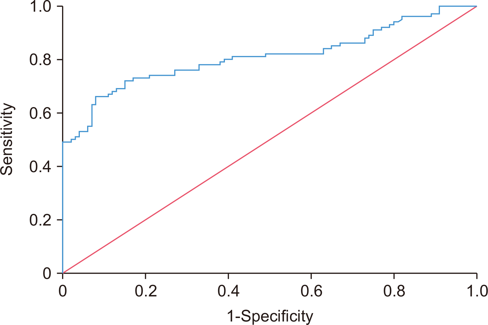
As shown in Table 2, serum IL-33 levels were significantly higher in RA patients than in controls (median 75.507 vs. 34.267 pg/mL, p<0.001). On ROC analysis, IL-33 demonstrated good discrimination (area under the curve=0.810, 95% CI=0.747~0.872, p<0.001), and a cut-off of 61.868 pg/mL yielded 66% sensitivity and 92% specificity (Figure 1).
There were no statistically significant differences in the distribution of IL-33 (rs16924159) genotypes or alleles between RA patients and controls. While the mutant allele and genotypes tended to be more frequent among patients, these differences did not reach statistical significance. Genotype distributions in both groups were in HWE. Detailed frequencies are illustrated in Table 3, and the amplification plot (Supplementary Figure 1) indicates clear allelic discrimination with the TaqMan assay for rs16924159 genotyping.
The outcome showed no statistically significant differences between responders and non-responders based on age, sex, familial history of RA, smoking status, or BMI. Although the mean age was marginally higher in non-responders (52.4±10.3 years) compared to responders (51.5±10.8 years), this was not significant (p=0.670). Most patients in both cohorts were between 40 and 60 years old. Male patients were more prevalent among responders (26%) than non-responders (14%), but this difference was not statistically significant (p=0.135). Neither positive family history for RA (p=0.674) nor smoking status (p=0.317) demonstrated any significant relationship with treatment outcomes. Both the mean BMI (29.4±5.3 vs. 28.4±6.0, p=0.385) and the proportion of obesity (48% vs. 30%, p=0.111) were higher in the non-responder group compared to responders, but these differences were not statistically significant. At TNF inhibitor initiation, baseline CDAI scores were comparable between future responders (median: 19 [14~29]) and non-responders (median: 20 [14~30]), with no statistically significant difference (p=0.909). Disease duration at the time of TNF inhibitor initiation was similar between responders and non-responders (median: 9 [1~27] vs. 8 [1~32] years, respectively, p>0.05) indicating balanced disease chronicity at baseline. The wide range of disease duration reflects real-world treatment trajectories, where some patients may remain on conventional therapies for extended periods before transitioning to biologic agents.
Use of methotrexate was higher among non-responders (76%) compared to responders (60%), but this was not statistically significant (p=0.132). Use of infliximab was significantly different, being higher in responders (50% vs. 28%, p=0.039), while etanercept use was higher in non-responders. Brand-specific analyses were not statistically significant (p=0.153), although a higher proportion of Enbrel use was recorded among non-responders. Serum IL-33 levels were significantly greater in non-responders compared to responders (median: 153.000 [15.105~783.105] vs. 63.394 [14.315~267.315] pg/mL, p<0.001).
The homozygous mutant (AA) genotype was significantly more frequent among non-responders to TNF inhibitors compared to responders (OR=5.26, 95% CI=1.07~25.77, p=0.040). Carriers of the non-disease allele (GG+GA) were significantly more common among responders (OR=0.18, 95% CI=0.03~0.92, p=0.040). However, no significant differences were observed for allele frequencies between the two groups. By design, 50 patients were responders and 50 were non-responders; this 50/50 split reflects sampling and not an empirical response rate. Detailed results are shown in Table 4.
Serum IL-33 levels were significantly higher in individuals with the homozygous mutant (AA) genotype of IL-33 (rs16924159), indicating a strong association between this polymorphism and IL-33 levels (p=0.010, Table 5).
The presence of GA and AA genotypes of IL-33 (rs16924159) was significantly associated with high disease activity (p<0.001), whereas the GG genotype was most frequently observed in patients in remission. Details are shown in Table 6.
Disease activity levels varied among patients receiving different infliximab and etanercept brands (p=0.037). Most patients on Remicade had low disease activity, while those on Enbrel were more likely to have high disease activity. Detailed distributions for each brand are presented in Supplementary Table 2.
DISCUSSION
In the present study, the mean age of the patients with RA was 52.0±10.5 years, and the highest prevalence was observed in the age group of 40~60 years. The same was found by Jumaah Fadhil et al. [11] and Lateef et al. [12] in their previous research, who also found similar age groups. RA predominantly affects middle-aged individuals, particularly females. The increased susceptibility during this age may be attributed to immunological changes with aging, psychological stress, thymic involution, and environmental factors, such as tobacco smoking, which has been shown to activate autoreactive lymphocytes that are the cause of RA development [13-15].
Beyond the IL-33/Serum STimulation 2 (ST2) axis, several host factors—particularly family history [16], smoking [17,18], and adiposity [19,20]—may shape RA biology; however, in our cohort these variables did not differ between TNF inhibitor responders and non responders (all p>0.10). Given their lack of association with treatment response in our data, extended discussion of these variables was removed to maintain focus on IL-33 concentration and rs16924159 genotype—the principal findings of this study.
The present work detected significantly higher serum IL-33 concentrations in RA patients than in healthy controls, consistent with earlier findings of IL-33 involvement in RA pathogenesis [21,22]. Elevated concentrations of IL-33 in synovial fluid and serum have been linked with unfavorable disease prognosis and possibly as a future biomarker of disease activity [3]. IL-33 has been shown to enhance immune responses, induce the production of autoantibodies, and functions as a pro-inflammatory cytokine induced by TNF-α and IL-1β [4]. Variable expression of IL-33 was noted in certain studies due to differences in sample type and immune microenvironment [23].
IL-33 herein was 66% sensitive and 92% specific in RA detection. These are similar to findings from previous studies, Abdel Salam et al. [22] reporting 93.3% sensitivity and 83.3% specificity, and Jawad et al. [21] reporting 83.3% sensitivity and 97.0% specificity. Although the sensitivity in the current study was lower, the high specificity provides support for the potential of IL-33 as a confirmatory biomarker of RA. The varying levels of diagnostic precision of this biomarker among studies may be attributed to differences in methodology, i.e., disparities in study design, patient population, and threshold values used in interpretation.
In the present study, no association between the rs16924159 SNP and RA risk was observed. Although the GA+AA genotype was more common among RA patients under the recessive model, the overall contribution appears to be modest. Because RA is a polygenic condition and the most significant contribution is made by human leukocyte antigen alleles to genetic susceptibility [24], it would not be surprising that IL-33 rs16924159 has little effect, which could be influenced by other genetic and environmental factors. Ethnic genetic heterogeneity and gene–environment interactions may also explain the inconsistent results across different populations [5].
This study found significantly higher amounts of IL-33 in the serum of non-responders compared to responders, as established by previous research pointing to IL-33 as a biomarker of adverse response to TNF inhibitors [6,7]. Higher levels of IL-33 have been associated with chronic inflammation and uncontrolled disease, implying that IL-33-dependent mechanisms may contribute to RA pathogenesis in TNF blockade-resistant patients [4,8]. While some studies have shown that there were no differences in the levels of IL-33 between responders and non-responders [25], the current data affirm that IL-33-induced inflammation may require specialized therapeutic approaches in some RA populations.
The differential elevation of IL-33 in TNF inhibitor non-responders may reflect not only increased disease activity but also a mechanistic contribution to therapeutic resistance. As demonstrated in Liu et al. [26], IL-33 sustains synovial inflammation through ST2-dependent activation of M1 macrophages, T helper 17 (Th17) cell responses, and pro-inflammatory signaling cascades involving nuclear factor kappa light chain enhancer of activated B cells and Mitogen Activated Protein Kinase pathways. These pathways remain active despite TNF blockade, suggesting that IL-33 may play an active role in maintaining inflammation in TNF-resistant disease phenotypes. This further supports IL-33 as a candidate for therapeutic targeting in refractory RA subgroups.
This research found that the IL-33 rs16924159 AA genotype to be linked to non-response to TNF inhibitor treatment in RA patients and found that having at least one G allele was associated with a better response to treatment. These results are consistent with earlier studies that indicated AA genotype carriers have increased disease activity and decreased clinical remission rates after TNF inhibitor treatment [27]. Mechanistically, the A allele was associated with increased IL-33 expression, increased Th17 cells/ IL-17 signaling, and decreased soluble ST2 levels, which together induce refractory inflammation in the face of TNF blockade. GG genotype carriers, by contrast, exhibit decreased IL-33 activity and enhanced therapeutic response. Taken together, our data support IL-33 (and rs16924159) as promising research biomarkers for risk stratification and monitoring; they are not yet ready to guide treatment selection without external validation and comparative, multi arm evidence.
This research confirmed that there was a highly significant positive association between CDAI scores and IL-33 serum levels. The highest values were observed in the high disease activity group compared to those in remission. These findings corroborate Matsuyama et al. [28], who recorded high levels of IL-33 in active RA patients, which provided evidence for its use as a potential inflammatory biomarker. Mu et al. [29], however, reported reduced levels of IL-33 in some patients with high disease activity, confirming that serum IL-33 is not necessarily predictive of joint-localized inflammation. IL-33 expression may be heterogeneous and is controlled by genetic, immune, or environmental factors. From the biological perspective, IL-33 stimulates Th17 and B cells activation with induction of RA pathogenesis and association of disease worsening severity [29].
In this study, it was established that there was a strong association between the IL-33 rs16924159 (G>A) polymorphism and IL-33 levels in the sera of RA patients, where AA genotype carriers showed the highest levels and GG carriers showed the lowest. These findings suggest that the A allele may upregulate IL-33 expression or stabilize the protein. Mechanistically, the A allele might enhance IL-33 production by increased nuclear factor kappa-light-chain-enhancer of activated B cells signaling or by precluding proteolytic degradation, possibly by resistance to cleavage by enzymes such as matrix metalloproteinases. To the best of our knowledge, this is among the first study to directly examine the influence of IL-33 rs16924159 on IL-33 serum levels in RA and provide new data on the genetic regulation and its pathogenic role in RA; nonetheless, confirmatory studies in independent populations are warranted. In the present research, we identified a strong association between IL-33 rs16924159 genotypes and disease activity in RA patients, as assessed by CDAI and AA genotype was most common in patients with high disease activity (63.64%). The GA and GG genotypes showed a more varied distribution across activity levels. This concurs with Iwaszko et al. [27], who also correlated the AA genotype with more severe disease and poorer treatment response to TNF inhibitor. This supports the potential of IL-33 rs16924159 as a susceptibility biomarker for RA in precision medicine, as well as a stratification biomarker for RA treatment.
Since IL-33 and disease activity were assessed after 6 months of treatment, the findings reflect post-treatment associations rather than baseline predictors. Thus, the data support the potential utility of IL-33 as a monitoring biomarker rather than a treatment-guiding tool.
Our findings—higher serum IL-33 and the rs16924159 AA genotype associating with greater disease activity and lower 6 month TNF inhibitors response—are biologically plausible and support stratification/monitoring. However, because all patients received TNF inhibitors and we did not test a biomarker by treatment interaction across alternative therapies, these data indicate associations rather than treatment guiding predictive utility.
IL-33’s diagnostic performance in our cohort was moderate, and we lacked external validation. Clinical implementation will require validation of cut-offs in independent populations and demonstration that IL-33 (and rs16924159) predict differential benefit across treatment classes. Pending such evidence, IL-33 should be regarded as an investigational biomarker for stratification and monitoring.
The modest sample size—particularly in the TNF inhibitors responder/non responder stratification (50/50)—limits statistical power and yields wide CIs for some estimates (e.g., non response OR=5.26; 95% CI=1.07~25.77). A post hoc two sided power analysis for AA vs. (GA+GG) indicated ~66% (65.8%) power (18% vs. 4%); ~70 participants per group would be needed for 80% power. Findings should be interpreted with caution and validated in larger cohorts. The single arm nature (all patients received TNF inhibitors), absence of external validation, potential confounding by TNF inhibitors type/brand, and two-center, single region recruitment limit generalizability and inferences about treatment guiding utility. Our findings indicate associations that warrant validation in independent and comparative cohorts. Additionally, baseline RF and anti-cyclic citrullinated peptide antibody titers were unavailable for most patients due to the retrospective nature of the study, in which biosample collection was performed 6 months after TNF inhibitor initiation, and historical immunological data were not consistently recorded.
CONCLUSION
This study elucidates the role of IL-33 serum level and rs16924159 polymorphism in RA and therapy response. Elevated IL-33 levels were significantly associated with high disease activity and lack of response to TNF inhibitor therapy, elevated IL-33 and the rs16924159 AA genotype were associated with higher disease activity and lower 6 month TNF inhibitor response. Given the moderate diagnostic performance, lack of external validation, and absence of a biomarker by treatment interaction analysis, IL-33 (and rs16924159) should be considered promising research biomarkers for risk stratification and monitoring rather than treatment guiding tools. Prospective, multicenter, and comparative studies are required to establish clinical utility, reflecting a genetic predisposition affecting clinical course.
SUPPLEMENTARY DATA
Supplementary data can be found with this article online at https://doi.org/10.4078/jrd.2025.0095
 XML Download