



 PDF
 Citation
 Print


Introduction
Microphthalmia and anophthalmia are serious eye development conditions that are often related to genetic causes. Over 30 genes have been associated with these conditions, and one of the most important is the Forkhead Box E3 (FOXE3) gene, which produces a protein important for the development of the lens and is located on chromosome 1p33. This gene was first linked to a condition called anterior segment dysgenesis, which is inherited in a dominant way, and later was found to be connected to recessively inherited conditions like primary aphakia, microphthalmia, and sclero-cornea [1]. The FOXE3 protein is crucial for the development of the eye, especially the lens.
Primary ciliary dyskinesia (PCD) is a group of inherited conditions that occur in about 1 in every 7,500 people. The Dynein Axonemal Heavy Chain 5 (DNAH5) gene is situated on chromosome 5p15.2, makes a subunit of the axonemal outer dynein arm (ODA), which is a key part of the cilia that help them move in a way that uses adenosine triphosphate [2]. Mutations in the DNAH5 gene are known to cause PCD.
In this report, we describe a case where a child from South India had both FOXE3 and DNAH5 gene mutations.
Case Report
A 3-month-old female infant, the firstborn of parents who are third-degree relatives, exhibited frequent respiratory tract infections and had small eyes on both sides. She could respond to light. There was no history of global developmental delay, seizures, hearing problems, or sleep issues. Furthermore, there was no indication of gestational diabetes, pregnancy-induced hypertension, or hypothyroidism. Her birth weight was recorded at 2.534 kg. She had dyspnea at birth and spent 25 days in the neonatal intensive care unit, where she was breastfed.
Family background
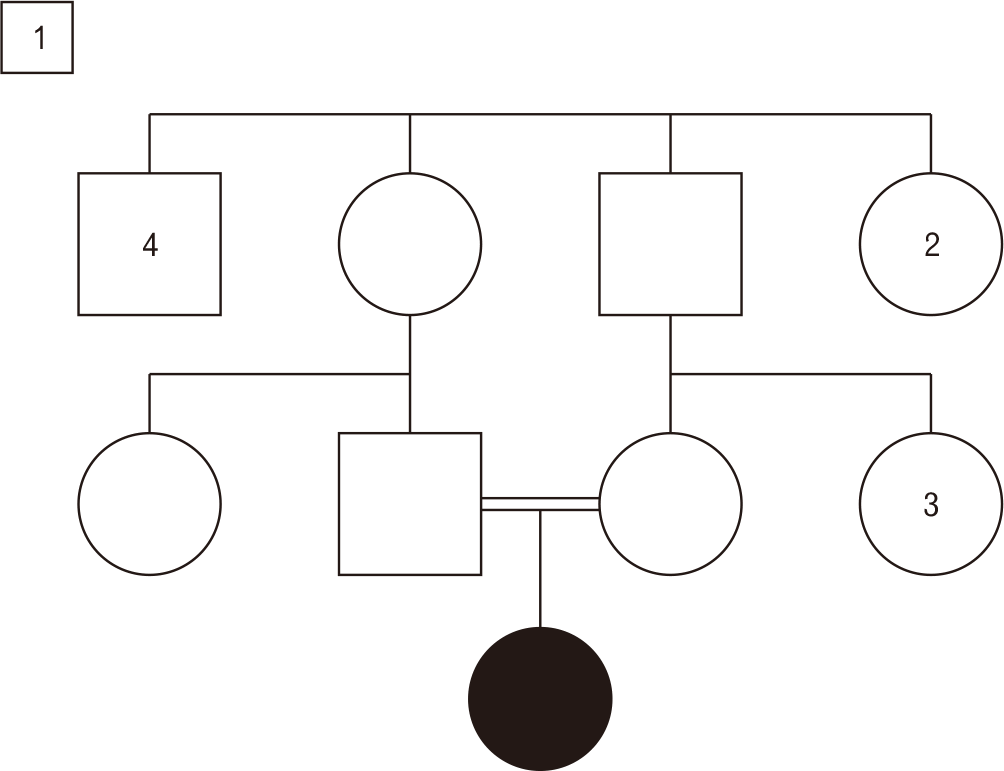
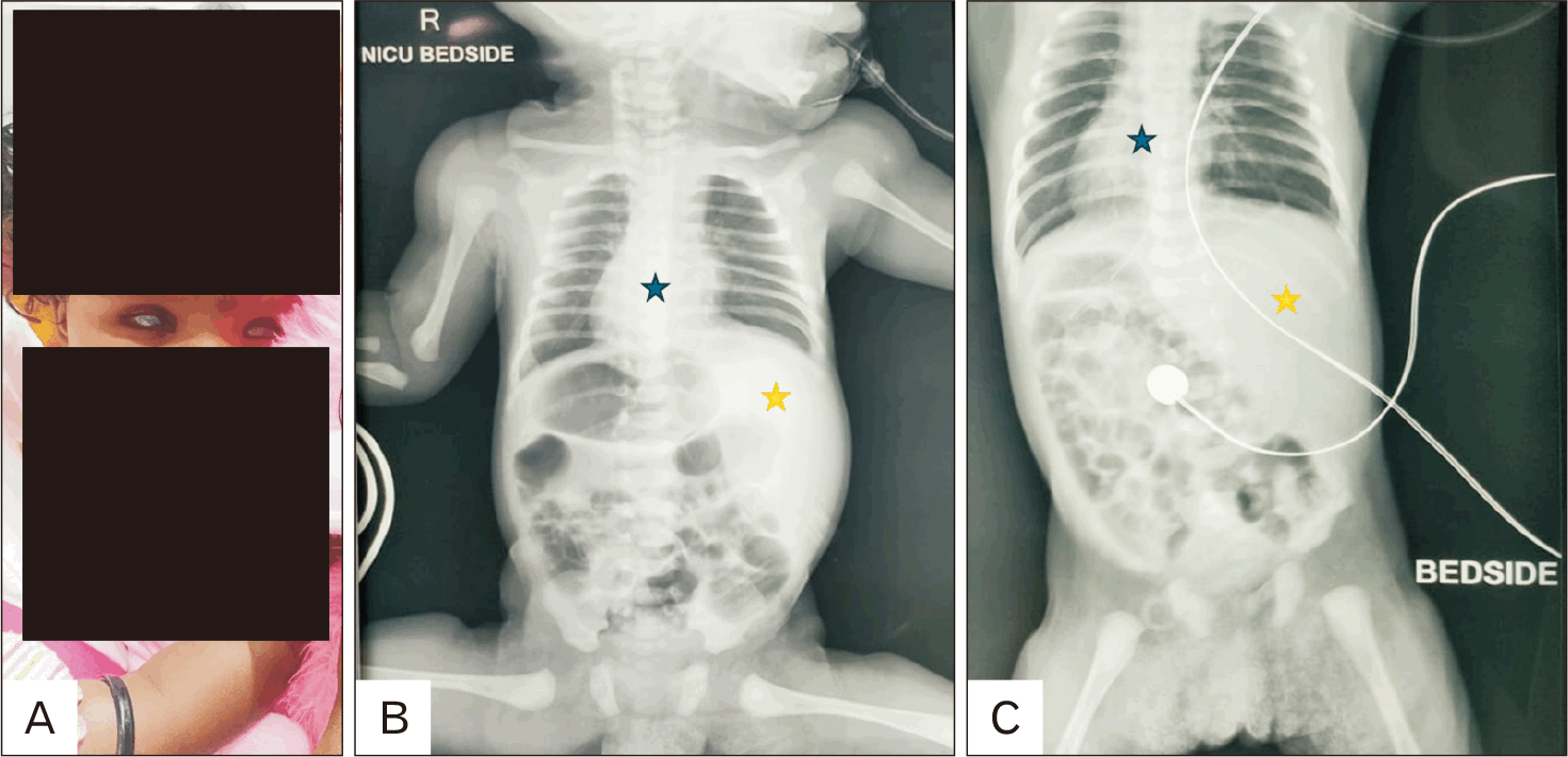
She was born to a couple with a third-degree consanguinity, and there were no other similar issues reported in the family (Fig. 1). Upon examination, microphthalmia was observed in both eyes. Dysmorphic features included hypertelorism, a depressed nasal bridge, a prominent philtrum, and a cupid bow upper lip (Fig. 2A). The respiratory system exhibited basal crepitations, while the cardiovascular system revealed the apex of the heart positioned on the right side. All other systems appeared normal. The clinical diagnosis was identified as PCD accompanied by autosomal recessive micro-ophthalmia.
Investigations
X-ray and ultrasound showed situs inversus with dextrocardia, right sided stomach, left sided liver (Fig. 2B, C). After pre-test counselling, whole exome sequencing was ordered.
Results
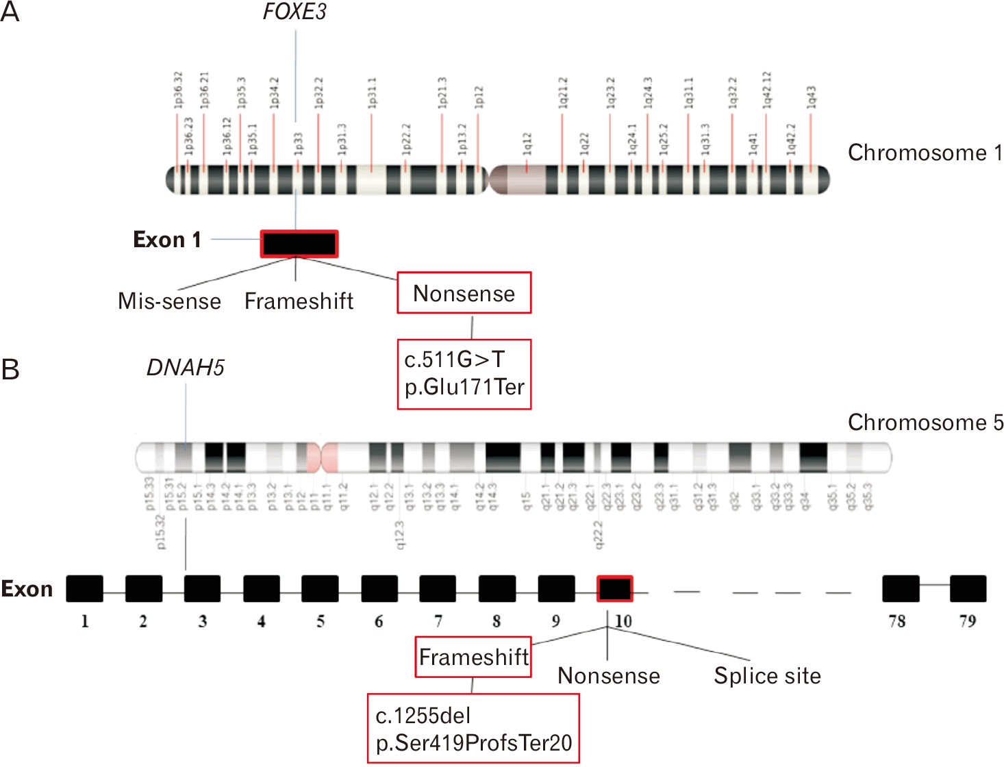
Whole exome sequencing showed a homozygous nonsense variant in exon 1 of FOXE3 gene that resulted in a stop codon and premature truncation of protein at codon 171 and a homozygous single base pair deletion in exon 10 of DNAH5 gene that resulted in a frameshift and premature truncation of protein 20 amino-acids downstream to codon 419 (Fig. 3) [3, 4]. Both the variants were pathogenic according to American College of Medical Genetics and Genomics guidelines (Table 1) [5].
Genetic counselling
Post-test counselling was conducted. Recommendations were provided to avoid upper respiratory tract infections. A comprehensive eye examination was suggested. Validation of the parents was requested as this was their first child. The couple was informed that there was a 25% likelihood of recurrence in each pregnancy. Additionally, they were identified as carriers of the aforementioned genes. The genetic counselling significantly influenced them, as it facilitated their understanding of the disease and improved their ability to cope with their child’s condition. This study was approved by the Institute Ethics Committee of All India Institute of Medical Sciences, Bhubaneswar (T/IM-NF/25-26/124). Informed written consent has been obtained from parents for publication of the clinical image.
Discussion
The FOXE3 gene produces a transcription factor consisting of 319 amino acids in length. This factor is essential for proper eye formation in vertebrates. This protein is crucial in controlling the growth of lens epithelial cell via processes related to growth, programmed cell death and cellular cycle [6, 7]. The FOXE3 protein has a highly conserved part that binds DNA called the forkhead domain, which covers amino acids 71 to 165. Most harmful changes in the FOXE3 gene result in the protein being cut short, which stops it from properly binding DNA and causes the eye-related problems seen. According to Dawood et al. [8], there are 28 harmful or likely harmful changes in the FOXE3 gene linked to microphthalmia and sclerocornea. Of these, 10 (35.71%) are frameshift changes that lead to premature stopping of the protein, while 18 (64.28%) are substitution changes, including 7 (25%) nonsense changes and 11 (39.28%) missense changes. All the missense changes are found in the fork-head domain. Frameshift and nonsense changes are expected to lead to shortened proteins that can’t bind DNA properly [8]. Valleix et al. [9] found a homozygous c.720C>A (p.C240*) FOXE3 mutation in a consanguineous family from Madagascar, where three children had sclerocornea, bilateral aphakia, microphthalmia, and other eye problems. Iseri et al. [10] studied two unrelated families from Pakistan, one with microphthalmia, aphakia, sclerocornea, and glaucoma, who had a c.244A>G (p.Met82Val) substitution change, and the other had cloudy corneas, aniridia, microphthalmia, and congenital aphakia, with a frameshift change of c.21_24del (p.Met7IlefsX216). Anjum et al. [11] confirmed the same c.720C>A (p.C240*) FOXE3 mutation in a large inbred family with similar conditions. Mouse studies suggest that FOXE3 is involved in iris development, and in mice with one copy of the FOXE3 gene, the lens is normal, but the iris is underdeveloped and there are unusual connections between the iris and the cornea [12]. Understanding how FOXE3 functions and how its mutations affect eye development is important for understanding these eye disorders and may help in developing new treatments.
The PCD condition, which affects multiple body systems, is due to the improper functioning of cilia and flagella. Recurrent respiratory infections happen because of a failure in mucociliary clearance, which is due to cilia that don’t move properly, leading to chronic inflammation and bronchiectasis [2]. Dysfunction of cilia in the embryonic node leads to the randomization of the body’s left and right sides leading to situs inversus [13]. Kispert et al. [14] found that the mouse version of this gene (MDNAH5) is expressed in ciliated cells in the upper and lower parts of the lungs. Mutations in DNAH5 are found in 28% of PCD cases, and this increases to about 50% when only cases with ODA defects are considered, showing how important DNAH5 is for PCD. About 40% of the mutations linked to immotile cilia are located in the C-terminal region of DNAH5. PCD is a condition that can have multiple genetic causes, and DNAH5 dysfunction is a key reason for PCD related to defects in the ODA [15].
In our case, there were two pathogenic variants in FOXE3 and DNAH5 genes. The co-existence of FOXE3 and DNAH5 together is most likely explained by their shared relevance in developmental biology and ciliopathy-related disorders, rather than a direct molecular interaction.
This case brings out the diagnostic importance of whole exome sequencing in identifying dual pathogenic variants contributing to complex phenotypes. The coexistence of FOXE3 and DNAH5 mutations illustrates the intersection of ocular malformations and ciliopathies, emphasizing the importance of genetic counselling for affected families. Early recognition and multidisciplinary management are crucial for improving prognosis and quality of life.
 XML Download