



 PDF
 Citation
 Print


Introduction
Michelangelo di Lodovico Buonarroti Simoni (1475–1564) stands as a monumental figure in Renaissance art, celebrated for his mastery of form, anatomy, and expressive storytelling through sculpture. His Moses (1513–1515), part of the tomb of Pope Julius II in San Pietro in Vincoli, has drawn attention not only for its psychological intensity but also for its anatomical precision. Previous studies have explored Moses from psychoanalytic [1-3], neuroaesthetic [4], and general art historical perspectives [5]. Yet, little attention has been given to the detailed anatomy of the forearm, particularly the brachioradialis, despite its prominence along the extensor surface.
Renaissance artists, including Michelangelo, often dissected cadavers to deepen their understanding of human anatomy [6]. Observing both living subjects and cadavers enabled artists to represent not only the structural details of muscles and tendons but also the dynamic interplay between form, movement, and psychological expression. The aim of this essay is to analyze the brachioradialis in Moses, examining its anatomical accuracy, biomechanical function, expressive significance, and implications for modern anatomy and surgical pedagogy.
Anatomical Observation of Michelangelo’s Moses
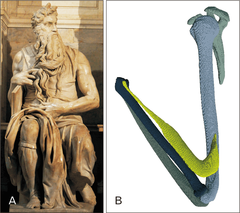
When approaching Michelangelo’s Moses, the right arm immediately draws attention. The limb rests upon the tablets of the Law, flexed yet imbued with latent energy. The extensor surface of the forearm reveals a strikingly bulging muscle mass, rendered with fidelity to both volume and contour (Fig. 1A) [7]. This swelling is not arbitrary; it reflects Michelangelo’s deep anatomical knowledge acquired through dissection and careful study of living models.
The prominent muscle on the lateral-dorsal forearm corresponds anatomically to the brachioradialis, originating on the lateral supracondylar ridge of the humerus and inserting on the distal radius (Fig. 1B). Its prominence is enhanced by the semi-pronated position of the forearm and partial elbow flexion. Secondary contributions may arise from the extensor carpi radialis longus and brevis, yet the brachioradialis dominates the visual impression. Michelangelo thus transforms a functional muscle into a sculptural instrument for expressing tension, readiness, and controlled power (Table 1).
Symbolic and Functional Significance of the Brachioradialis in Moses
Michelangelo’s emphasis on the brachioradialis conveys psychological and moral restraint. The semi-flexed forearm suggests suppressed motion: the muscle is coiled but intentionally restrained, reflecting Moses’ internalized discipline and righteous anger.
The bulging muscle functions as an emotional signal. Observers perceive the tension pre-reflectively, intuitively understanding Moses’ inner state. The anatomical accuracy reinforces the narrative: the muscle directs attention along the forearm to the hand and the tablets, integrating functional anatomy with theological symbolism.
Hilloowala highlighted Michelangelo’s dual focus on grandeur (Apollo) and pathos; the brachioradialis embodies this dialectic [8]. Its heroic form communicates strength and nobility, while its restrained contraction conveys human struggle and controlled emotion.
Biomechanical Analysis and Positional Study of the Right Forearm
Michelangelo’s rendering optimizes the brachioradialis’ visibility and expressive potential through precise biomechanical configuration:
1. Elbow flexion: the elbow is flexed to approximately 70°–80°, based on proportional angular estimation using high-resolution photographs taken orthogonally from three documented viewpoints. Forearm positioning was analyzed through visual triangulation aligned to the olecranon-lateral epicondyle axis.
2. Forearm rotation: semi-pronation enhances visual salience, balancing contribution of biceps brachii and extensors.
3. Grip force: the hand’s firm grasp of the tablets transmits tension along the forearm, highlighting the tendon-muscle interface.
This biomechanical orchestration ensures that the forearm communicates dynamic readiness despite static presentation. In contrast to generalized forearms in contemporary works, Michelangelo integrates functional realism, expressive intent, and anatomical accuracy (Table 2).
Comparative Analysis with Contemporary Artists
Michelangelo’s anatomical mastery surpasses his contemporaries:
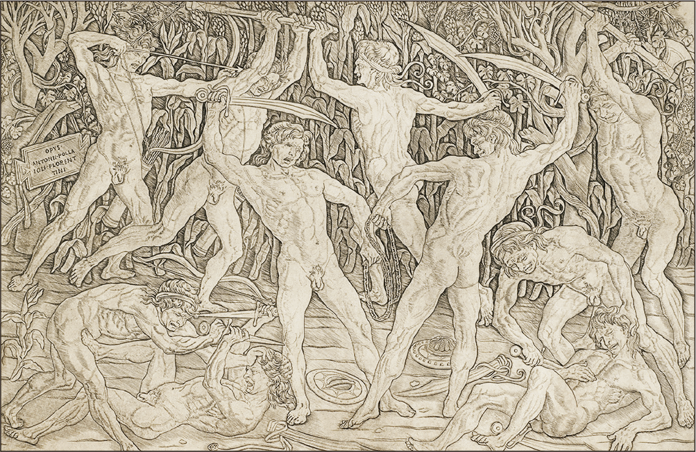
• Pollaiuolo: In Battle of the Ten Nudes (c.1470–1475), emphasized muscular volume in generic forms; lacked joint-specific accuracy (Fig. 2) [9].
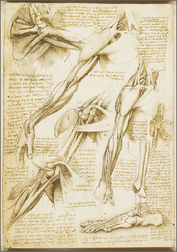
• Leonardo da Vinci: In Windsor anatomical drawings (c.1487–1515), produced precise anatomical drawings, but muscles were largely studied in isolation, rarely capturing tension in three-dimensional context (Fig. 3) [10].
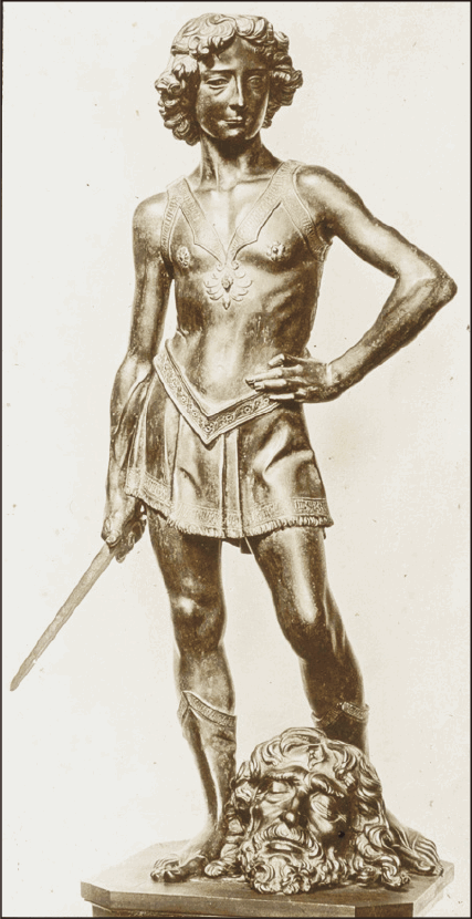
• Verrocchio: In David (c.1473–1475), focused on surface modeling; forearms were visually convincing but lacked functional specificity (Fig. 4) [11].
In Moses, the brachioradialis is rendered positionally accurate, functionally plausible, and psychologically expressive, representing a unique integration of anatomy and narrative in Renaissance sculpture (Table 3).
Psychoanalytic and Neuroaesthetic Implications
The brachioradialis in Moses exemplifies how anatomical details mediate emotional and psychological perception:
• Freud interpreted the sculpture as embodying suppressed wrath and moral authority, conveyed through muscular tension [1].
• Neuroaesthetic studies demonstrate that anatomically salient features elicit specific neural, cardiac, and galvanic responses, enhancing emotional engagement [4].
• Figueroa emphasized embodiment; the forearm allows observers to reconstruct possible sequences of movement, merging physical observation with mental inference [5].
Michelangelo’s work demonstrates that anatomy can serve as a conduit between inner psychological states and outer visual form (Table 4).
Implications for Anatomical Education and Surgical Perspective
The brachioradialis in Moses offers enduring lessons for anatomy and surgery:
• Observation of function: joint-specific positioning illustrates muscle activation and tendon orientation.
• Functional and expressive understanding: muscle form communicates both biomechanical and emotional information.
• Pedagogical value: sculptural representations enhance spatial reasoning and appreciation of anatomical interdependence.
Michelangelo’s synthesis of dissection-based knowledge, functional realism, and expressive intent anticipates principles relevant to modern reconstructive and orthopedic surgery, emphasizing anatomical precision in context.
Conclusion and Future Directions
Michelangelo’s Moses exemplifies the union of anatomy, expression, and psychology. The brachioradialis, prominently displayed along the extensor surface, conveys tension, potential motion, and moral restraint. Comparative and biomechanical analysis highlights Michelangelo’s singular achievement in integrating functional anatomy with expressive narrative.
From a modern perspective, the sculpture informs anatomical pedagogy, surgical observation, and neuroaesthetic understanding. Future research could examine other muscles in Michelangelo’s oeuvre, linking anatomical fidelity to expressive intent and exploring perceptual and emotional responses. The comparative schematic visualization provides anatomical and expressive context, demonstrating how Michelangelo’s rendering uniquely integrates functional biomechanics with psychological intention.
Ultimately, the brachioradialis in Moses illustrates how art, anatomy, and human perception converge, offering lessons that remain relevant across centuries.
 XML Download