



 PDF
 Citation
 Print


Introduction
Plantaris is one among the superficial muscles of the posterior compartment of the leg. It is often considered as vestigial muscle with the small fleshy belly that arises from the lower part of the lateral supraconylar line of the femur. Its long, slender tendon passes under the gastrocnemius and along the medial side of the tendocalcaneus to get inserted into the posterior surface of the calacaneus. Plantaris may be absent in 4%–20% of cases [1]. Duplication [2], triplication [3] and quadrifurcation [4] of the plantaris has also been reported. Variations in the insertion pattern of plantaris is also common. Plantaris is a muscle of importance in plastic surgery, as its tendon grafts can be used to reconstruct tendons and ligaments. It could compress neurovascular structures or undergo tendonitis and cause problems in athletes. Hence, it is worthwhile to record all unique variations of this muscle. We report a novel variation of plantaris and discuss its clinical implications in this report. The goal of this report is to document the information related to the novel variation of plantaris muscle. This variation could be of special interest to vascular surgeons, plastic surgeons and orthopedic surgeons.
Case Report
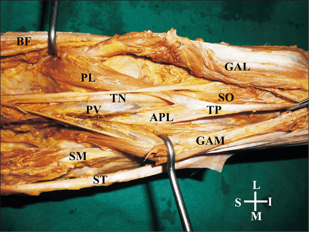
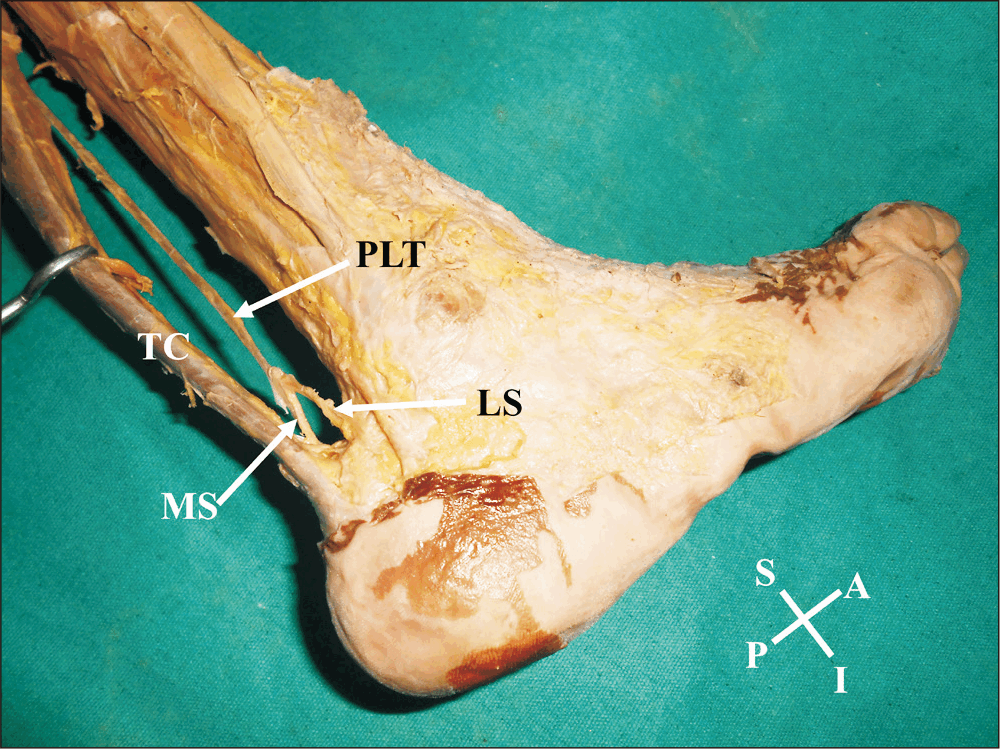
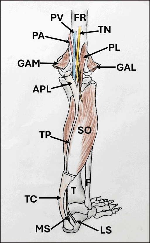
During dissection of a female cadaver approximately aged 60 years, an unique variation of the plantaris muscle was noted in the right lower limb. The variation was unilateral. The heads of gastrocnemius muscle were cut and reflected to expose the variant plantaris muscle. The plantaris had two heads of origin. The lateral head was fleshy and the medial head was aponeurotic (Fig. 1). The lateral head took origin from the lateral supracondylar line and was 7.9 cm long and 1.3 cm broad. The medial aponeurotic head took origin from the capsule of the knee joint, oblique popliteal ligament and the fascia covering popliteus muscle. It was 4.2 cm long and 1 cm broad. The two heads joined each other behind the main neurovascular bundle at an acute angle (like the cutting edges of a scissor) and the narrow gap between these two heads was traversed by tibial nerve, popliteal vein and popliteal artery. There was no sign of any narrowing or dilataion of the popliteal vessels either proximal, distal or at the site of their passage between the two heads of plantaris. The tendon of plantaris was 28 cm long and travelled down first between the gastrocnemius and soleus muscles and then along the medial side of the tendocacaneus until the ankle joint. At the ankle joint, it divided into medial and lateral slips. The medial slip merged with the anterior surface of the tendocalcaneus and the lateral slip merged with the deep fascia, lateral to the insertion of the tendocalcaneus. Nerve supply of the lateral fleshy belly was derived from the tibial nerve. The fat and fascia surrounding the variant muscle was cleared carefully and photographs were taken. The variations are shown in Figs. 1, 2. A simplified schematic diagram of the variation has been given as Fig. 3.
Discussion
Plantaris muscle is one of the phylogenetically degenerating muscles of the lower limb. It contributes less than 1% power of plantar flexors. However, it is invaluable beacause of increasing use in tendon and ligament repairs [5, 6]. Plantaris is a weak flexor of the knee and ankle joints. Its rupture could mimic deep vein thrombosis and, in cases of rupture of tendocalcaneus, it can still plantar flex the ankle and mislead the proper diagnosis [7]. Double headed, triple headed and four headed origins of the plantaris have been documented. However, in most of the reported cases, the fleshy bellies or the tendons were not in a dangerous position to the neurovascular bundle of the popliteal fossa. In addition, in most of the earlier reported cases, the additional heads were fleshy. In the current case, the medial head was almost entirely aponeurotic and took origin from the medial side of popliteal neurovascular bundle. Kotian et al. [8] (2013) have reported a case of bifurcation of the fleshy belly of plantaris, and each of the parts having a separate tendon. The two long tendons encircled the popliteal neurovascular bundle. However, there was a large gap between these two tendons [8].
Variations in the origin, number of bellies and course of the tendon of plantaris could compress/entrap tibial nerve and popliteal vessels, leading to neurovascular symptoms. Olewnik et al. [9] (2018) have reported muscle and tendon passing behind the popliteal vessels. They claim that it could compress these structures, if hypertrophied [9]. The commonest area of tibial nerve entrapment is at the ankle, under the flexor retinaculum, and is known as ‘tarsal tunnel syndrome’ [10]. Entrapment of tibial nerve and popliteal vessels in the popliteal fossa are rare. However, there are reports on its entrapment in the gastrocnemius muscle [11]. ‘Popliteal entrapment syndrome’ is a condition where the popliteal artery gets entrapped in anomalous slips of hamstrings, gastrocnemius, popliteus or some fibrous bands in the popliteal fossa. It might lead to complications like claudication in any age group [12]. In a case like the current case, the entire neurovascular bundle of the popliteal fossa could get compressed as there was a ‘scissor effect’ in the small gap between medial and lateral heads of the plantaris.
Knowledge of variations of plantaris tendon is also of importance to surgeons as it is used in the repair of tendocalcaneus, ligaments of the ankle and flexor tendons of fingers. There are reports on abnormal attachments of the plantaris tendon causing Achillis tendinopathy symptoms [13]. In the current case, the plantaris tendon was divided into medial and lateral slips before its final insertion. Similar reports where the tendon splits and shows variant attachments have been published and their involvement in tendinopathies or failure of tendon grafts have been documented [14, 15]. The current report is rare in having variations both in origin and insertion pattern of plantaris. This type of variation has to be kept in mind for the differential diagnosis of popliteal artery entrapment syndrome. Plantaris could also be used for tendon harvest in tendon graft surgeries.
Muscles of the lower extremity develop from the mesenchyme between 4th and 8th week of intrauterine life. The paraxial mesoderm forms the somites. Some of the cells of these somites migrate ventrolaterally into the lateral plate mesoderm, forming the lower limb and eventually give rise to the lower limb muscles. The additional part of the plantaris in the current case could be formed due to the formation of additional myoblast cells through mitotic division after their migration from the somites into the popliteal region.
In conclusion, though there are reports on duplication, triplication and other anomalies of plantaris, a case like the current case, with an aponeurotic additional head is not available in the published literature as per our survey. Since the popliteal neurovascular bundle was passing through the narrow gap between the medial and lateral heads of the plantaris, there is a high chance of them getting compressed at one or other stage of life. A knowledge of this muscle could be useful to radiologists, plastic surgeons and sport medicine specialists.
 XML Download