Journal List > Anat Cell Biol > v.59(2) > 1516095823

Watanabe, Matsuura, Kigata, and Kobayashi: Two cases with fenestrations in the left renal veins for the passages of the renal, gonadal, and suprarenal arteries

Abstract

Fenestrations in the left renal vein (RV) were detected in two Japanese cadavers: a 96-year-old male (Case 1) and an 81-year-old female (Case 2). In Case 1, four left RVs emerged anterior to the renal arteries at the hilum. Fenestration 1 (0.5 cm) was found in the second highest vein. Fenestration 2 (1.0 cm) was found in the third highest vein. Three left renal arteries originated from the abdominal aorta and the inferior branch passed through fenestration 1. The left testicular artery arising 3.5 cm distal to the second-highest renal artery traversed fenestration 2. In Case 2, a fenestration was observed 4.5 cm distal to the left RV drainage into the inferior vena cava. The left middle suprarenal and left ovarian arteries originating from the abdominal aorta at L1/L2 and L2, respectively, passed through the fenestration. These rare variations enhance understanding of the development of the renal, gonadal, and suprarenal arteries.

Introduction

The renal veins (RVs) are paired vessels that emerge from the renal hilus and drain into the inferior vena cava. Before draining into the inferior vena cava, the left RV collects blood from the ipsilateral suprarenal and gonadal veins and passes anterior to the aorta. By contrast, the right RV exits the renal hilus and drains directly into the inferior vena cava without tributaries.
The left RV shows frequent variations in its ramification patterns, including differences in its course, number, and tributaries [1]. These variations are often associated with variations in the gonadal and suprarenal vessels, resulting in variable positioning of these vessels [2-4]. Preoperative recognition of possible variations in the vascularity of the kidney, genital glands, and suprarenal glands is essential to avoid unexpected complications during retroperitoneal procedures.
Fenestration of the left RV is a rare anatomical variant with a reported incidence of 3.34% [5]. In most previously reported cases, the testicular artery (TA), which arises from the abdominal aorta (AA), passes through the fenestration [5-8]. The left renal artery can also pass through the fenestration [5, 9]. Fenestrations of the left RV represent complex variations involving arteries to the gonads and kidneys and have significant implications in diagnostic imaging and surgical procedures. Knowledge of these rare variants is clinically beneficial and valuable for understanding the morphogenesis of retroperitoneal veins, including the left RV.
Here, we report two cases (male and female cadavers) showing fenestration in the left RV, through which the renal and testicular arteries or the suprarenal and ovarian arteries (OAs) passed, respectively, observed in a routine gross anatomy class for undergraduate students at the National Defense Medical College.

Ethical statement

The cadavers used in this study were donated to our institution with written comprehensive consent from the donors and their families who agreed to their use in anatomical education and research. The research protocol and manuscript preparation were conducted in accordance with the “Guidelines for the research involving cadavers” published in 2021 by the Japanese Association of Anatomists.

Case Report

Case 1: a 96-year-old Japanese male cadaver

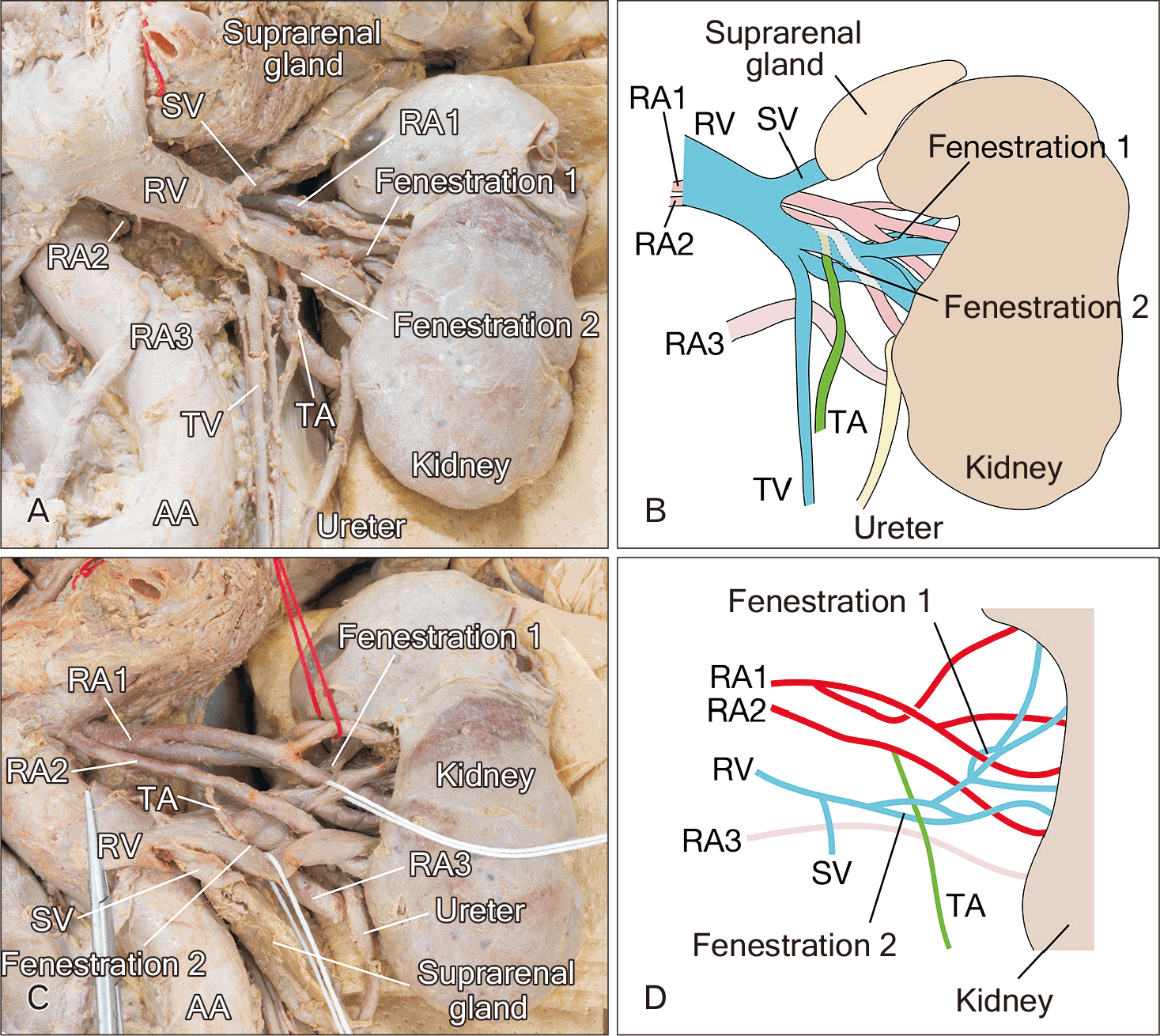
Four veins emerged from the left renal hilus anterior to the renal arteries to form the left RV (Fig. 1). One fenestration (fenestration 1), with a maximum diameter of 0.5 cm, was found at the confluence of the highest and second highest veins. Another fenestration (fenestration 2), with a maximum diameter of 1.0 cm, was located at the terminal confluence, where the lowest vein drained into the others. Three left renal arteries (RA1, 2, and 3) were identified in this case. The top (RA1) and second (RA2) originated from the AA at the L2 vertebral level while RA3 originated from AA at the L3 vertebral level. The branch from RA1 passed through fenestration 1 (Fig. 1), whereas the left TA originating 3.5 cm distal to the origin of RA 2, passed through fenestration 2.

Case 2: an 81-year-old Japanese female cadaver

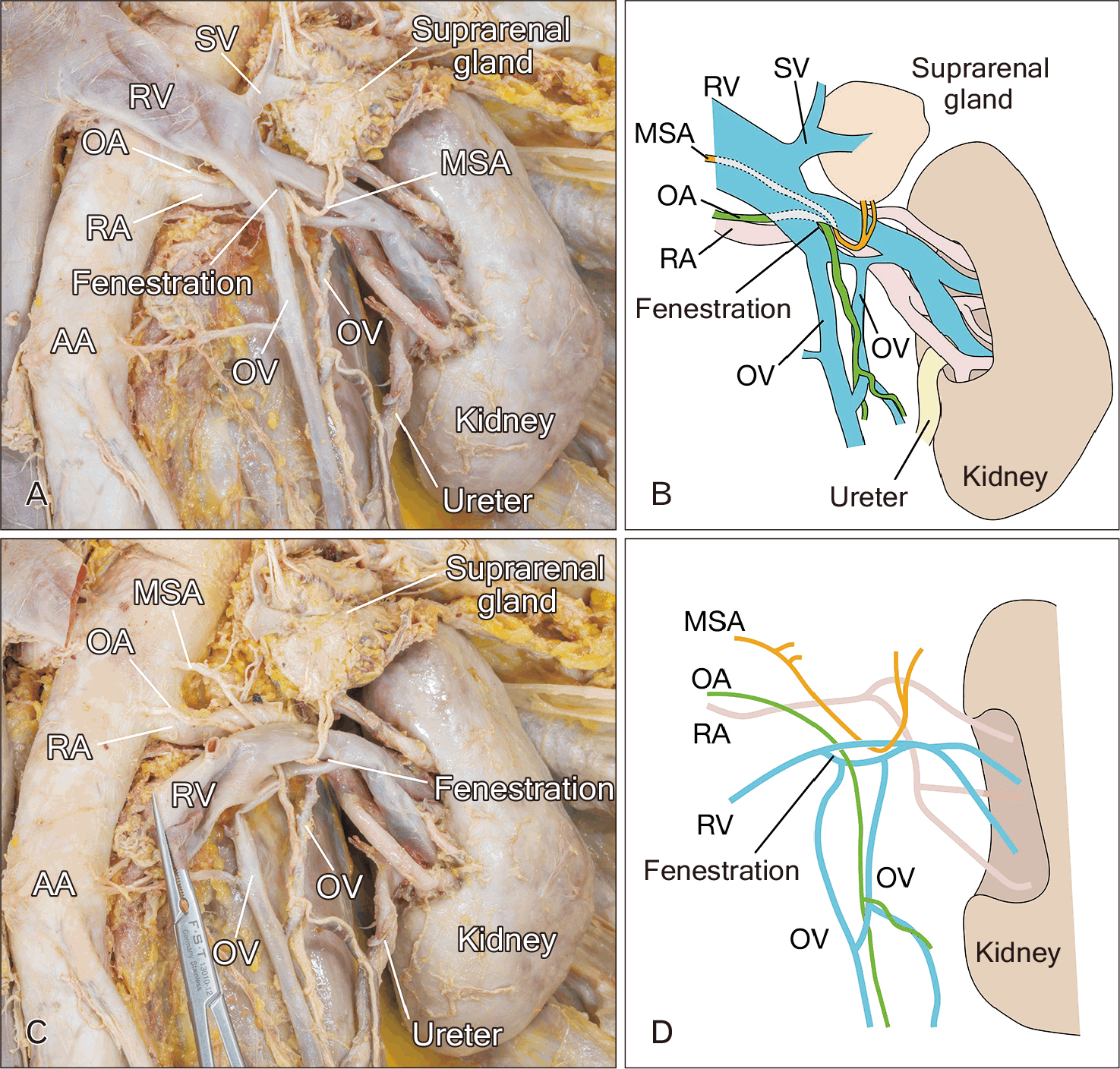
A fenestration with a maximum diameter of 1.5 cm, was observed in the left RV (Fig. 2). The left middle suprarenal (MSA) and OAs originated from the AA at the L1/L2 intervertebral disc and L2 vertebra, respectively, both passing through the fenestration.

Discussion

Here, the TA and RA or the OA and MSA passed through the fenestration of the left RV. Previous studies have reported that fenestrations in the left RV are usually singular and found near the confluence of the gonadal and suprarenal veins [5-9]. Moreover, only the RA and TA passed through the fenestration of the left RV [5-9]. Thus, the case of the MSA passing through a fenestration in the left RV as well as the incidence of two different arteries passing through the same fenestration, as detected in Case 2, is extremely rare.
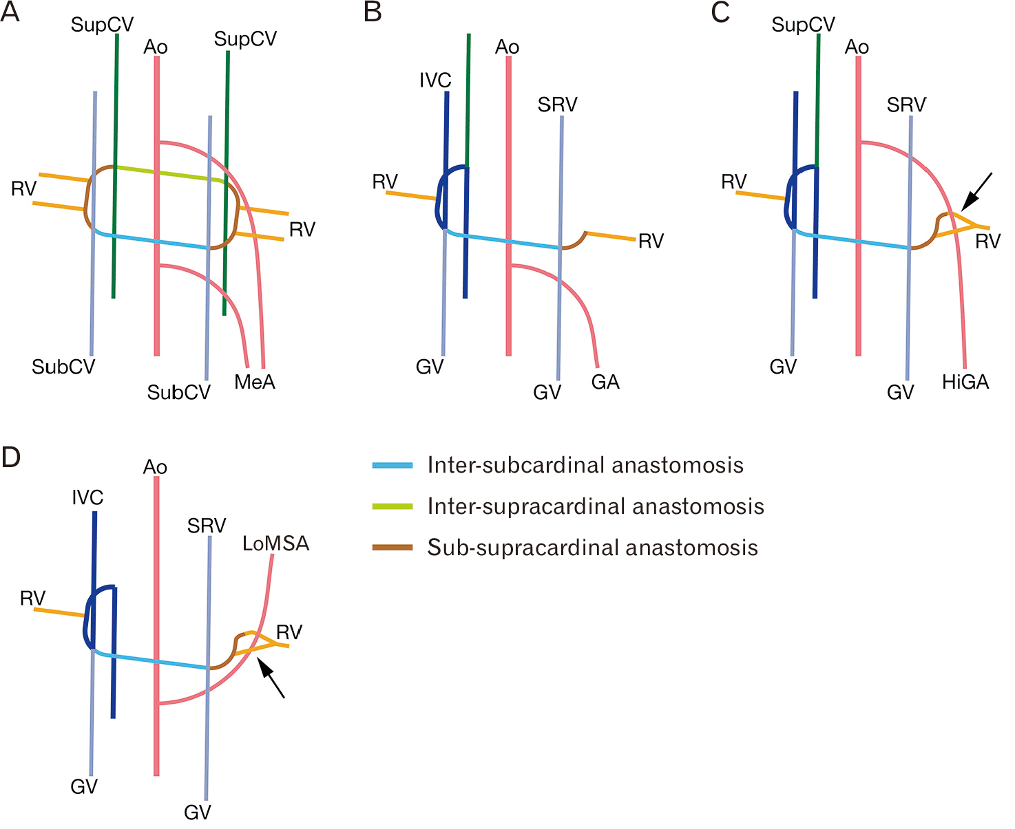
Fig. 3 illustrates the developmental process of the left RV and inferior vena cava. These veins are primarily formed through the persistence and regression of paired supracardinal and subcardinal veins and their anastomoses, namely the sub-supracardinal, inter-subcardinal, and inter-supracardinal anastomoses (Fig. 3) [10]. The segment of the RV near the renal hilus is thought to be formed by sub-supracardinal anastomosis, from which RVs emerge (Fig. 3) [7, 10]. The presence of an artery running between the ventral renal vein (VRV) and dorsal renal vein (DRV) is thought to be a factor in the formation of fenestrations, as it impedes complete venous fusion [7]. In the fetus, the RV drains into the inferior vena cava between T12 and L3 [10]. Consequently, arteries such as the gonadal artery or MSA which arise from the aorta at this branching level range, potentially become entrapped in the fusion of the VRV and DRV and sub-supracardinal anastomosis. In the present case, the gonadal artery and MSA arising either from the aorta at a level close to or arising directly from the RA may have been entrapped in the fusion of the VRV and DRV (Fig. 3).
Our cases are unique in the following ways. First, the testicular, renal, suprarenal, and OAs passed through a fenestration in the left RV. Second, in Case 1, the TA arose from the RA and passed through a fenestration in the left RV. Third, in Case 2, two different arteries passed through the same fenestration. The findings in our cases may contribute to a better understanding of the developmental processes of the left RV and its related vessels.
Acknowledgements

The authors sincerely thank those who donated their bodies for this field of anatomical research. The results of this study may increase the overall knowledge of humanity and improve patient care. Therefore, the donors and their families deserve the highest gratitude [11].

Notes

Author Contributions

Conceptualization: TK, YK. Data acquisition: TK, YW, MM. Data analysis and interpretation: TK, YW, MM. Drafting of the manuscript: TK, YW, MM. Critical revision of the manuscript: TK, YK. Approval of the final version of the manuscript: all authors.

Conflicts of Interest

No potential conflict of interest relevant to this article was reported.

Funding

None.

References

1. Satyapal KS. 1999; Infra-renal angles, entry into inferior vena cava and vertebral levels of renal veins. Anat Rec. 256:202–7. DOI: 10.1002/(SICI)1097-0185(19991001)256:2<202::AID-AR11>3.0.CO;2-B.
2. Jetti R, Jevoor P, Vollala VR, Potu BK, Ravishankar M, Virupaxi R. 2008; Multiple variations of the urogenital vascular system in a single cadaver: a case report. Cases J. 1:344. DOI: 10.1186/1757-1626-1-344. PMID: 19025604. PMCID: PMC2602998.
3. Nayak SB, Sirasanagandla SR, Shetty SD, Kumar N. 2013; Multiple vascular variations at the vicinity of the left kidney. Anat Sci Int. 88:230–3. DOI: 10.1007/s12565-013-0177-y. PMID: 23649479.
4. Nayak SB. 2025; Concurrent variations of left testicular vessels. Anat Cell Biol. 58:299–302. DOI: 10.5115/acb.24.198. PMID: 39788735. PMCID: PMC12178696.
5. Damen NS, Jianu AM, Rusu MC. 2025; The prevalence of the fenestrated left renal vein. Surg Radiol Anat. 47:100. DOI: 10.1007/s00276-025-03614-y. PMID: 40097643. PMCID: PMC11913925.
6. Mirapeix RM, Sañudo JR, Ferreira B, Domenech-Mateu JM. 1996; A retrocaval right testicular artery passing through a hiatus in a bifid right renal vein. J Anat. 189(Pt 3):689–90.
7. Padur AA, Kumar N. 2019; Unique variation of the left testicular artery passing through a vascular hiatus in renal vein. Anat Cell Biol. 52:105–7. DOI: 10.5115/acb.2019.52.1.105. PMID: 30984464. PMCID: PMC6449590.
8. Yılmaz E, Tatar C, Keskin A, Yalçın B, Gürses IA. 2024; Left testicular artery with high origin passing through a left renal vein fenestration and double right renal arteries in a male cadaver: a case report. Folia Morphol (Warsz). 83:947–51.
9. Gündoğdu E, Serçek M, Aşılıoğlu BK, Gündoğdu M. 2022; The first reported case of left renal vein fenestration. Surg Radiol Anat. 44:1181–4. DOI: 10.1007/s00276-022-03000-y. PMID: 35941238.
10. Satyapal KS, Kalideen JM, Haffejee AA, Singh B, Robbs JV. 1999; Left renal vein variations. Surg Radiol Anat. 21:77–81. DOI: 10.1007/BF01635058. PMID: 10370998.
11. Iwanaga J, Singh V, Ohtsuka A, Hwang Y, Kim HJ, Moryś J, Ravi KS, Ribatti D, Trainor PA, Sañudo JR, Apaydin N, Şengül G, Albertine KH, Walocha JA, Loukas M, Duparc F, Paulsen F, Del Sol M, Adds P, Hegazy A, Tubbs RS. 2021; Acknowledging the use of human cadaveric tissues in research papers: recommendations from anatomical journal editors. Clin Anat. 34:2–4. DOI: 10.1002/ca.23671. PMID: 32808702.

Fig. 1
(A, C) Ventral views of vessels distributed to the kidney, suprarenal gland, and testis on the left side in Case 1. (B, D) Schematic representations of (A) and (C) respectively. (C) Some branches of the renal artery are pulled upward with a red thread, and the left renal vein (RV) is sectioned and reflected downwards together with the left suprarenal gland to display the origin of the renal artery (RA) and testicular artery (TA). A white string is threaded through each fenestration to make them easier to see. AA, abdominal aorta; SV, suprarenal vein; TV, testicular vein.
acb-59-2-343-f1.tif
Fig. 2
(A, C) Ventral views of vessels distributed to the kidney, suprarenal gland, and ovary on the left side in Case 2. (B, D) Schematic representations of (A) and (C) respectively. (C) The suprarenal vein (SV) and renal vein (RV) have been sectioned, and the RV is pulled inferiorly to display the origin of the middle suprarenal artery (MSA) and ovarian artery (OA). AA, abdominal aorta; RA, renal artery; OV, ovarian vein.
acb-59-2-343-f2.tif
Fig. 3
Schematic drawings of the possible developmental process of the fenestration in the left renal vein (RV). (A) All the primitive vessels that can exist during the developmental process of the left RV, gonadal artery (GA), and middle suprarenal artery (MSA). (B) The typical development pattern. (C, D) The possible developmental process of the fenestration in the left RV through which the genital artery and MSA pass, respectively. The fenestration formed by an incomplete fusion of the ventral RV and dorsal RV and sub-supracardinal anastomosis (arrows). Ao, aorta; GV, genital vein; HiGA, genital artery with high origin; IVC, inferior vena cava; LoMSA, middle adrenal artery with low origin; MeA, mesonephric artery; SRV, suprarenal vein; SubCV, subcardinal vein; SupCV, supracardinal vein.
acb-59-2-343-f3.tif
TOOLS
Similar articles