



 PDF
 Citation
 Print


Introduction
Recently, Smith and colleagues [1-4] published a series of influential studies on the developing cranial base in non-human primates, offering important anthropological insights into midline growth mechanisms. In these reports, they characterized in detail the presphenoid–ethmoid synchondrosis (PSept), a distinct growth center located anterior to the basisphenoid (SPH) and beneath the pituitary fossa. Non-human primates consistently possess a separate presphenoid element, and bipolar ossification at the junction between the presphenoid and the ethmoid (ETH) or nasal septum was shown to be a major driver of facial and cranial-base growth.
In contrast, classical human embryology has regarded the presphenoid as absent in human fetuses, with the anterior part of the SPH believed to separate from the SPH and attach to the ETH only after birth [5]. Recent three-dimensional (3D) visualization studies have substantially advanced our understanding of sphenoid primordium morphology, but because segmentation in these studies relied largely on surface contours, the anterior condensation was labeled “presphenoid” without developmental evidence such as cartilage differentiation or a synchondrosis-like interface. Thus, while these 3D data provide an invaluable anatomical framework, additional histological criteria are needed to determine whether a true presphenoid element exists during human fetal development [6].
Our previous investigations of the developing human sphenoid and nasal septum [7-9] did not reveal any clear process by which the nasal septum detaches from the SPH and joins the ETH prenatally. Consistent with Baume [10] and Melsen [11, 12], we had assumed that such separation occurs only after birth. However, a structural transition may take place earlier at the SPH–ETH junction—before overt separation—through changes in cellular density and matrix characteristics.
Although we were unable to prepare new sections for this project, we re-examined available historical histological materials to reassess the development of the human SPH–ETH junction, with particular emphasis on late-term fetuses. Our aim was to reconsider the long-held assumption of a “missing presphenoid” in humans and to evaluate whether a human counterpart of the PSept may exist during fetal life.
Materials and Methods
Ethical considerations and specimen selection
This study was performed in accordance with the Declaration of Helsinki (1995, revised in 2013). We examined histological sections from 20 human fetuses (55–328 mm crown-rump length [CRL]; approximately 11–39 weeks of gestational age [GA]). All specimens used in this study were obtained between 1975 and 1985 through the institutional fetal donation program of the Department of Anatomy, Akita University. At that time, written consent for the educational and scientific use of the specimens was obtained from the families according to the institutional procedures then in place. The specimens had been preserved in 10% neutral buffered formalin for more than 30 years. Available records included only the date of donation and estimated GA, and no personal identifiers, obstetric details, or reasons for termination were retained.
The present study involved a re-examination of these long-preserved, anonymized specimens and was approved by the Akita University Ethics Committee (No. 1428), which permits the scientific use of historical anatomical materials under current ethical guidelines. No new specimens were collected, and no additional interventions were performed.
Grouping of specimens
For developmental comparison, the fetuses were divided into two groups based on well-established stages of cranial-base development.
(1) Midterm group (11–16 weeks; 58–125 mm CRL): a period in which the sphenoid and ethmoid primordia remain entirely cartilaginous and ossification of the spheno–occipital region has only begun.
(2) Late-term group (29–39 weeks; 240–328 mm CRL): a period characterized by advanced endochondral ossification of the SPH and the emergence of secondary structural changes at cranial-base interfaces.
This grouping reflects two morphologically distinct phases relevant to identifying the candidate SPH–ETH junction.
Exclusion of intermediate specimens
Specimens between 17 and 28 weeks of GA were not included because no adequately preserved cranial-base sections were available in this age range within the historical Akita University collection. Many of these intermediate specimens lacked intact midline cartilaginous skull-base regions, preventing reliable identification of the SPH–ETH junction. Because the aim of this study was to compare the early cartilaginous stage (midterm) with the advanced ossification stage (late-term), the absence of intermediate specimens did not affect the interpretation of developmental changes. Therefore, no new specimens were collected to fill this interval.
Histological processing and imaging
Before routine paraffin embedding, both midterm and late-term specimens were decalcified at room temperature in Plank–Rychlo solution (AlCl2·6H2O, 7.0% w/v; HCl, 3.6%; HCOOH, 4.6%) for 3–7 days. All sections were cut in the sagittal plane. Sections from small fetuses included the entire head, whereas larger heads were divided into 2–4 blocks prior to sectioning. Sagittal sections of the cranial base were prepared at 10 μm thickness using a rotary microtome and stained with hematoxylin and eosin (H&E) according to standard protocols. All images were captured using a Nikon Eclipse 80 microscope under identical illumination and magnification settings.
Chondrocyte density analysis
Chondrocyte density at the future SPH–ETH junction was quantified using H&E-stained sagittal sections. For each specimen, three non-adjacent sections clearly displaying the junction were selected, and the average of the three measurements was used for analysis. In each section, two 0.1 mm2 square regions of interest (ROIs) were placed:
(1) The SPH-side cartilage of the future SPH–ETH junction and (2) The ETH-side cartilage of the junction.
All chondrocytes within each ROI were manually counted. For comparison, an identical ROI was also placed in adjacent homogeneous cartilage regions of the SPH or ETH. Counting was independently performed by two observers, and measurements differing by more than 5% were jointly re-evaluated to ensure consistency.
Results
Midterm specimens showing homogeneous cartilage in the SPH–ETH junction
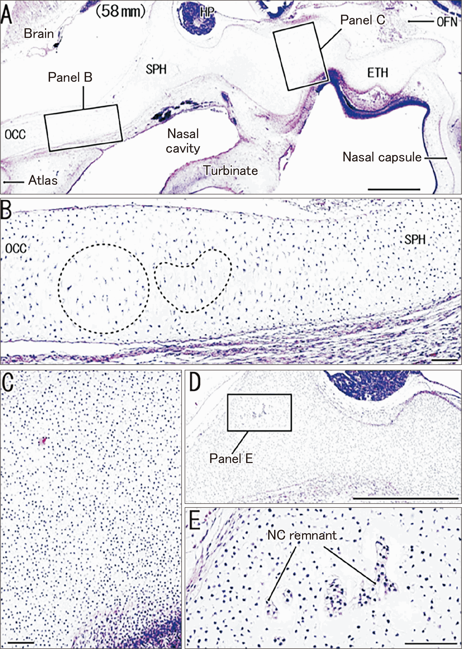
In all midterm specimens, the nasal capsule—including the cribriform plate and nasal septum of the ETH—was continuous with the anterolateral margin of the cartilaginous midline skull base (SPH). The future SPH–ETH junction consisted of homogeneous cartilage that was fully continuous with both the ETH and SPH (Fig. 1A). In contrast, the candidate occipito-sphenoidal junction contained slender or enlarged chondrocytes, clearly differing from the uniformly round cells of the SPH–ETH region (Fig. 1B vs. Fig. 1C). A notochord remnant was consistently identified in the SPH immediately posterior to the pituitary fossa (Fig. 1D, E), whereas no notochordal tissue was observed along the inferior aspect of the SPH. Together, these findings indicate that during the midterm period, the SPH–ETH junction remains structurally homogeneous and undifferentiated.
Larger midterm specimens showing early cartilage differentiation in the SPH–ETH junction
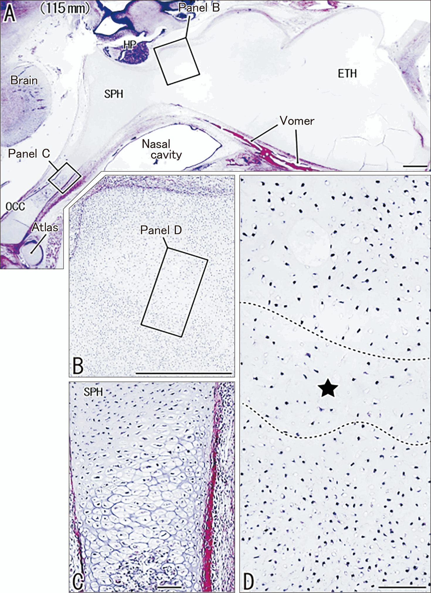
In two larger midterm specimens (120 mm and 125 mm CRL), a bipolar ossification center with columns of hypertrophic chondrocytes had already formed at the occipito-sphenoidal junction (Fig. 2A, C). At this developmental stage, the future SPH–ETH junction became histologically distinguishable from adjacent cartilage: sparsely scattered chondrocytes—often slender or irregular in shape—were present within the junctional region (Fig. 2B, D). Quantitatively, the SPH side contained >15 cells per 0.1 mm2, whereas the ETH side contained <8 cells per 0.1 mm2, representing the first clear asymmetric differentiation across this interface. These observations suggest that early specialization of the SPH–ETH junction begins during the later midterm period.
Late-term specimens: general features of the SPH–ETH junction
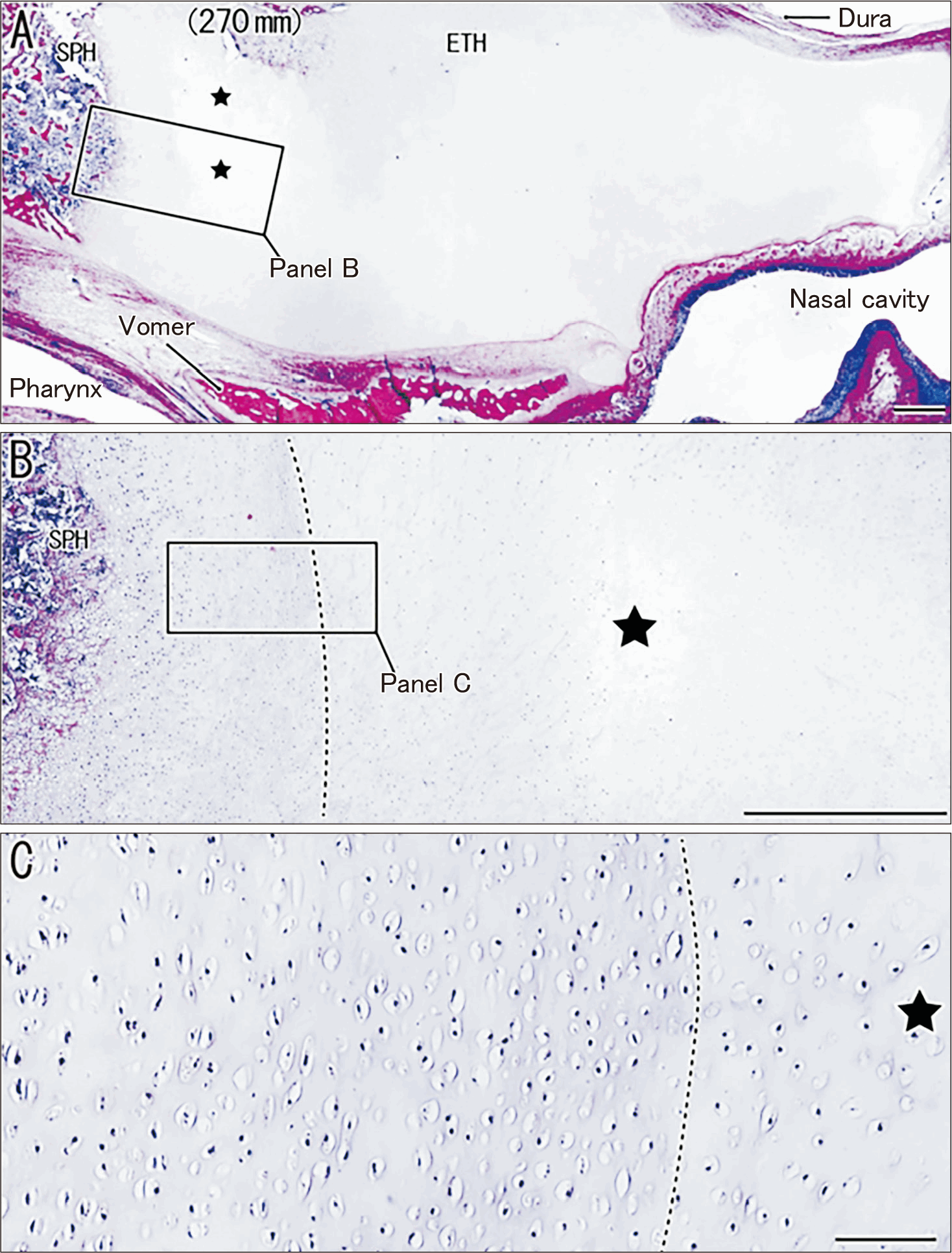
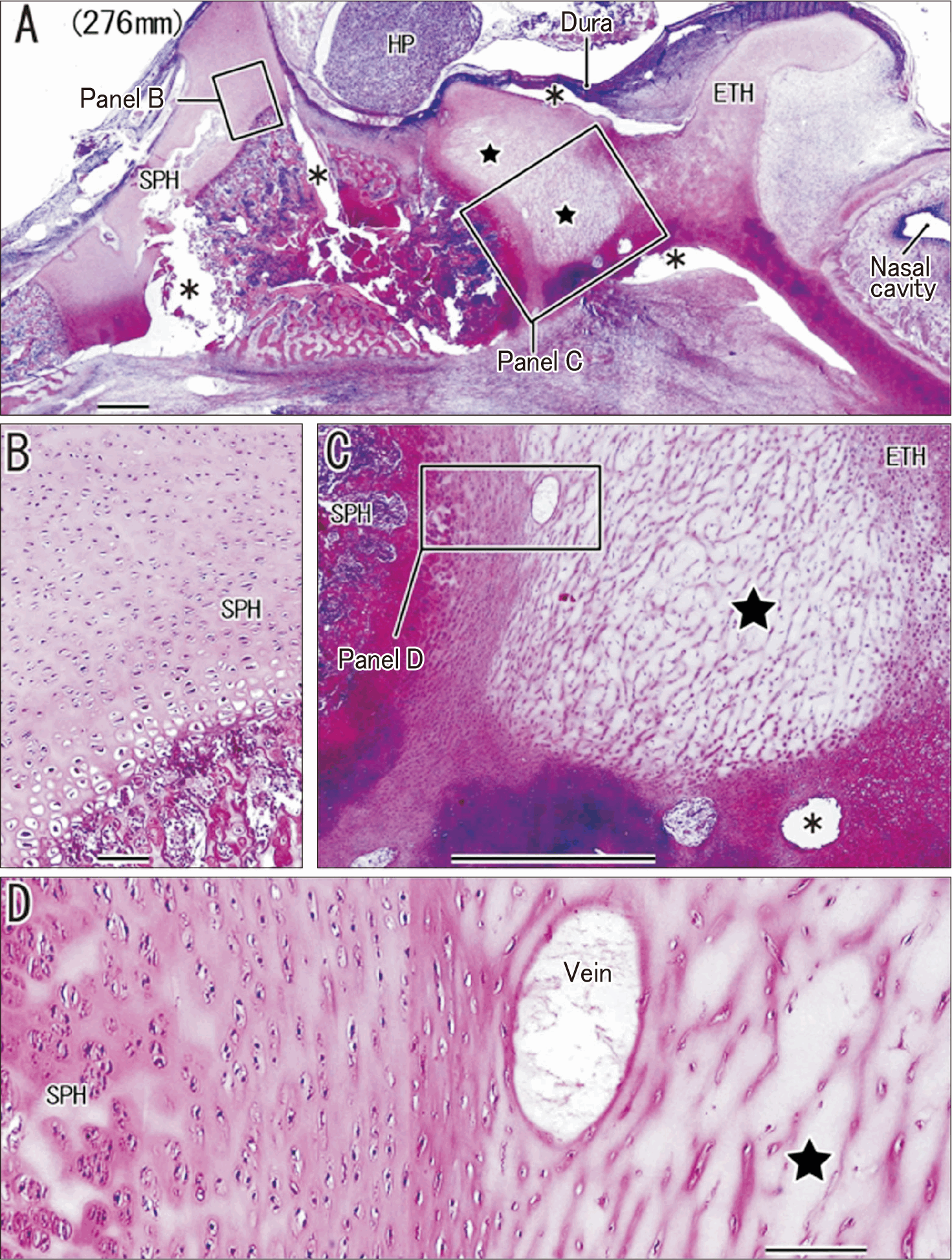
In all late-term fetuses (Figs. 3A, 4A, 5A, 6A), a second bipolar ossification center—distinct from the occipito-sphenoidal junction—was consistently present beneath the pituitary fossa within the SPH. Immediately anterior to this ossification center, the candidate SPH–ETH junction was characterized by sparsely distributed chondrocytes embedded in a less basophilic matrix (Figs. 3B, C, 4C, D, 5B, D, 6B). Cell-density measurements confirmed a clear asymmetry, with the ETH side containing 3–5 cells per 0.1 mm2 and the SPH side containing 10–15 cells per 0.1 mm2. These findings demonstrate that the SPH–ETH junction forms a distinct, low-density cartilage region that persists throughout late gestation. A summary of these observations is provided in Table 1. The sphenoid–ethmoid interface might correspond to the presphenoid-septal synchondrosis in Smith et al. (2017) [2].
Late-term specimens without anterior ossification in the ETH
In two late-term fetuses (268 mm and 276 mm CRL), the matrix of the SPH–ETH junction contained mesh-like fibrous structures, and no anterior ossification center was detected within the ETH (Fig. 4C, D). Although reduced basophilia may partly reflect long-term preservation, the organized architecture of these mesh-like fibers suggests a genuine structural feature rather than an artifact. Notably, these meshes differed from the basal-lamina–derived networks previously described in the early occipito-petrosal junction [13].
Late-term specimens with anterior ossification in the ETH
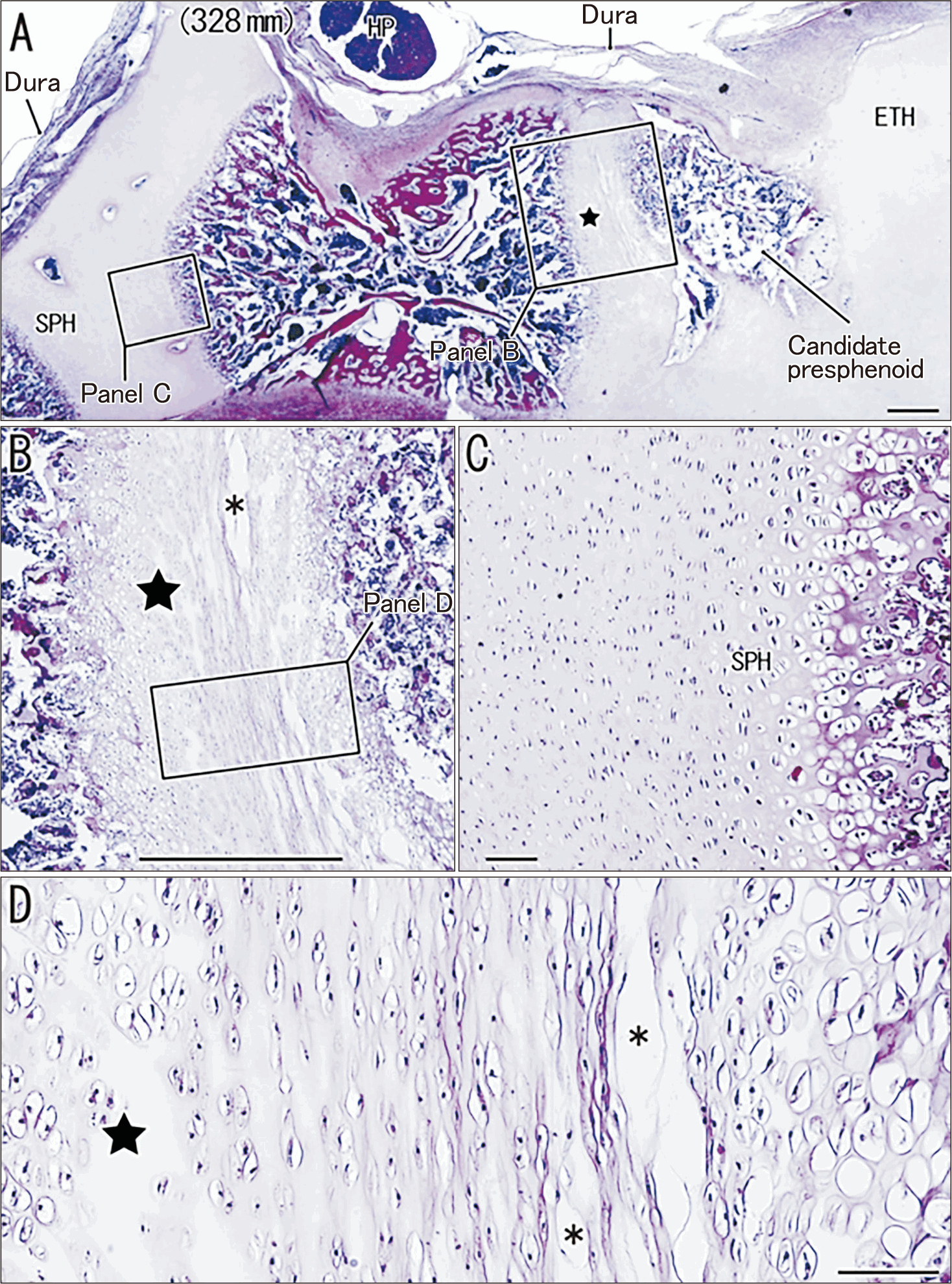
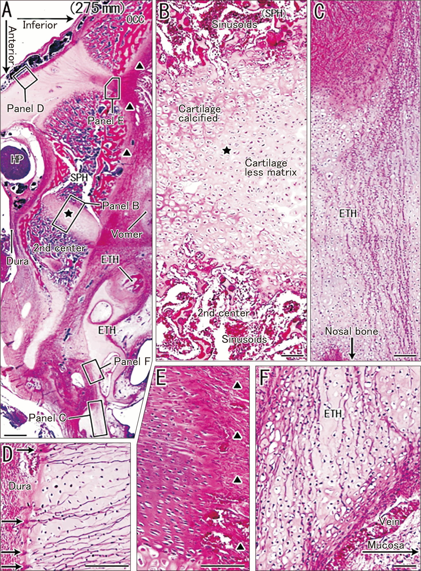
In three fetuses (275 mm, 305 mm, and 328 mm CRL), an anterior ossification center was observed at the posterior end of the ETH (Figs. 5A, 6A), representing a candidate presphenoid element in human fetuses—analogous to the presphenoid reported in non-human primates. The SPH–ETH junction exhibited a less eosinophilic matrix, and hypertrophic chondrocytes and calcified cartilage appeared at its margins (Fig. 6B). Matrix characteristics varied between specimens: the 305mm and 328 mm fetuses displayed fibrous bundles at the junction (Fig. 5B, D), whereas the 275 mm fetus did not (Fig. 6B). Despite these differences, the SPH–ETH junction remained consistently identifiable as a sparsely populated, round-celled cartilage distinct from the surrounding ETH and SPH cartilage (Figs. 5D, 6B).
Presence of fiber bundles within late-term cartilaginous regions
Distinct collagenous or basophilic fiber bundles—structures not present in midterm specimens—were observed in a subset of late-term fetuses. In the 305 mm and 328 mm CRL fetuses, supero-inferiorly oriented collagen bundles were found within the SPH–ETH junction (Fig. 5B, D), and their parallel arrangement produced linear artifactual separations during sectioning, reflecting their true anatomical organization. In contrast, the 255 mm and 275 mm CRL fetuses exhibited intensely basophilic bundles within the occipito-sphenoidal region (Fig. 6A, E, F). These bundles showed striking polarity, converging superiorly toward anchoring structures beneath the dura mater (Fig. 6A, D), while inferiorly becoming densely aligned along the thick external periosteum of the skull base (Fig. 6A, E). Within the ETH itself, bundles extended along the antero-posterior axis but lacked definite anchoring structures (Fig. 6C, F). Their consistent orientation and association with anatomical boundaries strongly suggest an organized architectural feature rather than a fixation artifact.
Discussion
This study provides the first histological evidence suggesting that human fetuses possess a developmental structure analogous to the presphenoid described in non-human primates. The SPH–ETH junction identified here consistently exhibited three characteristic features—(1) a distinct low-density cartilage region, (2) changes in chondrocyte morphology from round to slender or irregular forms, and (3) fibrous or mesh-like components within the matrix. Together with the frequent presence of an anterior ossification center, these observations strongly indicate that the anterior part of the SPH undergoes a transient differentiation pattern comparable to the presphenoid in non-human primates.
Comparison with the PSept of non-human primates
Smith and colleagues [2-4] demonstrated that the PSept in non-human primates is defined by bipolar ossification centers that sandwich a transitional cartilage zone with reduced matrix (“resting cartilage”). In our human specimens, only two late-term fetuses exhibited bipolar ossification. However, the low-density cartilage zone at the SPH–ETH junction, combined with an additional anterior ossification center, suggests a homologous developmental process. Unlike the “resting cartilage” described in primates, the human interface contained slender or irregularly shaped chondrocytes with sparse distribution, indicating a more dynamic or degenerative transition rather than maintenance of a resting state.
The anterior ossification center in humans has traditionally been assumed to form postnatally, accompanying the separation of the anterior SPH [5]. Our findings suggest that this separation may begin prenatally as mechanical stresses increase across the skull base. Indeed, in several specimens the SPH–ETH interface split readily during histological processing, likely due to tension generated by expanding ossification on both sides. Such splitting has not been reported in studies of nasal septum ossification [14], indicating that the SPH–ETH junction undergoes a distinct mechanical transition.
Fibrous components and mechanical implications
Two distinct fiber systems were identified. First, collagenous bundles running supero-inferiorly were present at the SPH–ETH junction in late-term fetuses. These bundles likely reflect intrinsic tensile forces generated during skull base elongation. Second, highly eosinophilic fiber bundles with dural anchorage were observed at the occipito-sphenoidal junction. Their staining pattern may be attributable to time-dependent formalin effects on collagen, a phenomenon known from silver staining of reticular fibers [15-17].
A comparable architecture is observed during early vertebral ossification, where basophilic bundles stretch longitudinally along the vertebral body, conferring resistance to twisting and bending. Likewise, the cartilaginous ethmoid contained longitudinal fiber bundles aligned with the antero-posterior axis, possibly reinforcing the anterior skull base against mechanical stress generated by deep invaginations of the developing nasal epithelium.
Developmental origin and possible identification of a human presphenoid
Our previous anatomical studies [18-20] established that the notochord runs along the inferior clival surface and enters the SPH immediately posterior to the pituitary fossa. The SPH–ETH junction described here, together with its associated anterior ossification center, lies anterior to the boundary of somitomere-derived mesoderm. This location implies that the anterior end of the SPH—eventually incorporated into the ETH—likely derives not from somitic mesoderm but from neural crest, similar to the nasal capsule.
Although comparative data from non-human primates are needed, the anterior ossification center identified in our specimens may represent a remnant presphenoid element. Therefore, the SPH–ETH junction we observed is a strong candidate for the human analogue of the PSept described in non-human primates [2]. The subsequent incorporation of this “transient presphenoid” into the definitive ethmoid may be driven by early pneumatization forces, including expansion of the sphenoid sinus [6, 7]. Notably, enlargement of ethmoidal air cells creates a suprabasal notch in the growing orbital roof in human infants [21], supporting the idea that early sinus development influences cartilage remodeling in this region.
Study limitations and future directions
The primary limitation of this study is the small number of suitable specimens, particularly in the mid-gestational period. Nevertheless, the consistent identification of a sparsely populated, fiber-containing cartilage zone—often associated with an anterior ossification center—strongly supports the presence of a human PSept analogue. Future studies using non-human primate fetuses, in combination with molecular profiling of chondrocyte subpopulations, will be essential to determine the degree of homology between the human SPH–ETH junction and the primate PSept.
 XML Download