



 PDF
 Citation
 Print


Introduction
Ischemic stroke remains a leading global cause of disability and mortality. While incidence and mortality rates have declined, treatment strategies remain largely empirical due to the incomplete understanding of its complex pathophysiology. Although restoring blood flow (reperfusion) after ischemia is critical, it paradoxically exacerbates brain tissue damage [1]. The mechanisms underlying neuronal injury in stroke involve multiple pathways, including calcium overload, excitotoxicity, free radical production, oxidative stress, and impaired protein synthesis [2, 3]. During cerebral ischemia/reperfusion (I/R), excessive reactive oxygen species (ROS) generation induces oxidative stress, causing damage to cellular components, including lipids, carbohydrates, proteins, and DNA.
Oxidative stress plays a pivotal role in neuronal injury following ischemic stroke. This pathological mechanism highlights the critical need for antioxidant-based therapeutic strategies to attenuate ischemic brain damage and improve functional recovery. While significant efforts have been devoted to identifying effective neuromodulators, few candidate therapies have successfully transitioned to clinical applications for stroke treatment.
Sirtuins, a class of histone deacetylases, represent a particularly promising cellular defense system against oxidative stress, functioning through multiple molecular pathways. Among these, Sirtuin 1 (SIRT1) is an NAD+-dependent histone deacetylase capable of regulating numerous non-histone proteins [4]. SIRT1 is widely expressed throughout the rat brain and plays a significant role in the pathophysiological mechanisms of I/R injury [5].
Extensive research has established that SIRT1 participates in multiple essential biological processes, including chromatin remodeling, transcriptional silencing, chromosome stability maintenance, cell cycle regulation, apoptosis, autophagy, metabolic regulation, growth inhibition, and oxidative stress response [6]. Of particular significance, SIRT1 plays a crucial role in maintaining mitochondrial function and integrity. Peroxisome proliferator-activated receptor gamma coactivator 1α (PGC-1α) is a central regulator of mitochondrial biogenesis and oxidative metabolism [7]. PGC-1α functions as a transcriptional coactivator for SIRT1, while SIRT1 reciprocally enhances PGC-1α activity, creating a positive feedback loop that critically regulates mitochondrial function [8]. Moreover, PGC-1α upregulates nuclear respiratory factor 1 (NRF-1) expression, which subsequently stimulates mitochondrial oxidative phosphorylation, modulates nuclear-encoded respiratory enzyme genes, and coordinates mitochondrial DNA replication, transcription, and protein expression [9]. Notably, activation of the SIRT1/PGC-1α pathway enhances oxidative stress resistance in chronic cerebral ischemia models and mitigates associated cognitive impairments [10].
Vitamin D, recognized as a neurosteroid, modulates multiple brain functions and has been associated with reduced stroke risk [11]. Its activation occurs through sequential hydroxylation, first in the liver to form calcifediol (25-hydroxyvitamin D [25(OH)D]), then in the kidneys where it is converted to its biologically active form, calcitriol (1,25-dihydroxyvitamin D) [12]. Calcitriol, an FDA-approved compound, has emerged as a promising neuroprotective agent. Its primary mechanism of action involves binding to the vitamin D receptor (VDR). Given the ubiquitous expression of VDR, vitamin D signaling appears to regulate diverse physiological processes, including immune system modulation, cell cycle regulation, and neurodevelopmental processes [13, 14]. These findings highlight the importance of developing novel therapeutic strategies targeting vitamin D pathways to improve post-stroke outcomes.
In multiple neurotoxicity models, vitamin D administration has been shown to enhance neuronal survival through dual mechanisms: (1) regulation of intracellular calcium homeostasis and (2) attenuation of free radical production. Both in vivo and in vitro studies demonstrate vitamin D’s ability to reduce oxidative stress markers and downregulate pro-inflammatory cytokine expression [15]. Nevertheless, the molecular mechanisms underlying vitamin D’s therapeutic effects in ischemic stroke remain insufficiently explored. Based on current evidence, we propose that vitamin D exerts neuroprotective effects in focal cerebral ischemia by mitigating oxidative stress damage, mediated in part through activation of the SIRT1-PGC1-α signaling pathway.
Materials and Methods
Animals
Forty-five adult male Wistar rats (weight range: 230–300 g) were procured from the Animal Research Facility of Arak University of Medical Sciences. The animals were housed under controlled environmental conditions with: A 12:12 hour light-dark cycle, ambient temperature maintained at 23°C±2°C, and relative humidity of 55%–60%. All animals received ad libitum access to standard laboratory chow and filtered water, except during specified experimental procedures. All experimental protocols were performed in full compliance with the institutional ethical guidelines of the Animal Ethics Committee of Arak University of Medical Sciences for animal research (Ethical Approval Code: IR.ARAKMU.AEC.1403.009).
Middle cerebral artery occlusion model
Focal cerebral ischemia was induced in rats using an established intraluminal filament model. Animals were anesthetized with 3% isoflurane (in 70% N2O/30% O2) and surgically prepared for left common carotid artery (CCA) exposure. A silicon-coated 4-0 monofilament (diameter: 0.28–0.30 mm) was inserted into the CCA and advanced 18–20 mm distally into the internal carotid artery to occlude the middle cerebral artery origin. After 60 minutes of occlusion, the filament was carefully withdrawn to initiate reperfusion.
Study design
Animals were randomly allocated into three experimental groups:
(1) Sham-operated group (n=15): animals underwent the same surgical procedure as the middle cerebral artery occlusion (MCAO) groups, but no monofilament was inserted into the CCA.
(2) Stroke-treated MCAO group (n=15): animals in this group received saline supplemented with ethanol, mirroring the treatment conditions of the calcitriol group but without the active drug.
(3) Calcitriol-treated MCAO group (n=15): rats were administered intraperitoneal injections of calcitriol (Cayman Company) at a dosage of 1 µg/kg body weight for three consecutive days after stroke induction.
The calcitriol was dissolved in ethanol and diluted with saline. Following ischemia induction, all animals were deeply anesthetized and decapitated 72 hours later for further experiments. Rats that achieved a full score in the behavioral test were included in the study, and those that died during model induction were replaced.
Neurobehavioral performance
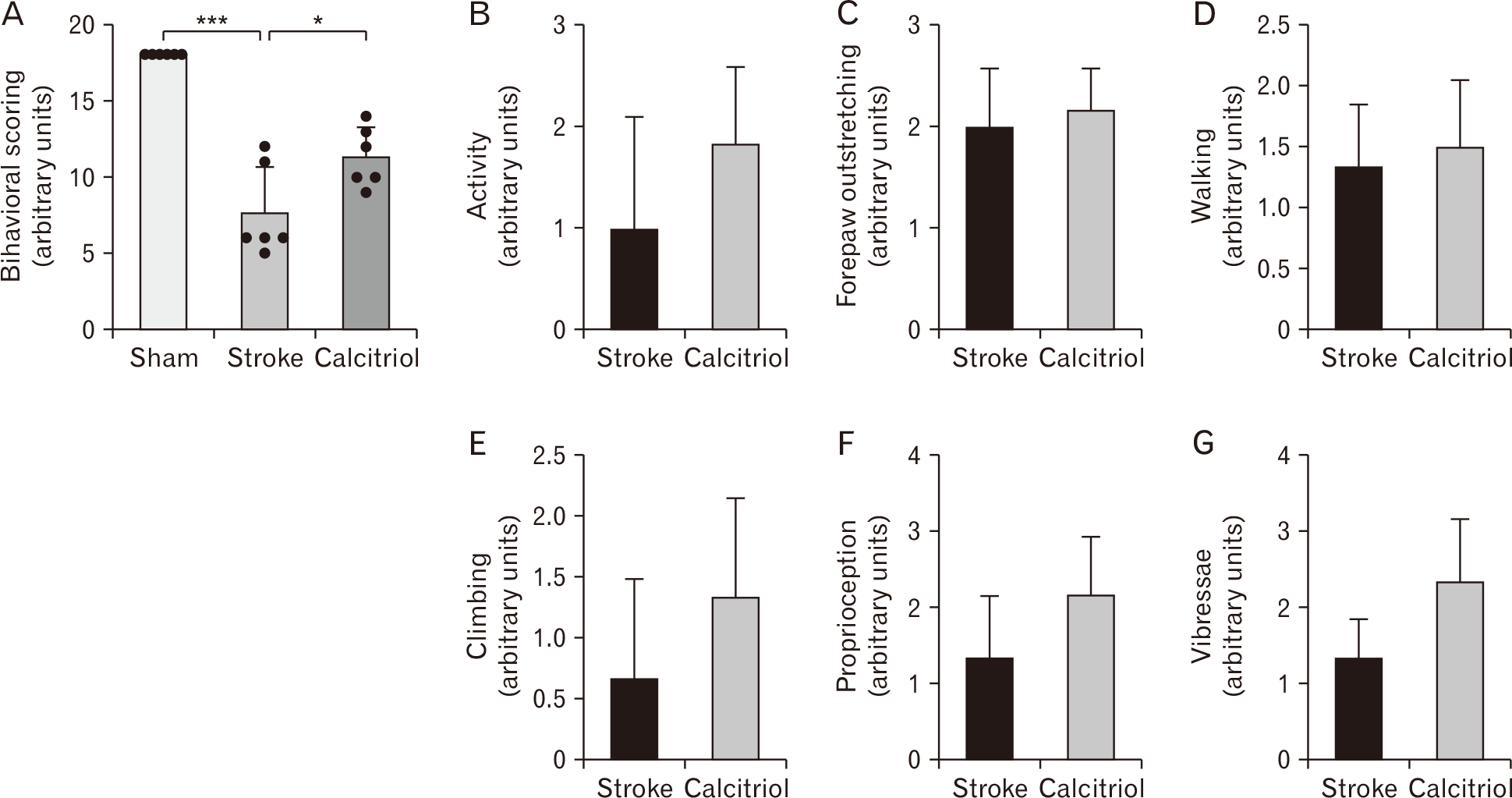
Neurological deficits in a MCAO model were evaluated using the Garcia score (an 18-point scoring system) before and 72 hours after occlusion for all groups (n=15) [16], with minor modifications. This test assesses six behavioral components: spontaneous activity [17], movement symmetry [18], climbing ability [19], forelimb extension [20], body awareness [21], response to vibrissae stimulation. Each component contributes to the total score, with lower scores indicating more severe neurological dysfunction. In this study, sham-operated animals consistently scored the maximum (18 points), confirming the absence of functional impairments.
Assessment of brain infarct volume
Following a 72-hour reperfusion period after ischemia, animals were euthanized (each group; n=4), and their brains were rapidly harvested and flash-frozen at −20°C for 10 minutes. Coronal sections (2 mm thick) were then prepared using a brain matrix (Zivic Instruments). These sections were immersed for 20 minutes in 1% 2,3,5-triphenyltetrazolium chloride (TTC; Merck) dissolved in phosphate-buffered saline (37°C). This staining process distinguishes normal brain tissue, which appears dark red, from infarcted areas that remain unstained. To account for edema-related volume changes, the corrected infarct volume for each brain section was calculated using the following formula: Infarct measurements were conducted by an investigator blinded to treatment group assignments to ensure unbiased assessment.
Tissue preparation
After the completion of behavioral assessments, the rats were sacrificed for histological analysis (each group; n=4). Anesthesia was first induced using 10% chloral hydrate, followed by transcardial perfusion—initially with 0.9% normal saline, and then with 10% neutral buffered formalin (NBF). The brains were then removed and fixed in 10% NBF at 4°C for 48 hours. Subsequently, the brain tissues were embedded in paraffin, sectioned coronally at a thickness of 5 micrometers, and stained using Nissl staining for examination under a light microscope (Nikon Eclipse Ti-U; Nikon).
Nissl staining
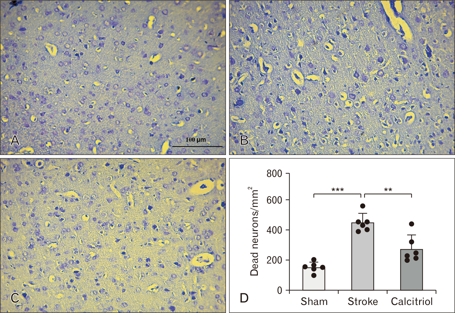
Brain tissue sections were deparaffinized in xylene and stained with 0.1% cresyl violet solution. Stained sections were visualized and imaged using a light microscope (Nikon Eclipse Ti-U). For quantitative assessment, neurons were counted based on specific criteria: healthy neurons exhibit well-defined cell bodies with prominent Nissl substance and visible nuclei. In contrast, dead or degenerating neurons appear shrunken, with darkly stained (pyknotic) nuclei and a loss of Nissl substance, reflecting cytoplasmic degradation.
Measurement of oxidant and antioxidant levels
Following brain extraction, the ischemic cortical tissue was dissected and homogenized in RIPA lysis buffer. The homogenate was centrifuged (10,000×g, 15 minutes, 4°C), and the supernatant was collected for analysis. MDA levels were quantified as an indicator of lipid peroxidation using the thiobarbituric acid (TBA) assay, following established methodology [22]. The supernatant was mixed with a stock solution containing: 15% trichloroacetic acid, 0.375% TBA, and 0.25 N hydrochloric acid (HCl). The reaction mixture was heated to 95°C (90°C–100°C range) for 15 minutes, then centrifuged at 5,000×g (10 minutes). Absorbance was measured at 532 nm using an ELISA reader (Garni Medical Engineering Co.). In biological systems, NO undergoes rapid oxidation to stable metabolites (nitrite and nitrate). Therefore, nitrite concentration serves as a reliable surrogate marker for assessing NO production. Nitrite concentration was determined using a modified Griess reaction. The assay protocol consisted of: mixing 100 µl supernatant with 100 µl freshly prepared Griess reagent containing: 0.1% N-(1-naphthyl) ethylenediamine dihydrochloride and 2% sulfanilamide in 5% phosphoric acid, incubating the mixture at room temperature (25°C) for 30 minutes protected from light and measuring absorbance at 540 nm using a microplate reader.
Total antioxidant capacity (TAC) was measured using the ferric reducing antioxidant power (FRAP) assay according to established methodology. The procedure was as follows: FRAP reagent preparation (combined the following preheated [37°C] components: 25 ml sodium acetate buffer [0.3 M, pH 3.6], 2.5 ml TPTZ solution [0.01 M in 0.04 M HCl], 2.5 ml FeCl3·6H2O solution [0.02 M] and final working volume: 1.5 ml FRAP reagent), assay protocol (mixed 50 μl supernatant with 1.5 ml FRAP reagent, incubated at 37°C for 15 minutes in the dark, measured absorbance at 593 nm against a blank).
Real time PCR
Fresh tissue samples (n=4) from the penumbra region of the injured cortex were collected in sterile microtubes and immediately flash-frozen in liquid nitrogen for storage at –80°C until processing. Total RNA was extracted using TRIzol reagent (MaxZol), with concentration and purity verified by spectrophotometric measurement of A260/A280 ratios. Complementary DNA (cDNA) was synthesized from extracted RNA using the cDNA Synthesis Kit (Parstous) under standard reaction conditions. For quantitative analysis, real-time PCR reactions were performed in 10 μl reaction volumes containing 1X Ampliqon RealQ Plus 2X Master Mix Green (Ampliqon), gene-specific primers (sequences listed below), and diluted cDNA template, with amplification conducted by an Applied Biosystems real-time PCR system (Thermo Fisher Scientific) using optimized cycling conditions. The primer sequences utilized in this experiment were as Table 1.
Statistical analysis
The data were reported as mean±SD, and statistical analyses were conducted using GraphPad Prism version 8.0 (Dotmatics). The t-test was used for comparing two samples, while comparisons among groups were performed using one-way analysis of variance (ANOVA) followed by Tukey’s post-hoc test. A statistical significance level of P-value <0.05 was considered.
Results
Therapeutic effects of calcitriol against I/R injury
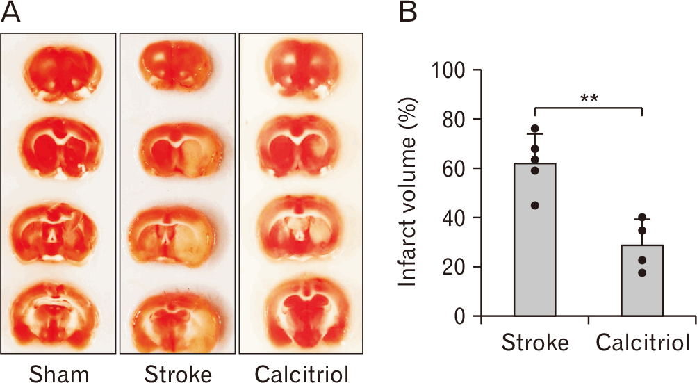
To calculate the volume of brain infarction, vital staining with TTC was used 72 hours after MCAO. As illustrated in Fig. 1, the sham group displayed no signs of cerebral injury, whereas the stroke group exhibited a significant infarct volume. Treatment with calcitriol led to a notable decrease in infarct volume compared to stroke group (P<0.01). Neurological scores and infarct size serve as critical biomarkers for evaluating ischemic brain injury severity. In our recent study, one-way ANOVA followed by Tukey’s post-hoc test demonstrated statistically significant intergroup differences (P<0.05). Neurological assessment using the 18-point Garcia scale revealed: sham-operated animals maintained normal scores, indicating intact neurological function, vehicle-treated controls showed significant deficit, and calcitriol-treated animals exhibited substantial functional improvement (Fig. 2). This suggests that calcitriol may have a therapeutic effect against neurological injuries induced by infarction.
Cell death in I/R injury
Nissl staining revealed significant ischemic damage in the cortical region, with I/R injury causing a 66% increase in neuronal cell death compared to sham controls (P<0.001). As demonstrated in Fig. 3, calcitriol treatment significantly attenuated this neuronal loss, reducing cell death by 61% versus the vehicle-treated I/R group (P<0.01). Quantitative analysis of Nissl-positive cells confirmed these findings (Fig. 3).
Therapeutic mechanism of calcitriol against I/R
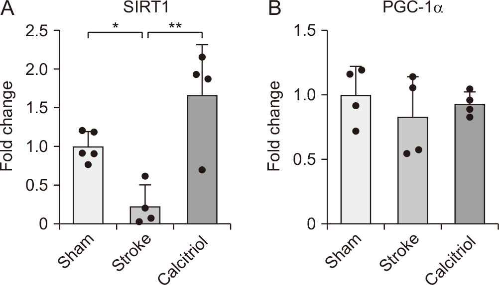
To elucidate the neuroprotective mechanisms of calcitriol following cerebral ischemia, we examined mRNA expression levels of SIRT1 and PGC1-α in the ischemic penumbra using quantitative RT-PCR. Statistical analysis by one-way ANOVA demonstrated significant intergroup differences in SIRT1 expression. Post-hoc analysis revealed that vehicle-treated ischemic animals showed decreased SIRT1 (P<0.05) and PGC1-α expression compared to sham controls. Importantly, calcitriol treatment further significantly enhanced mRNA expression of SIRT1 (P<0.01) relative to vehicle-treated animals, as illustrated in Fig. 4. Although PGC1-α expression increased following calcitriol, the difference was not statistically significant.
Therapeutic impact of calcitriol on markers of oxidative stress
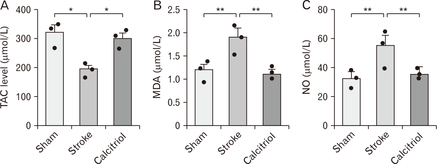
This study examines the pivotal role of oxidative stress in cellular survival pathways following cerebral ischemia, with particular emphasis on the neuroprotective potential of calcitriol. Through a comprehensive analysis of key oxidative stress biomarkers—MDA, NO, and TAC—we demonstrate that oxidative damage constitutes a fundamental mechanism in ischemic brain injury. One-way ANOVA analysis showed significant variations in MDA, NO, and TAC levels across the groups. Post-hoc Tukey’s test indicated that vehicle-treated rats exhibited increased MDA and NO levels alongside decreased TAC levels compared to the sham-operated group 72 hours after MCAO (P<0.05, P<0.01, respectively). In contrast, calcitriol treatment resulted in a notable reduction in MDA and NO levels while significantly enhancing TAC levels compared to the vehicle group (P<0.05, P<0.01, respectively) (Fig. 5). These findings demonstrate that the therapeutic potential of calcitriol may stem from its ability to simultaneously target multiple components of the oxidative stress cascade.
Discussion
The key discovery of this research indicates the therapeutic effects of calcitriol against brain injuries, which were confirmed through the improvement of neurological deficit scores and the reduction of lesion volume and oxidative stress. Mechanistically, these neuroprotective effects appear mediated through activation of the SIRT1 antioxidant pathway, suggesting a novel molecular target for stroke intervention. Ischemic stroke remains a leading global cause of mortality and long-term disability [23], with comparable burdens across developed and developing nations. Although thrombolytic therapy has proven effective, its use is limited due to a narrow therapeutic window and strict criteria [24]. These limitations underscore the critical need for complementary therapeutic approaches. Nutritional interventions like vitamin D supplementation offer distinct advantages, including a favorable safety profile, cost-effectiveness, and widespread accessibility. Emerging evidence supports their potential in both neuroprotection and functional recovery [25, 26].
Vitamin D, a fat-soluble secosteroid, is primarily synthesized in the skin through ultraviolet B radiation-mediated conversion of 7-dehydrocholesterol, with dietary intake serving as a secondary source. This essential nutrient plays critical roles in maintaining neural homeostasis through immunomodulation of neuroinflammatory pathways, enhancement of endogenous detoxification mechanisms, and regulation of neurotrophic factor expression [27]. Epidemiological data reveal a strong inverse correlation between serum vitamin D levels and cerebrovascular disease risk and neurological disorders, such as stroke [28, 29]. Studies in rats have demonstrated that a lack of vitamin D results in increased brain infarct sizes and more pronounced behavioral impairments following stroke induction [30].
The bioactivation of vitamin D occurs through two sequential hydroxylation reactions: (1) hepatic conversion: 25-hydroxylation in the liver produces 25(OH)D, the major circulating form and (2) renal activation: 1α-hydroxylation in proximal renal tubules generates calcitriol (1,25-dihydroxyvitamin D3), the biologically active metabolite. This tightly regulated metabolic pathway highlights the critical relationship between vitamin D homeostasis and neurological function. Given these mechanisms, ensuring adequate vitamin D status—whether through sensible sun exposure (15–30 minutes daily) or supplementation (1,000–4,000 IU/day)—represents a potentially crucial modifiable factor for cerebrovascular and neurological disease prevention [31].
Accumulating evidence demonstrates that vitamin D exerts significant neuroprotective effects following ischemic brain injury. Experimental studies have shown that calcitriol administration can reduce total infarct volume in rat MCAO models, preserve blood-brain barrier integrity, and attenuate neuronal apoptosis. These protective effects have been consistently observed across multiple studies [32, 33], suggesting vitamin D’s potential as a therapeutic agent for stroke. However, the precise molecular mechanisms underlying these benefits remain incompletely characterized. Current gaps in understanding include: the relative contribution of genomic vs. non-genomic signaling pathways, potential interactions with other neuroprotective systems, and optimal dosing and administration timelines. Further investigation is needed to elucidate these mechanisms and translate preclinical findings into clinical applications [34]. This investigation employed a comprehensive approach to evaluate ischemic stroke outcomes through quantitative measurement of cerebral infarction volume and systematic assessment of functional neurological deficits.
Our findings demonstrate that calcitriol treatment provides substantial neuroprotective effects, significantly reducing neurological deficit scores and decreasing cerebral infarction volume. These protective effects occur against the complex pathophysiology of cerebral I/R injury, which is characterized by excessive ROS generation [35] leading to widespread oxidative damage of cellular macromolecules (lipids, proteins, and DNA) and subsequent activation of apoptotic pathways. Importantly, we established calcitriol’s potent antioxidant properties through three key measurements: enhancement of TAC, reduction in lipid peroxidation, and decrease in nitric oxide metabolites, collectively demonstrating its ability to mitigate oxidative stress following ischemic insult. Vehicle-treated animals demonstrated significant oxidative stress, characterized by reduction in TAC versus sham controls, increase in NO metabolites, and elevation in MDA levels. Calcitriol administration markedly attenuated these oxidative disturbances, producing: recovery of TAC activity, reduction in NO levels, and decrease in MDA concentration. These findings demonstrate calcitriol’s potent antioxidant effects in mitigating IR-induced oxidative damage.
SIRT1 modulates PGC-1α activity through post-translational modifications (deacetylation and phosphorylation), critically regulating mitochondrial biogenesis and oxidative metabolism in neural tissue [36, 37]. Under hypoxic conditions, PGC-1α upregulation serves as an adaptive response, with studies demonstrating its neuroprotective effects against oxidative stress-induced apoptosis [38, 39]. Our findings reveal that calcitriol treatment significantly elevated SIRT1 expression in the ipsilateral cortex 72 hours post-MCAO, suggesting its involvement in calcitriol-mediated neuroprotection. In contrast, PGC-1α expression exhibited an increasing trend; however, this change did not reach statistical significance. The ischemic cascade initiates when cerebral blood flow interruption (≤30% of baseline) creates an energy crisis, with reperfusion paradoxically generating cytotoxic ROS levels [40]. Mitochondria, while essential for ATP production, become mediators of cell death when compromised. PGC-1α regulates energy metabolism by promoting mitochondrial gene expression and biogenesis. Notably, we observed a strong positive correlation between PGC-1α and SIRT1 expression levels, indicating that their coordinated upregulation may represent a key mechanism in calcitriol’s therapeutic effects against ischemic injury.
To our knowledge, this study provides the first evidence of calcitriol’s interaction with the SIRT1/PGC-1α pathway in cerebral ischemia. Our RT-PCR analysis revealed two key findings: vehicle-treated animals showed decreased SIRT1 expression in the peri-infarct cortex, suggesting an endogenous neuroprotective response to ischemic stress, and calcitriol treatment significantly amplified this effect by further increasing SIRT1 expression. These results indicate that calcitriol potentiates the brain’s intrinsic antioxidant defenses through SIRT1 activation. While previous research has identified K610 deacetylation as a mechanism for SIRT1 activation by other compounds, our study uniquely demonstrates the temporal pattern of SIRT1/PGC-1α regulation post-ischemia, calcitriol’s specific enhancement of this pathway, and a correlated reduction in oxidative stress markers. These novel findings position the SIRT1/PGC-1α axis as a key mediator of calcitriol’s neuroprotective effects, opening new avenues for targeted stroke therapies.
Although SIRT1 is known to activate PGC-1α through deacetylation, the lack of significant changes in PGC-1α mRNA expression following calcitriol treatment in our study may be explained by post-translational regulation mechanisms. SIRT1 can enhance PGC-1α activity via deacetylation without necessarily affecting its transcriptional levels. Moreover, SIRT1 exerts its effects through multiple downstream targets beyond PGC-1α, including FOXO transcription factors, p53, NF-κB, PPARγ, LXR, and AMPK, influencing diverse cellular processes such as apoptosis, inflammation, oxidative stress, lipid metabolism, and mitochondrial biogenesis. Therefore, the observed neuroprotective effects of calcitriol-induced SIRT1 upregulation may involve alternative SIRT1-mediated pathways independent of PGC-1α transcriptional activation.
In conclusion, our findings demonstrate that calcitriol exerts significant neuroprotection against cerebral I/R injury by activating the SIRT1 pathway, resulting in a reduction in lipid peroxidation (MDA) and an increase in TAC. This pathway activation reduces infarct volume and improves neurological recovery on standardized deficit scales. Calcitriol’s therapeutic potential is further supported by its established safety profile, blood-brain barrier permeability, and multimodal mechanisms of action. The robust neuroprotection mediated through SIRT1 activation highlights this pathway as a novel therapeutic target that merits clinical translation for ischemic stroke treatment.
Limitation
Although our findings indicate an upregulation of SIRT1 following calcitriol treatment, further studies employing pharmacological inhibition or genetic silencing (e.g., SIRT1 knockdown) are required to confirm the causal role of SIRT1 in mediating the neuroprotective effects of calcitriol.
 XML Download