



 PDF
 Citation
 Print


Introduction
In the interforaminal region of the mandible, the inferior alveolar nerve divides into the mental and incisive branches. The anterior portion of the mandible on the lingual side has numerous bone canal structures through which the incisive branch of the inferior alveolar nerve passes [1]. Furthermore, the midline lingual foramina are located in the midline of the lingual cortical bone of the mandible, which is important for the blood supply around the mental spine [2, 3]. The mandibular canal exists in the mandible that connects the mandibular foramen on the inside of the ramus to the mental foramen on the outside of the mandibular body. It runs through the mandible to a position 2–3 mm in front of the mental foramen, then bends backwards and connects to the mental foramen, and is called the anterior loop [3-6]. The inferior alveolar nerve is a sensory nerve derived from the mandibular nerve of the trigeminal nerve, and its incisive branch innervates the first premolar, canine, incisor, and their labial gingiva. The mandibular central incisors receive nerve fibers from the incisive branches on both sides [1, 7]. Preventing surgical complications such as paresthesia of the anterior teeth and surrounding mucosa is important [8]. The lingual foramen is found on the lingual side of the mandible, the sublingual artery enters above the mylohyoid muscle, whereas the submental artery enters below the mylohyoid line [9, 10]. In a case in which a cortical bone perforation was found during implant placement on the lingual side of the mandibular canine, the bleeding site was found 4 cm posterior to the mandible [11]. Furthermore, the development of a hematoma in the floor of the mouth can lead to airway obstruction [12]. The McGill consensus states that 2-implant mandibular overdentures are the standard of care for the treatment of edentulous patients [13]. Having an accurate understanding of the anatomical structures involved in placing implants between the mental foramina to prevent nerve paralysis and subcutaneous bleeding from the floor of the mouth is crucial.
In this study, the anterior loops of the donated bodies were dissected and morphological measurements were performed. In addition, the anterior loop, mandibular incisive canal, and lingual foramen were segmented from three-dimensional (3D) image data acquired using cone-beam computed tomography (CBCT) to observe the interior of the mandibular interforaminal region.
Materials and Methods
The samples used were 21 sides of 13 specimens (6 males, 7 females, average age 78.3±9.7 years) from the anatomy training course at the School of Dentistry, Ohu University. This study was approved by the ethics committee of Ohu University (approval number: 234). The head was fixed, and the Frankfort horizontal plane of the specimen was parallel to the floor. The CBCT images were obtained by setting the imaging area to include the interforaminal region of the mandible. CBCT was performed using a MORITA 3DX Multi-Image Micro CT FPD (Morita Co.). The CBCT was performed at 80 kV, 5 mA, and 18 seconds. Thereafter, the mental nerve was clearly visible around the mental foramen of the specimen and cut. After removing the surrounding soft tissue, the mandible was cut behind the mental foramen and extracted. Furthermore, after a midline incision, the cortical bone in front of the mental foramen was removed with a dental engine, and the anterior loop of the mandibular canal and the incisive branch bifurcated from the inferior alveolar nerve and distributed to the anterior incisor region were dissected out.
Morphometric measurements around the mental foramen
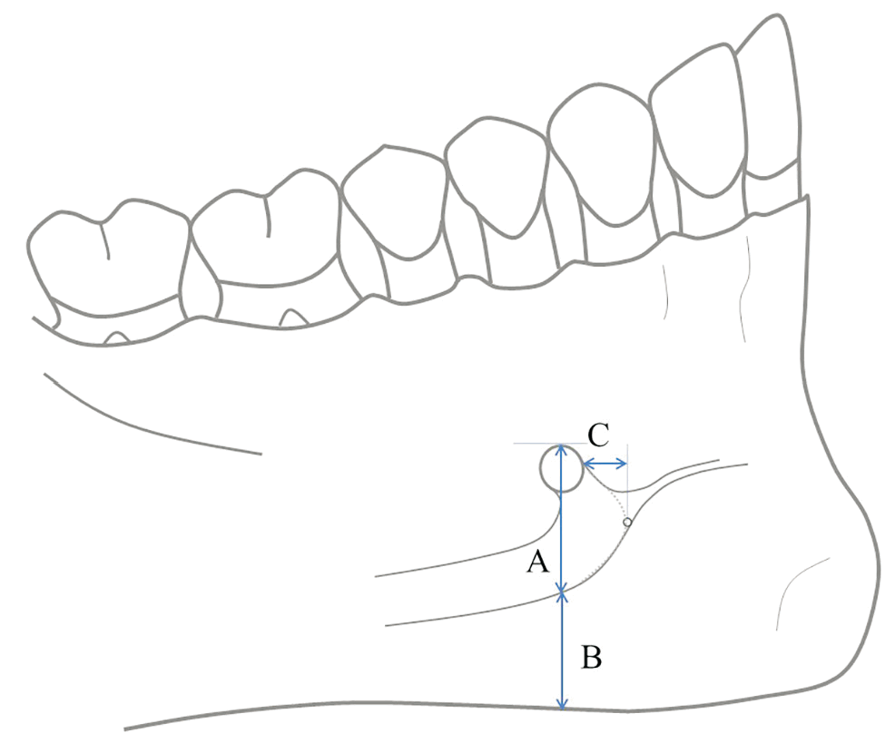
After dissection, the specimens placed on mandibular plane horizontal were photographed using a digital camera; Canon EOS 7D Mark II (Canon) after a ruler was placed at a standard distance. The captured image data were measured using the public domain software Image J (National Institutes of Health) [14] for three items: “A,” the vertical distance from the superior margin of the mental foramen to inferior border of the mandibular canal; “B,” the vertical distance from the inferior border of the mandibular canal to inferior border of the mandible on the same straight line as in “A”; and “C,” the horizontal distance from the anterior margin of the mental foramen to the anterior border of anterior loop (Fig. 1).
Three-dimensional observation of the mandibular body between the mental foramen using CBCT
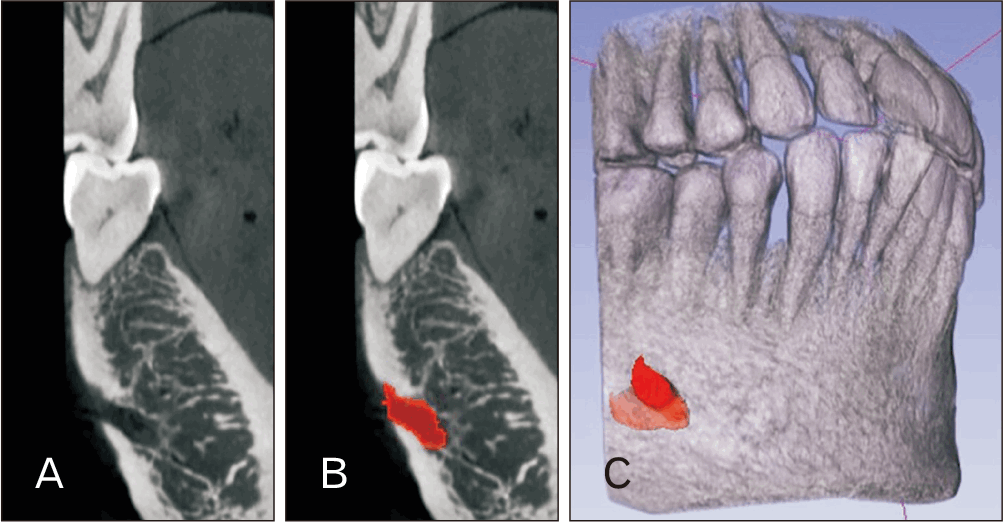
Using DICOM data from CBCT scans of four sides of four bodies, 3D observations were performed using 3D Slicer v5.6.1 (https://www.slicer.org) on the mandibular canal around the mental foramen and the canal structure continuing from the lingual foramen in the lingual cortical bone [15]. The segmentation editor module was used to segment the pores in the cortical bone and connect the tubular structures from consecutive two-dimensional slice images, and a 3D reconstruction image was created together with the entire data with increased transparency (Fig. 2).
Results
The vertical distance from the superior border of the mental foramen to the inferior border of the mandibular canal was 7.34±2.11 mm overall, 8.06±2.30 mm in male and 6.67±1.78 mm in female. The vertical distance from the inferior border of the mandibular canal to the inferior border of the mandible was 8.90±1.57 mm overall, 9.05±1.73 mm in male and 8.76±1.48 mm in female. The horizontal distance between the anterior margin of the mental foramen to the anterior border of anterior loop was 3.42±1.16 mm overall, 3.39±1.43 mm in male and 3.44±0.92 mm in female, and no sex differences were observed (Table 1).
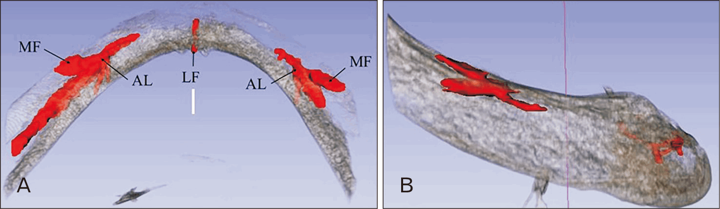
In some segmented CBCT images of the interforaminal region, the mandibular incisive canal continuously extends anteriorly from the anterior loop (Fig. 3). Although the mandibular incisive canal was discernible at the base of the anterior loop, it became indistinguishable anteriorly from the trabecular structure of the cancellous bone. Similarly, on the lingual side of the interforaminal region, a canal structure from the lingual foramen was observed in the cortical bone; however, no course could be identified in the cancellous bone.
Discussion
At a symposium held at McGill University in Montreal, Canada, in 2002, the restoration of the edentulous mandible with a conventional denture was stated to no longer be the most appropriate first-choice prosthodontic treatment. A 2-implant overdenture (IOD) should be the first choice of treatment for edentulous mandibles [13]. Since 2014, the Republic of Korea has reflected public opinion and made it possible for people aged 75 years and over to have up to two implants inserted using health insurance, although they will have to pay a copayment. Furthermore, the applicable age range was 75 years or older in 2014 but was gradually lowered to 70 years or older in 2015 and 65 years or older in 2016 [16]. A study utilizing data from the Korean National Health and Nutrition Examination Survey reported that the expansion of insurance coverage was associated with both a reduced need for dentures and an increased utilization of dentures and dental implants [17]. Therefore, the application of IOD with implants placed in the interforaminal region of the mandible is expected to expand.
The anterior loop is a recognized anatomical structure where the mandibular canal runs forward beyond the mental foramen within, curves back, and then exits through the mental foramen [3-6]. This structure develops as the child grows, and after the age of 5, the direction of the mental foramen shifts in a posterior and superior direction [18].
Dissection results showed that the horizontal distance between the anterior margin of the mental foramen to the anterior border of anterior loop was 3.42 mm. A Korean study also found that the horizontal distance between the anterior edge of the anterior loop and anterior edge of the mental foramen was 3.05 mm [4]. On the other hand, a study from New Zealand found an average of 1.20 mm [5], while a CBCT study from Greece found an average of 0.89 mm [6]. Differences in the morphology of the anterior loop are related to gender and race. In the present study, no sex differences were found in the length of the anterior loop; however, previous studies have shown a greater tendency in males. Regarding racial differences, the anterior loop showed larger values in Asians [19]. The limitations of this study include a limited sample size, the advanced mean age of the donated cadaveric specimens, and the absence of categorization based on the status of the remaining dentition. Therefore, no sex differences were observed in the results.
3D Slicer is an open-source software platform that allows image segmentation and quantification of the regions of interest [15]. In this study, we performed a 3D reconstruction of the anterior loop of the mandibular canal in front of the mental foramen and the luminal structure connecting to the lingual foramen. From the segmented 3D reconstruction images generated by 3D Slicer, determining the continuity of the luminal structure within the cancellous bone was difficult. Similarly, in CBCT observations of the mandibular canal, evaluation of the continuity of the canal structure within the bone in all specimens was difficult [20-22]. A study using micro-CT to observe the zygomatic facial foramen and zygomatic canal found that smaller foramina could be detected with high accuracy and were observed more frequently than in previous studies [23]. Observation of the segmented CBCT images revealed that it was difficult to identify the mandibular incisive canal connecting to the anterior loop of the mandibular canal, and the luminal structures extending from the lingual foramen. Therefore, it is necessary to observe the CBCT slice images of the surgical field.
However, the observation of the accessory mental foramen using CBCT has been reported [24]. In the present study, observing the opening of the lingual foramen was also possible.
Artificial intelligence–based segmentation of CBCT images can automatically segment the mandibular canal [25]. However, in the present study, although structures such as the mental and lingual foramens were detected within the cortical bone, their continuity within the cancellous bone was unclear.
In a case in which perforation of the cortical bone was observed on the lingual side during implant placement in the canine region of the mandible, the source of bleeding was found 4 cm posterior to the mandible [11]. The damage to the anterior loop inside the mandible may cause damage to the mental nerve that is distributed to the lower lip [8]. In addition, a hematoma on the floor of the mouth may cause airway obstruction [12]. To prevent these complications, careful attention must be paid to the anterior loop, mandibular incisive canal, and lingual foramen when implants are placed between the mental foramina.
When placing an implant in the interforaminal region of the mandible, examining the surgical field in detail using slice images in the preoperative diagnosis with CBCT is important to reduce the risk of bleeding and nerve paralysis.
 XML Download