



 PDF
 Citation
 Print


Introduction
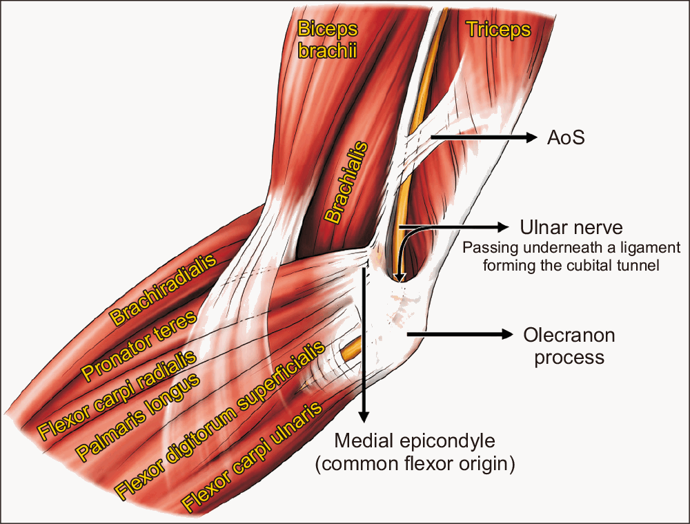
One of the important upper-limb peripheral nerves, the ulnar nerve (UN), is susceptible to compression at several anatomical sites along its course. Although the elbow’s cubital tunnel is commonly acknowledged as the most frequent location of entrapment, occurrences of proximal UN compression have also been linked to other anatomical structures, including the arcade of Struthers (AoS) [1]. The AoS is a thickening of the brachial fascia or an aponeurotic or musculoaponeurotic structure that extends from the medial intermuscular septa to the medial head of the triceps brachii muscle at a variable distance above the medial humeral epicondyle (Fig. 1) [2-4] and was first explained by Kane et al. (1973) [1].
The existence and relevance of the AoS have long been debated in the anatomical and clinical literature [5-11]. While some studies highlight its role as a consistent anatomical structure contributing to UN compression, others question its clinical significance due to variations in its morphology and prevalence [11-13]. As the UN passes through the AOS, it is susceptible to compression due to the arcade’s variable fibromuscular architecture. Excessive fibrotic changes in this region can lead to chronic nerve entrapment, necessitating surgical consideration during decompression [14]. In anterior UN transposition, the AOS must be meticulously identified and released to prevent recurrent nerve compression. Neglecting this anatomical landmark is frequently associated with persistent symptoms and unsatisfactory clinical outcomes [14-17]. While the gross anatomical course and nerve intimacy within the AOS are well-recognised, a significant deficit persists in the literature regarding its quantitative microarchitecture. Specifically, the precise distribution of fibrous versus muscular constituents and their internal structural arrangement remains poorly characterised, leaving the mechanical basis of entrapment at this site largely theoretical.
This study used cadaveric specimens to examine the detailed morphology, histological architecture, and anatomical interaction of AoS with the UN. By providing detailed morphometric data, this research seeks to clarify the significance of the arcade as a site of nerve entrapment that produces features suggestive of chronic compressive changes and to offer insights for surgical practice.
Materials and Methods
In this study, 46 upper limbs from 23 formalin-fixed adult cadavers (17 male and six female), aged between 55 and 75 years, were dissected. These cadavers had been donated to the Department of Anatomy for medical education and research. Limbs showing anatomical deformities, evidence of prior surgery, traumatic injuries, or pathological conditions affecting the medial aspect of the arm were excluded. As none of the specimens met these exclusion criteria, all were included in the study.
Standard dissection procedures were used to expose the medial aspect of the arm, with the upper limb in a supine position [18]. From the axilla to the medial epicondyle of the humerus, a longitudinal incision was made along the arm’s medial side. The skin, subcutaneous tissue, and deep fascia were meticulously dissected to reveal the underlying structures. The AoS was identified as a musculoaponeurotic structure connecting the medial head of the triceps brachii to the medial intermuscular septum. Naked eye observations were made to determine the structural composition of the arcade (muscular, aponeurotic, or mixed/muscular-aponeurotic). Later, histological analysis of these arcades was done to confirm their microscopic composition. Subsequently, the following morphometric observations were made.
Extent of the arcade: a digital vernier calliper (Mitutoyo) was used to measure the arcade’s length from its proximal to distal attachment with a precision of 0.01.
Distance from medial epicondyle: it was noted how far the medial epicondyle was from the arcade’s proximal and distal ends. Each measurement was recorded twice by two independent observers to minimise intra-observer variability.
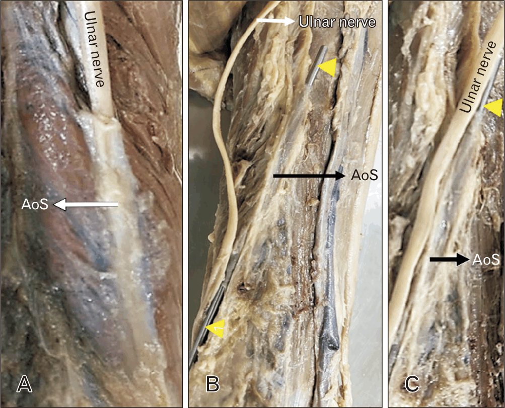
The UN was traced proximally from its origin in the medial chord of the brachial plexus in the axilla to the cubital tunnel near the elbow distally to determine the nerve’s location with respect to the arcade, including whether it was underneath (Fig. 2A), superficial to (Fig. 2B), or lying next to (Fig. 2C) the arcade. When the nerve was located beneath the arcade, it was moved proximally and distally to assess its mobility. The nerve cross-section was collected at the level of the narrower proximal aperture of this arcade and subjected to further microscopic analysis in cases where the nerve was not freely mobile. Furthermore, a special histological stain, Masson’s Trichrome, was employed.
Microscopic analysis of the arcade
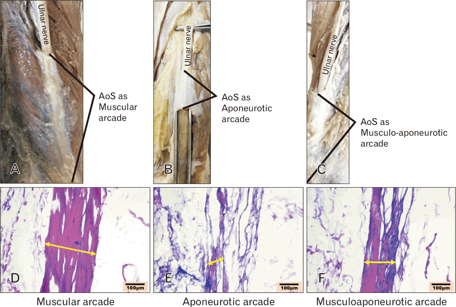
Following gross morphometric measurements, a 5 mm cross-sectional tissue sample of the AoS was excised from the mid-point of the structure. After fixation in 10% formalin and paraffin embedding, the samples were sectioned at 5 µm. Masson’s Trichrome was used to stain the sections, distinguishing between muscle fibres (red) and collagen (blue/green). Stained slides were imaged using a light microscope (Olympus BX51; Olympus) at 10× magnification. Quantitative analysis was performed using ImageJ software. For each section, three representative fields were captured, and colour-segmented regions were quantified to determine the proportions of muscle and collagen fibre areas, expressed as percentages (Table 1, Fig. 3D–F). Similarly, histological examination of the UN, trapped beneath the AoS (all three variants), was performed to visualise changes resulting from compression. Following the gross and histological examination, an objective criterion was defined to identify features suggestive of chronic compressive changes in the UN due to AoS:
(1) Lack of proximal–distal nerve mobility beneath the arcade,
(2) Visible tethering at the proximal aperture,
(3) Histological thickening of epineurium and perineurium, and
(4) Increased endoneurial collagen deposition
Statistical analysis
Statistical analysis was performed using Microsoft Excel 2024 (Microsoft). The prevalence of the AoS was calculated using the total number of dissected limbs as the denominator, while proportions of morphological subtypes were calculated using only AoS-positive limbs. Continuous variables, including arcade length and distance from the medial epicondyle, are presented as mean±SD after assessment of normality.
We classified and examined differences in the arcade’s structure (muscular, aponeurotic, or mixed) and its location in regard to the UN. Comparisons of continuous variables between muscular and aponeurotic arcades were performed using the independent-samples t-test when data were normally distributed; otherwise, the Mann–Whitney U test was used.
Associations between categorical variables, including arcade structural type and UN position, were assessed using the chi-square test when expected cell counts were ≥5. Fisher’s exact test was used when expected cell counts were small, particularly for analyses involving the mixed subtype. A P-value <0.05 was considered statistically significant.
Ethical considerations
The cadaver was donated voluntarily for medical education and research under the institute’s voluntary body donation program. Research involving cadaveric data was granted a waiver by the Institutional Ethics Committee of All India Institute of Medical Sciences, Bathinda, Punjab, India (waiver No. IEC/AIIMS/BTi/2026/04/06), provided that donor anonymity was maintained throughout data collection. All specimens were handled with respect and in compliance with institutional policies.
Results
The AoS was identified in 37 of 46 dissected limbs, corresponding to an overall prevalence of 80.43%. Among AoS-positive limbs, the arcade was muscular (Fig. 3A) in 23 cases (62.16%), aponeurotic (Fig. 3B) in 11 cases (29.72%), and mixed (Fig. 3C) in 3 cases (8.11%). The distribution of structural subtypes did not differ significantly (χ2=3.67, P=0.16).
Morphometric measurements
The mean arcade length was 5.23±1.13 cm (range: 3.0–7.5 cm). There was no statistically significant difference in mean arcade length between muscular and aponeurotic types (5.28±1.08 cm vs. 5.12±1.23 cm; independent samples t-test; P=0.63).
The mean distance of the arcade from the medial epicondyle was 4.82±1.25 cm (range: 2.5–7.0 cm). This distance did not differ significantly between muscular and aponeurotic arcades (4.85±1.28 cm vs. 4.79±1.21 cm; independent-samples t-test, P=0.86).
Position relative to the UN revealed that in 27 arms (72.97%), the nerve passed beneath the arcade; in seven arms (18.92%), it was found superficial; and in three arms (8.11%), it ran alongside without being subjected to direct compression/entrapment. As per Fisher’s exact test, no statistically significant correlation between the arcade’s structural type and the gross position of the UN (P=0.41).
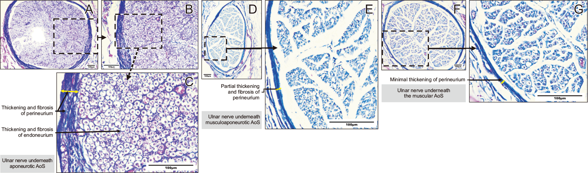
Quantitative histological analysis demonstrated marked differences in tissue composition across arcade subtypes. Muscular arcades showed a predominance of muscle fibres (84.6%±3.2%), whereas aponeurotic arcades exhibited significantly higher collagen content (89.3%±3.5%). Mixed arcades demonstrated intermediate proportions of muscle and collagen (Table 1). Owing to the small number of mixed arcades, no formal statistical comparison was performed for this subgroup. A crucial finding was that histological features of chronic compressive changes were observed in the UN, exclusively in cases with an aponeurotic arcade. In seven specimens, the UN exhibited macroscopic tethering and significantly restricted longitudinal excursion during proximal-to-distal manipulation. Histological analysis corroborated these findings, revealing chronic compressive stress characterised by perineural fibrosis and a dense accumulation of endoneurial collagen (Fig. 4A–C). These data suggest that the histomorphological composition and resultant tissue stiffness of the arcade are more reliable predictors of pathological entrapment than the mere anatomical proximity of the nerve to the AOS. Comparatively, the histological examination of the UN underneath the musculoaponeurotic (Fig. 4D, E) and the muscular (Fig. 4F, G) AoS presented with partial to minimal thickening of the perineural sheath of nerve fascicles and negligible changes in the endoneurial coverings.
Discussion
When Kane et al. [1] discovered a fibrous canal on the medial aspect of the mid- and lower-third of the arm formed by an extension of the medial head of the triceps muscle and its aponeurotic expansion in 70% of the dissected limbs in 1973, they introduced the concept of the AoS. Since then, various authors have reported the existence of the AoS [19, 20]. Later, the findings of Kane et al. [1] were confirmed by Amadio and Beckenbaugh [19], who found the arcade in each of the 20 dissected limbs. According to Gonzalez et al. [7], 26 out of 39 arms (67%) had the arcade. al-Qattan and Murray [3] found the arcade in 68% of cadavers. In the present study, we observed AoS in 80.43% of cases, a rate significantly higher than that reported in previous studies. On the contrary, Siqueira and Martins [20] reported the arcade in only 13.5% of cases. According to Bartels et al. [21], they reported brachial fascia in ten limbs; however, none of these were thickened to take the shape of AoS. In his analysis of 104 arms from 64 cadavers and 300 UN surgeries, Dellon [6] failed to find a structure resembling the AoS. The discrepancy in reporting the AoS might be due to differences in its definition. The definition employed in this research substantially resembles that of von Schroeder and Scheker [22]. According to von Schroeder and Scheker [22], AoS is a fibrous canal on the medial aspect of the mid- and lower thirds of the arm, connecting the intermuscular septum to the internal brachial ligament and partially covering the UN. von Schroeder and Scheker [22] identified the AoS in every cadaver they had dissected and claimed that compressive neuropathy of the UN may be clinically exacerbated by the canal’s reduced proximal opening. According to Caetano et al. [23], the arcade may be involved in nerve compression when the proximal entrance of the arcade forms a V-shaped canal between the internal brachial ligament and the medial intermuscular septum, or when the medial head of the triceps is highly developed and covers a sizable portion of the nerve.
During dissection, we noticed variations in the morphology of the AoS, which might be due to their structural composition. Variations in its composition were grossly evident, with a predominantly muscular arcade observed in 62.16% of the cases. The likelihood of nerve compression is lower in this variation, because a muscular arcade compresses the nerve only during muscle contraction [21]. In contrast, aponeurotic arcades were present in 29.7% of cases and may exert a more compressive force on the nerve. Additionally, we observed in this study that the proximal aperture of the AS was smaller, particularly in aponeurotic AoS cases. In cases with ulnar compressive neuropathy, such variations may be more hazardous.
The mixed (musculoaponeurotic) arcade, identified in 8.1% of cases, combines features of both structural types, which may account for the varied clinical presentations observed in patients with chronic compressive changes in UN due to entrapment. This diversity highlights the importance of considering anatomical variations during surgical interventions, particularly during UN transposition surgery.
This is the first study to undertake a quantitative histological analysis of the arcade’s fibre composition. Prior descriptions of the AoS have been purely gross‐anatomical, categorising it as muscular, aponeurotic, or mixed based solely on macroscopic appearance [21, 24]. By applying digital image analysis to stained cross‐sections, we have objectively confirmed and refined these categories, providing precise percentage ranges for each tissue component.
The predominance of muscle fibres in the muscular arcade suggests greater elasticity and contractile capacity, which may reduce static mechanical compression on the UN but introduce dynamic changes in tunnel dimensions during triceps contraction [25]. In contrast, the high collagen content of aponeurotic arcades likely renders them stiffer, potentially increasing the risk of entrapment neuropathy during repetitive elbow flexion or extension. This structural composition provides a mechanistic explanation for the higher propensity of aponeurotic arcades to induce chronic UN compression, as further supported by histological findings of perineural and epineural fibrosis [26]. These insights emphasise the clinical importance of assessing the tissue quality of the AoS during surgical procedures, highlighting that release is particularly warranted when the arcade is firm and fibrous.
Mixed arcades with an intermediate fibre balance may combine both elastic and rigid properties, possibly explaining the wide variability in clinical presentations of ulnar neuropathy at the distal arm [27].
Although our statistical analysis did not reveal a significant association between arcade type and nerve position (beneath, superficial, or adjacent), the histological data suggest that tissue stiffness and compliance could influence the degree of contact or frictional forces exerted on the nerve. Future biomechanical studies, such as tensile testing of excised arcades, could elucidate whether the quantified fibre ratios predict the arcade’s tensile strength, elasticity, and propensity to constrict the nerve.
Our results further illustrate the connection between the arcade and the UN. The UN ran beneath the arcade in the majority of cases (72.97%), whereas it was superficial to the arcade in the remaining cases (18.92%). These findings are consistent with research by O’Driscoll et al. (1991) [27] and Apfelberg and Larson (1973) [28], which highlights the arcade as a crucial site of UN compression, especially when the nerve runs immediately beneath it.
The arcade is a crucial structure during UN surgery, according to a small number of researchers [1, 10, 29], especially during anterior disposition of the nerve. Hence, morphometric measurements and their topographic locations relative to fixed bony points, such as the medial humeral epicondyle, are of paramount importance. The morphometric investigation showed that the arcade’s length was 5.23±1.13 cm on average, with a range of 3.0 to 7.5 cm. The mean distance from the medial epicondyle was 4.82±1.25 cm, with a range of 2.5 to 7.0 cm. These measurements align with those found in previous research [10, 30]. Our results extend this research from a surgical perspective by providing accurate measures. These results are significant because they offer precise cues for locating the arcade in UN decompression procedures. In order to detect and release the arcade, intermuscular septum, and internal brachial ligament during UN anterior transposition, surgeons should carefully palpate the area proximal to the medial epicondyle.
The cubital tunnel is the most frequent site of UN compression [28], entrapment at the arcade, on the other hand, is distinct since it takes place close to the elbow, which could affect clinical symptoms such as paraesthesia, intrinsic hand muscle weakness, and altered UN distribution feeling [31]. Campbell et al. (1991) [31] highlighted the potential difficulties in identifying ulnar neuropathy at the elbow in their study, noting differences in the UN’s architecture within the cubital tunnel.
The current study offers important insights into the anatomical considerations and clinical implications of chronic compression of the UN proximal to the elbow, especially at the AoS, given the arcade’s close proximity to the medial intermuscular septum. During surgical exploration, the combined contribution of the arcade and septum to nerve compression should be thoroughly assessed.
The AoS may serve as a secondary compression site for the UN, which would explain its clinical relevance. Although proximal compression at the arcade might worsen symptoms and result in double-crush syndrome, the cubital tunnel is still the most well-known entrapment site [32]. More severe clinical symptoms result from double-crush syndrome, which happens when compression at one location makes the nerve more susceptible to compression at another location. The term “double crush syndrome” was initially used by Upton and McComas [32] in their 1973 paper, “The double crush in nerve entrapment syndromes.” According to their theory, nerve compression at one location may make it more vulnerable to harm at a later location. Patients may experience increased paraesthesia, weakening of intrinsic hand muscles, and altered sensation in the UN distribution as a result of this dual-site compression.
In cases of persistent ulnar neuropathy despite decompression at the cubital tunnel, the AoS should be considered as a contributing factor. Moreover, anatomical variations in the arcade (muscular vs. aponeurotic) may influence the degree of compression and the resulting clinical manifestations. As such, preoperative imaging modalities, including ultrasonography and magnetic resonance imaging, can aid in identifying anatomical variations and planning surgical strategies [33, 34]. While the present study focuses on cadaveric morphology and histology, the findings provide a structural basis for understanding potential UN constraint at the distal arm. The observed differences in collagen content suggest that tissue composition may influence mechanical behaviour, a finding that could be further explored using collagen subtype analysis and biomechanical testing in future studies.
Limitations
While sufficient to detect broad compositional trends, the cadaveric sample remains limited in number and may not capture the full spectrum of variation across ages or pathological states. In addition, formalin fixation can slightly alter tissue staining properties, although our standardised protocols and internal comparisons mitigate this effect. Additionally, Masson’s trichrome highlights fibrosis, but the technique is not specific for neuropathy. Moving forward, correlating histological composition with in vivo imaging (e.g., ultrasound elastography), electrophysiological data, and clinical data from patients with ulnar neuropathy could help bridge anatomical findings with clinical practice. Finally, molecular analyses of collagen subtypes and muscle fibre characteristics may further refine our understanding of the arcade’s role in nerve entrapment.
Conclusions
This study provides a critical update to understanding the AoS by demonstrating that its histological composition is a key determinant of its pathological role. Our quantitative data suggests that aponeurotic arcades, rich in collagen, are the most likely to be associated with features suggestive of chronic compressive changes. This highlights the importance for surgeons to identify the AoS and carefully assess its texture and composition, as a dense, fibrous structure warrants careful release during UN decompression to prevent persistent neuropathy.
 XML Download