



 PDF
 Citation
 Print


Introduction
The mammalian tooth is a classic model for studying epithelial-mesenchymal interaction, tooth formation and organogenesis because it combines accessible embryology with clearly staged morphological landmarks and conserved signaling pathways.
The tooth formation proceeds through an ordered stage, the dental lamina, bud, cap, and bell morphogenesis, cytodifferentiation of odontoblast and ameloblast, and followed by crown mineralization and root formation. Moreover, each phase is controlled by histological, molecular, and genetic reciprocal interaction [1].
Historically, the laboratory mouse has dominant genetic and developmental studies of odontogenesis due to its extensive genetic toolkit, numerous inbred strains and the availability of transgenic and reporter lines. Depending on the mouse has produced a detailed molecular atlas of tooth development and extensive literature on the main signaling pathways (BMP, FGF, Shh, and Wnt) that establish the framework for tooth development through continuous reciprocal interactions between the dental epithelium and the underlying mesenchyme [2, 3].
Rats, by contrast, offer a complementary advantage that are particularly relevant to anatomy and translational work, the larger tooth size (which simplifies the dissection and histological preparation), physiological and pharmacological similarities with human that are valuable in toxicology and preclinical testing [4]. Furthermore, the upcoming revolutionary clustered regularly interspaced short palindromic repeats (CRISPR)/Cas9 increasing genetic tractability in rat models [5]. However, despite these strengths, a side-by-side comparison of mandibular M1 development across the two species is unavailable.
This review provides a practical and anatomy focused comparison of mouse and rat M1 development. Moreover, summarize morphologic and histologic staging for embryonic and postnatal M1 formation, compare molecular regulators with attention to where species diverge or scale, discuss methodological and translational implications for experimental design and identify specific gaps where rat-specific molecular data are limited and further work is needed.
Search Strategy
Methods and search strategy
A comprehensive literature search was performed using PubMed, PubMed Central, and Google Scholar databases to identify studies related to mouse and rat first molar (M1) development. The search strategy using the following keywords: “mouse molar development,” “rat molar development,” “comparative odontogenesis,” “tooth morphogenesis,” “BMP,” “FGF,” “Shh,” and “Wnt signaling.” Reference lists of key papers were further examined to identify additional relevant publications.
Inclusion and exclusion criteria
Studies were included if they (1) directly compared molar development between mice and rats; (2) investigated molecular, genetic, or morphological parameters relevant to odontogenesis; (3) provided primary experimental data; and (4) contributed to understanding developmental timelines or signaling mechanisms. Studies were excluded if they focused exclusively on non-molar teeth, the non-dental tissues, or unrelated species.
Morphological and Histological Stages of M1 Development
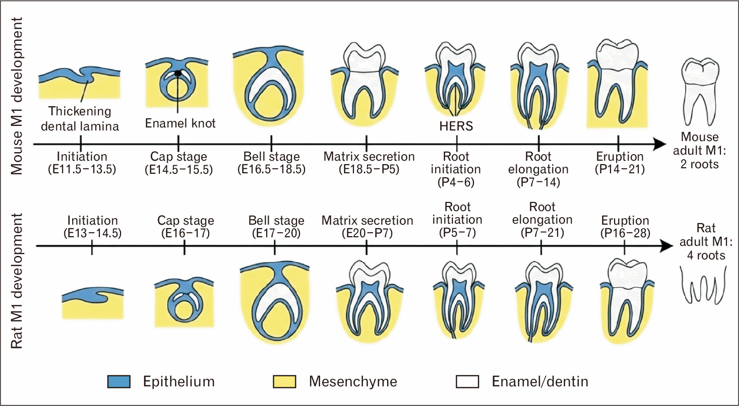
Development of M1 (lower first molar) in mouse occurs precisely 1–2 days earlier than the rat, due to short mouse gestation period.
The initiation (dental lamina and bud stage)
Tooth development initiated with the formation of dental lamina in the oral epithelium. In mice the lower first molar dental lamina appear by ~E11.5–E12 [6, 7]. Followed by the invagination of epithelium within the underlying neural crest derived mesenchyme (bud stage) at E12.5–E13.5 [8, 9]. However, in rats, the bud stage is appear around E14.5 [10, 11]. During this early phase of development the epithelial gene such as Foxi3 are more prominent expressed in the rat lamina and the initiation is marked by Shh [12]. Although the initial dental lamina is similar between species but the regions of genes activation in rats are more noticeable than the mice.
From cap to bell stage
The epithelium bud grows around the condensed mesenchyme of dental papilla to form a cap like structure. For the mice mouse ~E14.5–E15 [7, 15] and the rat ~E16–E17 [12, 16]. The important structure that is recognized at this stage is the enamel knot. A signaling center that control the cusp patterning through multiple pathways which include the BMP, FGF, SHH, and WNT signals [17, 18]. Further, as the tooth proliferated and transformed from cap to bell stage in mouse around ~E18.5 and rat ~E20 at this time the cusp became defined but with no mineralization [19, 20].
Histologically, at the late cap/early bell stage, the enamel organ has four recognizable cell layers; outer enamel epithelium, stellate reticulum, stratum intermedium, and inner enamel epithelium. At this stage of development, the similarity in shape between mice and rats is visible; however, the difference in tooth germ size is noteworthy. In particular, the rat M1 germs are larger, with the gene expression analysis confirm that IGF1 are more abundant in rat tooth germs, which responsible for increasing the growth rate, with no alteration of cusp shape [12, 21].
From late bell to crown formation
The cell differentiation and matrix secretion occur at late bell stage which start at E18.5 in mouse. According to micro-computed tomography (micro-CT) studies the dentin secreted P0–P5 [22, 23] and the M1 enamel with cusps largely complete within first two weeks postnatal [24, 25]. However, for the rat, the late bell stage period at E20 [26, 27]. The crown formation is slightly late but follows the same stages. Moreover, the dentin secreted P1–P5. Subsequently, the ameloblast laid down full enamel thickness about the end of first postnatal week [28, 29]. In both species after the crown mineralization, the root starts to develop (Table 1).
Root formation and eruption
Root elongation begins round P7 in mouse [30, 31] and P8–P9 in rat [32, 33]. The cervical loops give rise to Hertwig’s epithelial root sheath (HERS) guiding the root dentin and cementum deposition. In mice the M1 root typically forms two roots and by P19–P21 the tooth erupts into the occlusal plane [25, 30]. In rats, the first mandibular molar penetrate the gingiva at day P18 (Fig. 1) [34].
Molecular Mechanisms
The signaling pathways
Both mouse and rat share the same canonical signaling pathways that orchestrate tooth development. The reciprocal signals BMP, FGF, Shh, and Wnt are responsible for the epithelial mesenchymal interaction [17]. Initially, BMP4 is expressed in dental mesenchyme and FGF8 in the epithelium initiate the bud formation in both species [35]. Then, the cusp patterning in the enamel knot controlled, in both species, by Shh and BMPs [36]. In fact, many studies of dental signaling depend on mouse data as a representation of rat. Conversely, IGF1 signaling is an interesting species-specific finding, based on the cytogenetic finding it shows that IGF1 is expressed at higher levels in rat M1 compared to mouse which cause the rat tooth large in size [12]. However, inadequate rat single-cell atlases and loss of function genetics studies for key odontogenesis regulation are limiting the ability to assume species-specific molecular pathways. Therefore, the differences based on abundance or timing of signals as provisional unless confirmed by functional perturbation in both species.
The transcription factor
The transcription factors that regulate tooth development are almost similar between the mouse and rat. The key factors; Pitx2, Pax9, Msx1, Lef1, and Barx1, play an important role from initiation to patterning in both species [8, 37]. For example, the homeobox genes Barx1 and Dlx2 determine the molar rather than incisor fate in mouse [38], no evidence that an additional factor is needed in rat. Moreover, odontogenic transcriptional gene regulatory system appears to be the same in mice and rats and there is no known example of tooth developmental TF which is species-specific.
The stem cell roles
Rodent molars have a transit progenitor niches compare to continuous growing incisors teeth [39, 40]. In both species the M1 cervical loop proliferate to form HERSs and contribute to root formation. The progenitor stem cells are similar, and no major species-specific stem cell type has been identified in the M1. In general, the teeth in both species appear to have the same stem cells populations during development [41, 42]. Moreover, in vitro studies prove that adult dental pulp stem cells from both mice and rats can be induce to form tooth like structures (Table 2) [43-45].
Practical and Experimental Considerations
Genetic toolbox
Mice have an extremely varied genetic setup for tooth related research; knockout, knock in and transgenic lines all available for studies of genes affect the odontogenesis [46]. However, genetic engineered rats are historically limited. Although, the invention of genome editing CRISPR/Cas9 expand the rat models [47, 48]. Accordingly, almost any gene manipulated in mouse can now be applied in rat. Despite the mouse model still offers more immediate options and well-marked genome with less cost.
Physiological and pharmacological implication
The rats provide more mimic to human physiology in metabolism and pharmacokinetic. Rats exhibit metabolic pathways and hormonal regulation patterns that closely mirror those in people. Their blood composition, response to stress hormones, and energy metabolism follow similar trends, though with faster rates due to shorter lifespans [49, 50]. These similarities provide an effective model for understanding human nutrition, aging, and systemic diseases, while also clarifying where differences must be taken into account. There is extensive data on rat developmental responses to chemicals. On the other hand, mouse teeth development is faster but may not fully represent to rat or human pharmacodynamics. Researchers should consider these differences if a drug that disrupts mouse tooth development by P1 might have different effects on the more mature rat tooth at birth [51].
Technical handling
The larger size of rat embryos and pups can simplify some experimental operations. Dissecting of the P7 rat mandible or the intervention by injecting molecules into the developing jaw in postnatal periods is technically easier than the comparable mouse procedures [52]. Similarly, in vivo imaging and micro-CT of rat skulls provide higher resolution relative to mouse On the other hand, smaller mouse size can be advantageous for high-quantity work provide more samples per litter and requires less material per assay a fewer cells for molecular analysis [53]. Standard breeding and embryological techniques are almost identical in both species so choice often depends on the specific operation scale rather than feasibility (Table 3).
Disease Modeling and Translation Application
Translating to human odontogenesis
Rodent M1 studies provide information that covered several aspects of human tooth development. The similarity in signaling pathways (BMP, FGF, SHH, WNT) with the key transcription factors provide the base to understand the epithelial-mesenchymal interaction and how these pathways control the cell fate and cusp pattern. Mouse genetic models reproduce many human enamel and dentin related defects such as amelogenesis imperfecta, dentinogenesis imperfecta have been reproduced in mutant mice. For example, the Amelx and Enam mutant mice display hypoplastic and hypomineralized enamel analogous to human amelogenesis imperfecta which enabling genotype–phenotype correlations [54]. Another study demonstrates that dentin sialophosphoprotein (DSPP)-null mice exhibit tooth defects similar to human dentinogenesis imperfecta type III, including enlarged pulp chambers, increased width of the predentin zone, hypomineralization, and pulp exposure [55]. Moreover, a research highlights how mutations in the DSPP gene can lead to both dentinogenesis imperfecta and dentin dysplasia, underscoring the utility of mouse models in studying these conditions [56]. However, rat models of specific dental mutation are recently beginning to appear, but the data is still limited. Overall, mice remain the foremost model for gene related dental abnormality related studies.
Importantly, for translational work to human, the researchers are recommended to mapping rodent developmental stages to human gestational/postnatal time points using morphological landmarks (enamel knot or onset of matrix deposition) rather than chronological age alone. Moreover, human odontogenesis take months to years when compared to rodent crowns which form whin days to weeks, so the difference in timing and scale mean that direct chronological alignment must be done cautiously [57].
Toxicology and teratology models
Rats are the common model in safety testing of environmental toxin related studies. Therefore, rat more appropriate choice to evaluate the dental teratology and examine the rat pup dentition for defect [4, 58]. The rat M1 enamel and dentin chemically and structurally similar to human which serve as indicators of developmental toxicity. While the mouse study is available, the toxicology data are based on rat model.
Tissue engineering and regeneration
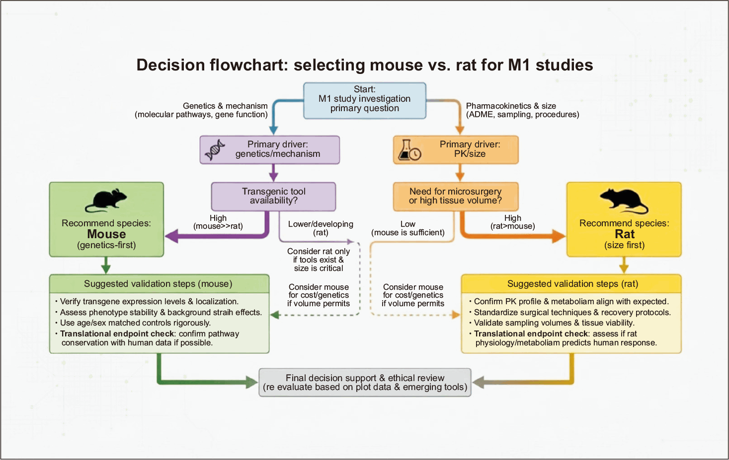
Both mice and rat are used in tooth regeneration related research. Mouse dental stem cells design for bioengineering studies. Moreover, biomimicry of mouse molar and incisor stem cells recently generated capable of differentiating into ameloblast- and odontoblast-like cells [59]. The rats large tooth size provides an advantage for preclinical testing of biomaterials or cell therapies for tissue regeneration [60, 61]. In summary, mice currently dominate the basic science of tooth engineering, while rats may offer practical model for translational implants or grafts (Fig. 2).
Discussion
Fundamentally during study designing the researcher should select the more appropriate model for answering the right research question. Choosing mouse vs. rat depends on specific experimental needs and on the key differences. In fact, the variation between two species start from the gestational timing, mice gestate ~19 days vs. ~21 days in rats [62, 63]. A newborn mouse pup’s M1 is at an earlier developmental stage than a newborn rat. Therefore, the mouse offers a larger early postnatal window (P1–P7) for studying crown maturation [23, 64], while rats are born with more advanced tooth development. However, the tooth size which is for the rat M1s are ~2× larger than the mouse [12]. Substantially, the larger teeth allow easier dissection, imaging, and microinjection, which is ideal for microsurgery or histology. Another point is the genetic tools, mice are dominant models with a huge range of genetic lines and reagents [65, 66]. Although, the rat genetics has improved but it is still limited.
A key aim in any comparative review is to separate difference of timing/scale from the differences of mechanism. Many mouse-rat differences are explained by heterochrony (shifted timing) and scaling (tooth size) rather than fundamentally distinct molecular pathways. For example, the development signaling pathways of BMP, SHH, FGF, and WNT with the core transcription factors (Pitx2, Pax9, Msx1, Barx1, Lef1) in both species shared the same regulatory function. However, the quantitative differences such as higher IGF1 pathway activity are associated with larger rat tooth germs size which modulate the growth rate and organ size without changing cusp patterning. Therefore, experiments that investigate the pathway presence or absence (e.g., loss-of-function of Shh or Bmp4) will produce a mechanistic conclusion that generalized across species, while experiments that focus on dose sensitive growth changes e.g., tooth size, eruption timing, rate of matrix deposition) may produce species-specific quantitative effects.
Practically, the researcher should match the research question to the phenotype classify, or research that require genetic manipulated, lineage tracing or the interfere with the signaling pathways at the level of presence or absence, then the mouse is the best choice. On the other hand, the rat used for techniques that required a larger tissue size, microsurgery or pharmacokinetics related investigation.
When the translation to human structure or physiology is the goal of study then researchers should perform parallel validation experiments in both species (for example: a targeted comparison of IGF activity, enamel matrix deposition rate and response to a candidate teratogen) to distinguish the mechanism from species-specific kinetics or scaling.
Despite these comparisons many gaps remain. In particular, the quantitative gene expression atlases for rat tooth development are rare and most molecular developmental studies have been done only in mouse. Single cell sequencing of rat M1 development could reveal subtle species differences in cell lineage progression. Moreover, the functional genetics in rat tooth by revolutionary tool the CRISPR knockouts of odontogenic genes remain relatively unexplored. Finally, more cross-species toxicology and regeneration studies would validate how well mouse findings generalize to rat (and eventually human). To summarize, future research should exploit the strengths of each rodent model and aim to directly test whether observations in one apply to the other.
Conclusion
Mouse and rat first molars share fundamentally similar developmental programs and both species display a signaling and transcriptional control of tooth patterning. The key differences are in timing, size, and experimental setting. Mice are unrivaled for genetic studies of tooth development, on the other hand, the rats offer advantages in size and physiology. By choosing the best species for a particular research question, researchers can make maximal use of both models. This comparative overview provides a systematic framework for researchers to understand the relative merits and limitations of mouse vs. rat in dental developmental studies.
 XML Download