



 PDF
 Citation
 Print


Introduction
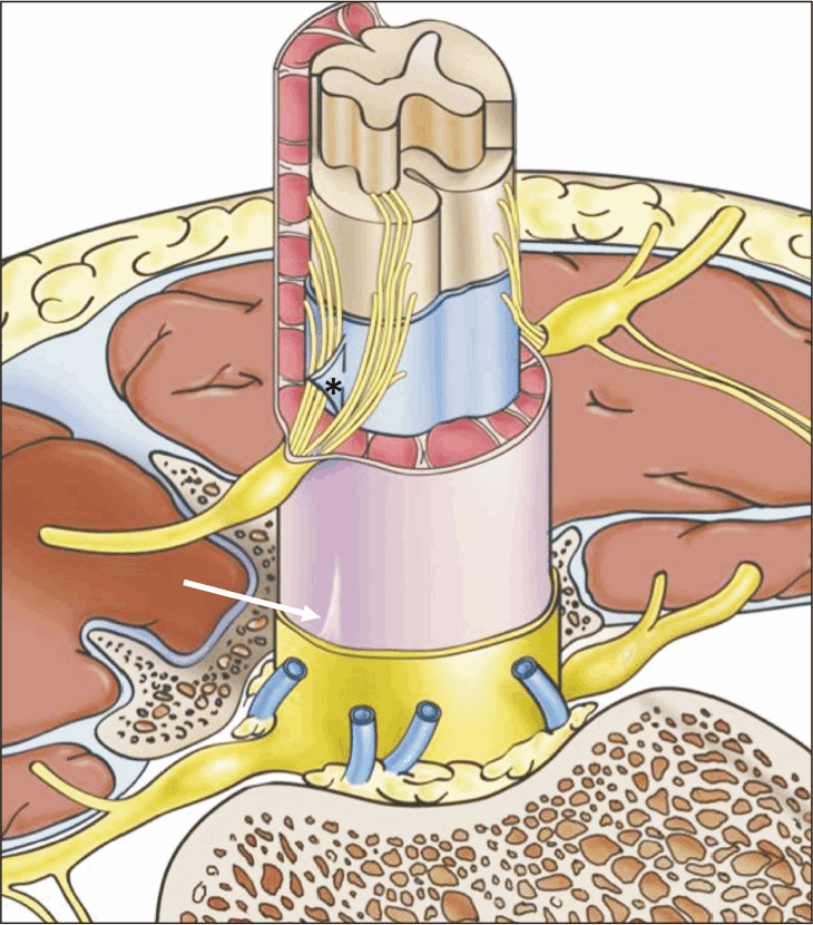
The denticulate ligaments (DLs) are bilateral lateral projections from the spinal cord to the inner aspect of the dura (Fig. 1) [1-4]. These structures are positioned between the anterior and posterior roots of the spinal nerves [1]. They were first reported in 1739 by the Swiss scientist Johann Jacob Huber [5], but there has been increasing controversy regarding their anatomical depiction (number, shape, attachments, and function). While limited, the histological findings regarding the DLs are important for our understanding. Research suggests they comprise collagen fibers arranged in longitudinal, transverse, and oblique positions [6]. The thickness of those fibers differs among spinal levels [1]. Clinically, the DLs serve as valuable landmarks for identifying the anterior and posterior rootlets of the spinal cord, the vertebral artery (VA), and spinal accessory nerves at the foramen magnum [1, 7].
The protocol of the study did not require approval by the ethical committees or informed consent. The study followed the Declaration of Helsinki (64th WMA General Assembly, Fortaleza, Brazil, October 2013).
Gross Anatomy
Traditionally, the DLs are described as 20–21 fibrous spinal projections that stabilize the spinal cord within the spinal canal [2]. Each DL is covered by a leptomeningeal layer that continues with the arachnoid and pia mater of the spinal cord [8]. These ligaments consist of 21–22 triangular pairs that extend from the lateral aspect of the spinal cord and attach to the inner aspect of the dura mater (Fig. 2) [9]. Others report the DLs as long bilateral ribbon-like ligamentous structures that suspend the spinal cord within the dural sac [10].
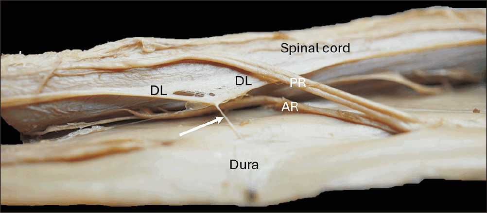
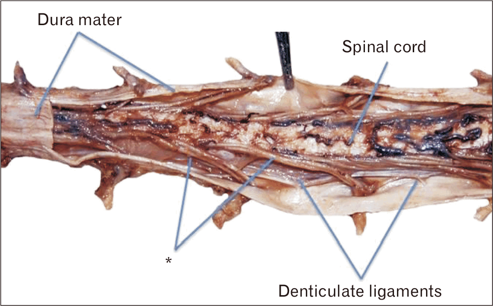
Following formalin-fixed dissections of five adult cadavers (four males and one female), Ceylan et al. [2] found that the DLs were only present in the cervical and thoracic regions of the spine. They described no DLs in the lumbar or sacral levels. Their most significant observations were that each DL was a single narrow fibrous strip ranging from the craniovertebral junction to T12 (Fig. 3) [2]. This is consistent with the finding of Tubbs et al. [4] in 12 cadaveric specimens: the last DL was present at the level of the conus medullaris between the spinal roots of the T12 and L1 nerves. Each ligament comprised triangular extensions perpendicular to the vertebral column [2]. The base of each extension was projected from a narrow fibrous strip of the DL in which the apex was attached to the dura mater.
The apex of each projection varied with the spinal region. Each apex in the cervical region was attached to the dura by a 3–5 mm long fibrous band or bands (single or double) (Fig. 4). In the upper thoracic region, no such fibrous bands were present as the apices of the extensions attached directly to the dura. The fibrous bands were also present in the lower thoracic region, but they were longer than those in the cervical region (21–26 mm) [2]. This is probably because of the motion capacities of different regions of the spinal column. The cervical and lower thoracic portions contain fibrous bands that attach to the dura, allowing mobility within their respective areas. However, the apices can connect directly to the dura due to the lack of mobility in the upper thoracic regions [2]. In both those levels of the vertebral column, the apical attachments presented superomedially to the intervertebral foramen of the inferior vertebrae. As dissection progressed inferiorly, the distance from the dural attachment to the intervertebral foramen increased (10 mm at the cervical levels, 10–15 mm at the upper thoracic levels, and 15–20 mm at the T11 and T12 levels) [2].
Comparable literature supports Ceylan et al. [2] in reporting that the DLs in the cervical region are attached to the dura approximately 2 mm posteriorly and 3 mm cephalad to the nerve root foramen [11, 12]. Each DL becomes closer to the intervertebral foramen caudal to its attachment than to the more cephalically placed DL [4, 13].
Intracranial Denticulate Ligament
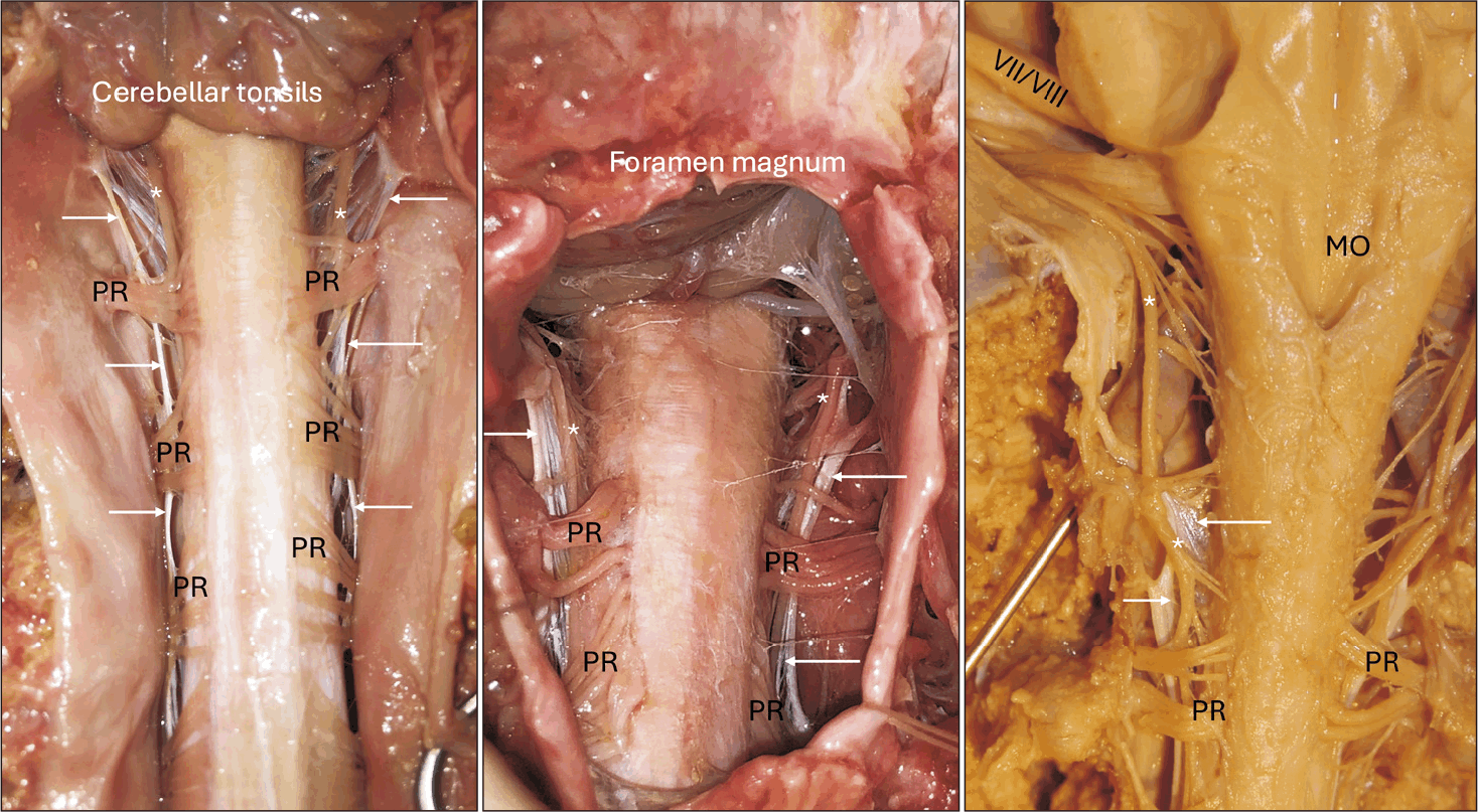
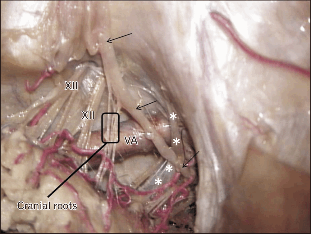
Uniquely, the intracranial DL, the first or highest DL, is often the largest of the DLs (Fig. 5) [6]. Among the five cadavers examined by Ceylan et al. [2], the mean length of the intracranial DL was 12 mm, and the mean width 8 mm. This DL was found to be attached laterally to the dura over the marginal sinus and just above the entrance of the VA as it pierced the dura. Through its course, the first DL was closely related to the spinal accessory nerve posteriorly and to the VA anteriorly [2]. In 15 cadavers, Tubbs et al. [14] studied the intracranial DL and found that on 20% of sides, the ligament is also attached to the adventitia of the VA. These authors found that the posterior spinal artery traveled posterior to this ligament on 93% of the sides, and following unilateral transection, rotation of the spinomedullary junction was increased by about 25%.
Antenatal Denticulate Ligaments
There was no study of antenatal DLs until that of Elvan et al. in 2019 [3]. Those authors aimed to establish the morphological features of the DLs during the fetal period. Following a laminectomy of 12 formalin-fixed fetuses (six males and six females) with a mean gestational age of 27 weeks, the presence and number of DLs were recorded at each spinal level. DLs were more prominent in the cervical and upper thoracic regions when the posterior roots were retracted medially. They ranged in number from 20 to 23, including five double DL processes within the same interval.
In contrast, no DL processes were present in 14 other intervals, where they were recorded as absent [3]. Elvan et al. [3] thus established that DL processes can present unilaterally or as double intervals within the same antenatal period. As in previous studies [6, 15], they observed that the distance between the DL processes increased, coursing inferiorly to the conus medullaris. This is believed to be because the vertebral bodies become larger from the cervical to lumbar sections of the vertebral column [1].
Histology
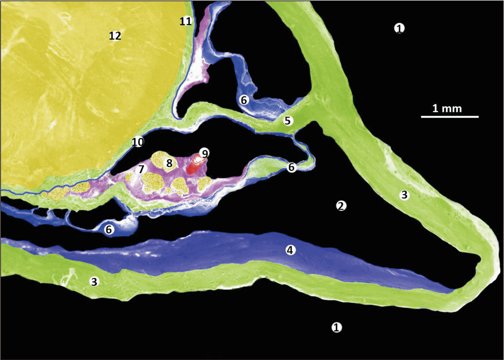
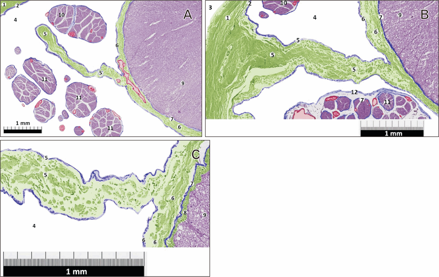
Few studies have investigated the histological nature of the DLs (Figs. 6, 7). We can see in Figs. 6, 7 a continuity between the collagen fibers (green) located in the dura mater, the collagen fibers (green) located inside the denticulate ligament, and the collagen fibers (green) of the subpial compartment. There is also a continuity of the leptomeninges between the arachnoid cells (blue) that form the arachnoid layer, the arachnoid cells (blue) that cover the denticulate ligament and the arachnoid trabeculae, and the pial cells (blue) of the pia mater. Elvan et al. [3] analyzed ten histological DL specimens from C5 and ten from T6 in five cadavers (five right-sided and five left-sided specimens at each respective level). Each specimen was stained with Masson Trichrome. Their analysis showed that the DLs were composed of collagen fibrous bundles. Histological examination revealed that the fibrous strip of the DL consisted of longitudinal collagen fibers, while the triangular projection contained transverse and oblique collagen fibers [2, 16]. The fibrous strip portion was also attached to the spinal cord by loose connective tissue [2]. The DLs penetrated the substance of the spinal cord at various cervical intervals to form a stronger attachment. The DLs at the cervical spinal levels were thicker than those at thoracic levels [2]. Nicholas and Weller, using scanning electron microscopy, found that the DLs consist of a collagenous core covered by a layer of leptomeningeal cells, continuous with the pia and arachnoid. These authors also demonstrated that the DLs fuse with the subpial collagen of the spinal cord [17].
Pathology
Polak-Kraśna et al. [16] explored the properties of the DLs in the cervical spine because of its higher incidence of injury, which is attributed to its high mobility. Sawada et al. [18] reported a meningioma arising from the DL at the C1 level. No dural attachment was identified. Meningiomas of the cervicomedullary junction are often classified based on their relationship to the DL [6].
Within the myelomeningocele population, the upper DLs are variable in length, thickened, and abnormally taut [19, 20]. The DLs have been linked to the kinking of the medullary spinal junction in patients diagnosed with Chiari II malformation [2]. Bedford et al. [21] proposed that the patient in their case report diagnosed with spondylosis had additional spinal cord injury due to hypertrophic DLs. The so-called Gingko leaf sign is seen in spinal meningiomas and is due to the interaction of the tumor and DL [22].
In 147 cases of spasmodic torticollis, Aksik found two cases where the spinal accessory nerve was compressed by a hypertrophied DL. A case of cervical brachialgia from an abnormally attached DL has been reported [9].
Anatomical Variations
There is little literature about variations of the DLs. However, the ligaments are usually at the most lateral point of the spinal cord. Patel et al. [23] reported an abnormal arrangement of the intracranial DL around the posterior inferior cerebellar artery (PICA). In their case report, the right PICA, arising from the VA, took an extreme lateral course toward the entrance of the VA into the posterior cranial fossa, where it turned around the lateral edge of the intracranial DL and then reversed and traveled medially toward the brainstem. As the PICA was tethered to the intracranial DL, there was slight compression against the floor of the posterior cranial fossa [16]. Notably, this DL appeared duplicated into two slips, the horizontal portion connecting at the first cervical nerve distal to the turn of the PICA.
As mentioned above, in fetuses, Elvan et al. [3] found an absence of the ligament at some levels and two ligaments at one spinal interval in others. Such variants have also been reported in adults [3]. The DLs can be fenestrated [4] and use one to three smaller prongs to attach to the inner surface of the dura mater. The tips of the DLs in the thoracic region can be heavier and more blunted [24]. These prongs can be longer in the cervical region.
Biomechanics
The DLs have been described as stabilizing or suspending the spinal cord within the vertebral canal. Although the function is debatable, some have suggested that the ligaments restrict cranial-caudal motion and others anterior-posterior motion [26]. In human cadavers, Tubbs et al. [4] found that the DLs were stronger in the cervical region and decreased in strength moving caudally. These authors also found that DLs became less resistant to anteriorly directed forces as the spinal cord coursed caudally; inferior to T8, the spinal cord contacted the anterior spinal canal before the DLs became taut. They also compared the forces necessary to produce tautness in the DLs with posterior distraction at different spinal cord levels; the DL in the upper thoracic region seemed more resistant to the same posterior distraction [4]. When cranial and caudal stresses were compared, the DLs tended to be more resistant to caudal stress owing to the decreased motion of the spinal cord (6–7 spinal segments caudally and 10–11 spinal segments cranially) once they were made taut [4]. In dogs, Tunturi concluded that the elasticity of the spinal cord and its investments was due primarily to the DLs and pia [27].
Surgical Significance
The DLs are significant landmarks for identifying spinal pathways during cordotomy, e.g., the dorsal root entry zone, and serve as the barrier between the anterior and posterior compartments of the spinal canal [2]. In specific intramedullary or intradural extramedullary tumors, they provide a reliable landmark for identifying a midline entry point to the spinal cord despite any distortion caused by those tumors [2]. The DLs are frequently cut for access to more anterior structures, and a suture can be placed through them to retract the spinal cord. One study suggested sectioning the DLs in the lower lumbar levels superior to the conus medullaris could yield better results when transecting the filum terminale in patients with a tethered spinal cord [2].
Although sectioning of DLs to treat cervical spondylotic myelopathy was abandoned because they have little effect on stabilizing anteroposterior directional movements of the spinal cord [8], cutting the DLs on the approach side allows surgeons to manipulate the spinal cord with minimal retraction and to release adhesions through the anterior dural defect [2]. The upper cervical DLs, including the intracranial, are significant in isolating contributions, e.g., the nerve of McKenzie to the spinal accessory nerve for denervation for spasmodic torticollis [3]. Tubbs et al. [28] found that McKenzie’s nerve was found in 70% of 30 cadaveric sides, and the highest DL was pierced on 11 sides (52.4%). On three sides (14.3%), the nerve traveled through two prongs of the DL. Bedford et al. [21] reported that hypertrophic DLs can compress the C2 root of the spinal accessory nerve.
Seichi et al. [29] reported that in three patients diagnosed with myelomeningocele, temporary recovery was achieved by transecting the DLs of the spinal cord in the lumbosacral region. Lastly, arteries to the lateral spinal cord have been classified based on their relationship with the DL, i.e., preligamentous or post-ligamentous [11]. Nagata et al. [12] reported two patients with spasmodic torticollis who responded to microvascular decompression of the spinal accessory nerve as it was compressed between the C1 DL and VA.
The DLs continue to be largely unappreciated despite their significance in neuroanatomy. This is the first comprehensive review of the DLs, consolidating established information about them to expedite further research. As this structure can be an important landmark during spine and craniocervical procedures, its anatomy should be well known by neurosurgeons.
 XML Download