



 PDF
 Citation
 Print


Introduction
With a growing number of users worldwide since they came onto the North American market in 2007, e-cigarettes have quickly become extremely popular among younger generations [1]. One reason for the rapid growth in e-cigarette usage is the presence of flavorings added to the e-liquids available in the marketplace [2, 3]. These flavorings make the e-cigarettes taste better and create a public perception that e-cigarettes are less hazardous to one’s health than traditional tobacco-based cigarettes [4]. The majority of the products being used today contain flavored e-liquids, thus creating an opportunity to study the potential harm caused by flavored e-liquids [5, 6]. Oral tissues are among the first biological areas affected by e-cigarette aerosols when a user inhales e-cigarette vapors [7, 8]. Oral fibroblasts are integral to the maintenance of the structural integrity of the gingiva and oral mucosa through the remodeling of the extracellular matrix (ECM) and through the process of wound healing [9-11]. Research has recently shown that the inhalation of e-cigarette aerosols can induce oxidative stress, inflammation, and cellular senescence in oral fibroblasts, which can negatively affect the overall integrity and ability to repair oral tissues [12, 13]. Many of the flavoring chemicals used in flavored e-cigarettes, such as cinnamaldehyde, vanillin, and ethyl maltol, were designed to be ingested, and therefore, there is limited information regarding the safety of these compounds when inhaled [14, 15]. Many studies have shown that these flavoring agents are cytotoxic to and promote inflammation in respiratory epithelial cells; however, few studies have examined their effects on oral fibroblasts [16-18]. This lack of research with respect to the toxicological effects of flavoring agents in oral fibroblasts represents an important knowledge gap given the widespread use of flavored products. Therefore, the objective of this review is to determine the contribution of flavoring additives to the toxicity associated with the inhalation of e-cigarette aerosols on oral fibroblasts. It is hypothesized that the inhalation of e-cigarette aerosols containing flavoring will decrease the viability of fibroblasts and increase oxidative stress and inflammatory responses to a greater extent than unflavored aerosols or no exposure [19].
Methods
We performed a comprehensive literature search using PubMed/PMC, ScienceDirect, and Google Scholar to identify studies examining the cytotoxic effects of e-cigarette flavoring compounds on cultured oral fibroblasts. The core search terms used for this review included: “vape flavoring chemicals oral fibroblasts,” “e-cigarette aerosol cytotoxicity fibroblast,” “cinnamaldehyde oral fibroblast toxicity,” “menthol e-cigarette cell viability,” “vanillin vape cytotoxicity,” “flavored e-liquids oral cells,” and “electronic cigarette flavor cytotoxicity gingival fibroblasts.” The time frame searched spanned from 2010 to October 2025, corresponding to the period during which modern e-cigarette products became widely available.
Eligibility criteria
In order to access the eligibility of the comprehensive research, included studies were documents that contained either peer-reviewed or original experiments that examined how flavored e-liquids, e-aerosol (or vapor), or an individual flavor chemical affected human oral fibroblast cells, including gingival, periodontal ligament, or buccal fibroblast cells. Included studies also had to have been written in English and published during the years 2010 through October 2025. Studies were not included if they examined cells other than oral-related cells, used animals that were pre-exposed subjects to nicotine or other toxins that may influence the data, were observational only, abstracts of conference presentations, or did not measure some aspect of fibroblast viability using biologically viable methods.
Risk of bias and study quality assessment
The risk of bias and the quality of the studies were determined by an adaptation of a framework developed for assessing in vitro toxicology studies; each study was examined in terms of its clarity of the research question, transparency of the experimental design, and characterization of the e-liquids and flavoring compounds that were being studied. As in vitro toxicology studies are particularly prone to bias due to the inadequate characterization of flavoring compounds, nicotine levels, solvents, and aerosol-generating techniques, these areas were scrutinized.
Additionally, studies that had adequate biological and technical replicates were evaluated. The outcome measures, such as cell viability, oxidative stress, inflammatory markers, apoptosis, and senescence, were reviewed for their methodological validity and consistency. Overall, most studies demonstrated moderate methodological rigor; however, variability in aerosol generation protocols, insufficient reporting of flavoring compound concentrations, and inconsistent control designs were identified as significant recurrent limitations.
Data extraction
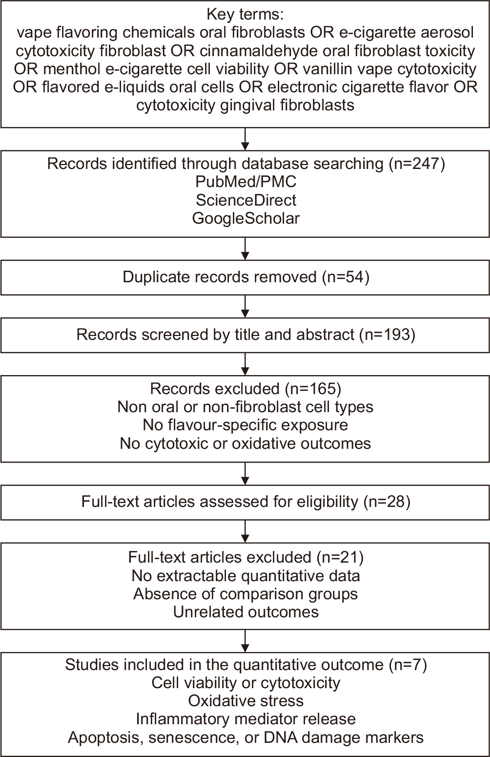
To ensure accurate study identification and selection, the literature search, screening, exclusion, and final inclusion process is summarized in Fig. 1. Two independent reviewers separately obtained information from each of the included studies on the experimental design, the type of oral fibroblast (used) in the study, the flavoring chemicals or e-liquids studied, the method of exposure, and the outcomes measured because of cytotoxicity, oxidative stress, inflammation, apoptosis, or senescence.
Results
Oxidative stress and cellular stress signaling
Oxidative stress emerged as the main biological outcome associated with exposure to flavored and unflavored e-cigarette aerosols. In the included studies, the generation of reactive oxygen species (ROS) was one of the most consistently reported endpoints. Several studies demonstrated increased intracellular ROS following exposure to flavored e-liquids or specific aldehyde-containing flavoring chemicals. Despite differences in assay types and exposure duration, ROS elevation remained a reproducible indicator of oxidative stress in oral fibroblast models [7]. Protein carbonylation, which is a stable biomarker of macromolecular injury, was reported prominently in the study conducted by Sundar et al. [7] who demonstrated significant increases in protein carbonylation following exposure to cinnamon-flavored aerosols. This oxidative protein damage coincided with activation of inflammatory pathways and early markers of cellular dysfunction. Sundar et al. [7] also identified DNA damage responses, including γ-H2AX upregulation, indicating that oxidative stress extended to genomic instability under exposure conditions involving flavored aerosols.
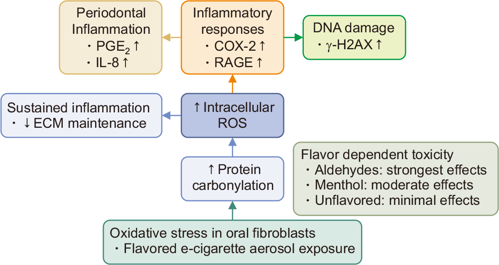
The reproducible elevation of ROS across studies is biologically significant because ROS is a key upstream driver of cellular stress responses. Oxidative stress is known to activate NF-κB and MAPK signaling, which promotes the release of IL-6, IL-8, and prostaglandin E2 (PGE2). In the context of vaping-related toxicology, these pathways are particularly relevant, as many studies reported that flavored aerosol condensates activate NF-κB and increase downstream inflammatory signaling in oral cell models. The detection of ROS, DNA damage (γ-H2AX), and protein carbonylation indicates that flavoring-chemical exposures induce both genotoxic and proteotoxic stress [7]. As outlined in Fig. 2, early oxidative stress from flavored aerosols triggers sustained inflammatory signaling and DNA damage.
Oxidative and genotoxic damage
A key oxidative stress outcome that was consistently identified across the studies was the buildup of protein carbonyls. Sundar et al. [7] performed a controlled air-liquid interface exposure using human periodontal ligament fibroblasts (HPdLFs) and revealed that exposure to flavored e-cigarette aerosols, such as classic tobacco (16 mg nicotine) and menthol (0 nicotine), led to a marked elevation of protein carbonylation. Densitometric analysis of OxyBlot bands revealed a significant increase in carbonyl-modified protein levels compared with air controls, indicating that flavored aerosols induced lasting oxidative modifications rather than transient ROS fluctuations. In addition to carbonyl buildup, fibroblasts displayed a broad oxidative inflammatory response. IL-8 secretion (pg/ml) significantly increased following exposure to other flavor types of tobacco and menthol, and PGE2 levels also increased under menthol aerosol conditions [7].
Parallel Western blot analysis displayed an increase in expression of COX-2, S100A8, and receptor for advanced glycation end products (RAGE), which are proteins associated with oxidative and inflammatory signaling. The increase in γ-H2AX levels signifies DNA double-strand break formation. This was further supported by Comet assay results, where the percentage of DNA in the tail was statistically higher in aerosol-exposed fibroblasts compared with air controls, confirming genotoxic stress [7]. However, these oxidative and genotoxic changes were not limited to fibroblasts. Gingival epithelial progenitors revealed similar elevations in COX-2, RAGE, and γ-H2AX, and a three-dimensional gingival tissue model also reproduced these responses, with tissues displaying increased IL-8 and PGE2 secretion as well as elevated γ-H2AX and COX-2 expression.
Flavor-dependent cytotoxic and inflammatory outcomes
Formulations containing aromatic aldehydes demonstrated the greatest reductions in viability. Sancilio et al. [13] showed a dose and time-dependent decline in gingival fibroblast survival. With ROS elevation at 24 hours and apoptosis at 48 hours. Lucas et al. [19] similarly reported that a mixed aldehyde flavor significantly reduced fibroblast cell counts, whereas propylene glycol/vegetable glycerin (PG/VG) or nicotine alone produced minimal effects. Ween et al. [17] observed flavor-dependent apoptosis ranging from 16.1% to 64.5%, with the highest toxicity from aldehyde-dominant flavors. Menthol and simple fruit flavors produced moderate cytotoxicity with smaller biochemical changes than aldehyde-rich aerosols. In Sundar et al. [7] menthol aerosol produced only a 1.1–1.2-fold increase in protein carbonylation, which was not statistically significant, whereas Classical Tobacco generated a 1.6–1.8-fold increase in carbonyl-modified proteins. Menthol also induced an increase in DNA damage markers, a rise in γ-H2AX phosphorylation compared to the Classic Tobacco. Table 1 summarizes the available in vitro evidence on e-cigarette-induced oxidative and inflammatory effects in oral models [7, 8, 12, 13, 17, 19, 20].
Vermehren et al. [12] displayed that unflavored aerosols produced the lowest cytotoxic impact as gingival fibroblasts exposed to e-cigarette vapor showed no reduction in viability, and metabolic activity was significantly increased at 1 hour, 24 hours, and 48 hours post-exposure. The evidence indicates that flavor chemistry is a principal determinant of cytotoxic potency, with aromatic aldehydes inducing the highest level of cytotoxicity, menthol inducing more moderate responses, and unflavored aerosols producing minimal toxicity [12].
Pro-inflammatory cytokine responses
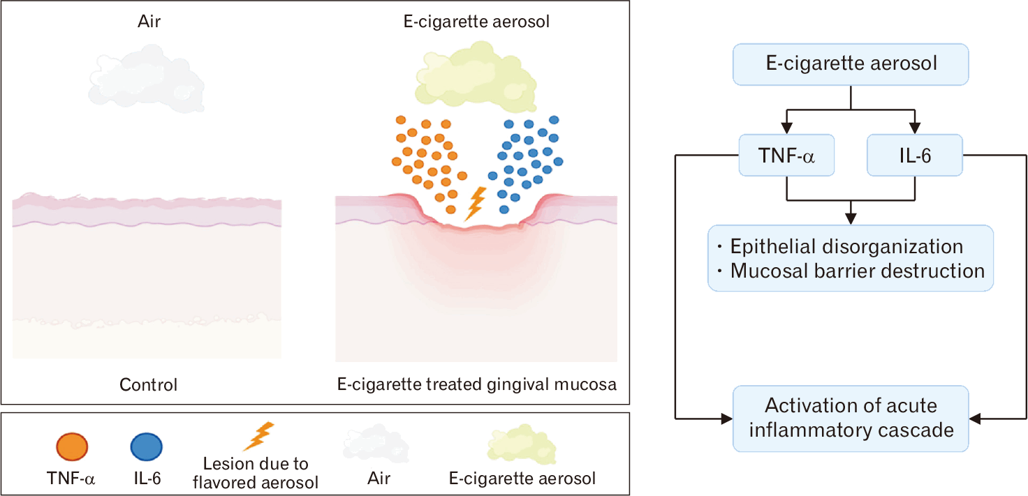
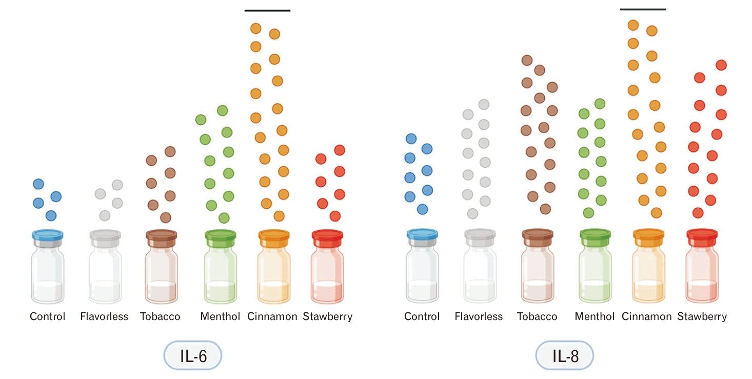
Across these modes, exposure to flavored or unflavored e-cigarette aerosols produced measurable increases in pro-inflammatory mediators such as TNF-α and IL-6, which are both central regulators of early innate immune activation. Repeated exposure to e-cigarette aerosol using a reconstructed human gingival mucosa tissue model demonstrated a clear, pro-inflammatory shift. In this study, aerosol-treated tissues exhibited significant expression of TNF-α and IL-6 compared with air-exposed controls, accompanied by epithelial disorganization and compromised mucosal barrier structure. The resulting release of cytokines leads to epithelial disorganization and mucosal barrier destruction, as shown in Fig. 3. The increase of these cytokines displayed an activation of an acute inflammatory cascade within the gingival epithelial layers and was consistent with multiple exposure durations [8]. However, in an oral epithelial cell line (OKF6/TERT 2), only IL-8 showed significant increases following flavored chemical exposure, whereas IL-6 showed no significant changes, and TNF-α was undetectable. This indicates that cytokine responsiveness is cell-type dependent and flavor dependent, showing that IL-6 and TNF-α upregulation is primarily observed in tissue-level mucosa models, rather than isolated epithelial monolayers [20]. This inflammatory response is highly flavor-dependent, with cinnamon causing the greatest cytokine increase in Fig. 4.
Lipid peroxidation and antioxidant enzyme
Lipid peroxidation and antioxidant enzyme activity were observed using primary studies to review oxidative stress pathways relevant to oral fibroblast toxicity. Two key indicators of oxidative injury are malondialdehyde (MDA) and superoxide dismutase (SOD). Evidence from human salivary biomarkers and in vivo/in vitro inhalation models demonstrates that exposure to e-cigarette aerosols can alter MDA and SOD [21, 22]. These findings help contextualize the oxidative-stress pathways identified in fibroblast models.
Recently, a human multi-omics study evaluating exclusive electronic nicotine delivery systems (ENDS) users reported significant shifts in salivary oxidative-injury biomarkers. Participants (ENDS users, n=13; non-users, n=5) demonstrated a puff-volume-dependent increase in MDA, which is consistent with elevated lipid peroxidation in oral biofluids. The same study also documented altered SOD activity in comparison with non-users, suggesting impaired antioxidant defenses associated with vaping exposure. These oxidative shifts happened alongside increased inflammatory mediators (IL-1β, IL-6, TNF-α), reinforcing coordinated oxidative-inflammatory responses in ENDS users [21]. Complementary findings were observed in a mouse inhalation and endothelial-cell model, which evaluated nicotine-free e-liquid base components. Continuous exposure to aerosolized PG/VG led to an increase in mitochondrial ROS accumulation and induced oxidative injury in lung tissue. Endothelial cells treated with e-liquid extract in vitro displayed significantly higher MDA levels and reduced SOD activity compared with untreated controls. Pharmacological inhibition of the mPTP opening partially restored SOD activity and reduced MDA elevation, which further linked both biomarkers to e-liquid-induced mitochondrial dysfunction [22].
Discussion
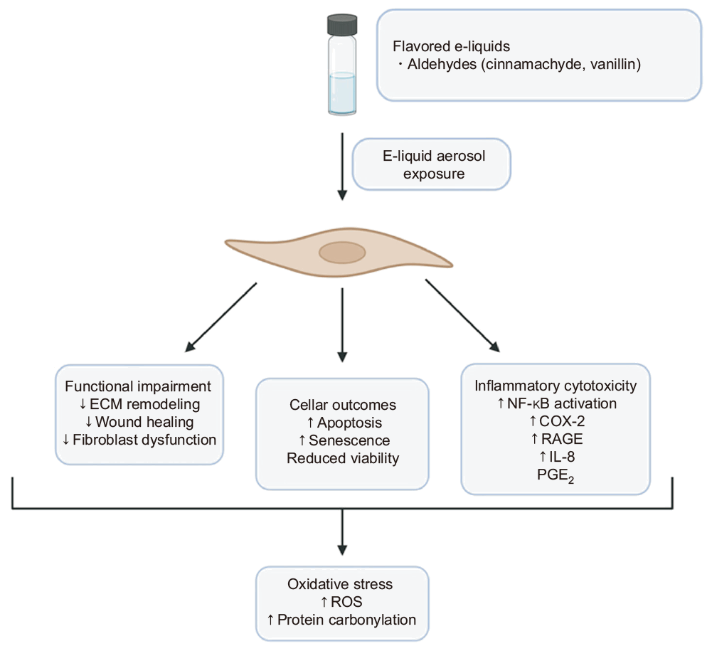
This review synthesizes evidence showing that flavored e-cigarette aerosols induce a consistent oxidative stress signature in oral fibroblast models, characterized by increased ROS generation, oxidative protein carbonylation, and downstream inflammatory mediator release. Collectively, these findings support a coherent mechanistic framework in which flavor-dependent oxidative stress contributes to fibroblast dysfunction and impaired ECM maintenance. Ultimately, these cascading responses lead to severe cellular outcomes and fibroblast dysfunction, summarized in Fig. 5.
According to National Surveillance data, approximately 6% of Canadians aged 15 years and older and 17% of students in grades 7–12 report vaping within the past 30 days, with youth demonstrating a preference for flavored products [23]. Likewise, recent U.S. data indicate that e-cigarettes are the most commonly used tobacco products among middle and high school students [24]. The high prevalence of flavor-driven vaping among adolescents and young adults, together with the available in vitro evidence, indicates that flavoring additives may represent an under-recognized oral health concern in this population.
Oral fibroblasts serve as first-line responders to inhaled aerosol constituents before systemic dilution or pulmonary filtration occurs. Gingival and periodontal ligament fibroblasts are consistently engaged in ECM turnover, wound repair, and modulation of inflammatory signaling. Unlike epithelial cells, fibroblasts are embedded within connective tissue matrices, where oxidative and inflammatory perturbations can directly disrupt collagen synthesis, matrix remodeling, and regenerative capacity. Research in fibroblast biology demonstrates that these cells actively modulate cytokine release and matrix turnover rather than serving merely as passive structural elements [9].
Under sustained oxidative or inflammatory stress, fibroblast-mediated connective tissue maintenance may become compromised.
Overall, these findings suggest that flavor-dependent fibroblast dysfunction may contribute to periodontal instability. Although in vitro findings cannot be directly equated with clinical disease, the observed convergence of oxidative stress and inflammatory dysregulation supports a biologically plausible link between flavored aerosol exposure and diminished periodontal resilience. These observations underscore the need to evaluate e-cigarette flavoring chemicals not only for pulmonary or systemic toxicity, but also for their potential consequences on oral tissue health.
A defining pattern shown in the results of this review is the separation between early cellular stress responses and later cell outcomes. These findings suggest that oxidative stress represents an early functional disturbance in oral fibroblasts rather than an immediate trigger of apoptosis.
The significance of this early oxidative injury lies in its impact on fibroblast function. Reports of increased protein carbonylation and γ-H2AX activation show that flavored aerosol exposure damages intracellular proteins and DNA in fibroblasts that remain metabolically active. These findings indicate disruption of molecular integrity and reflect sustained cellular injury beyond a transient stress response [7, 13, 17].
Several studies enhanced the translational relevance of their findings by using an air-liquid interface or whole aerosol exposure systems, which more accurately reflect inhalation-related exposures compared to submerged liquid extract methods. Sundar et al. [7] and Vermehren et al. [12] exposed oral cell models directly to generate e-cigarette aerosols or vapor under controlled puffing conditions. This preserves volatile and thermally generated constituents that are often lost or altered during solvent-based extractions.
However, this may also become a limitation with strong reliance on in vitro cell culture models to emulate the toxicity of flavored e-cigarette aerosols and e-liquids. Many studies exposed isolated oral fibroblasts or epithelial cells to liquid extracts or aerosol condensates for specific durations. While these models are valuable for identifying cellular mechanisms, they do not fully capture the complexities of the oral environment. As a result, findings from short-term in vitro exposure may overstimulate acute toxicity or fail to predict long-term adaptive responses that occur in vivo [7, 12, 13, 19]. Another limitation is the absence of key oral modifiers such as saliva and the oral microbiome in most experimental designs. Saliva contains antioxidants, enzymes, and buffering agents that can alter the chemical reactivity of aerosol constituents, while the oral microbiome can metabolize flavoring chemicals and influence inflammatory signaling [25].
Future investigations should prioritize advanced oral tissue models, including human gingival and oral mucosa constructs, over monolayer epithelial cell lines and non-oral tissue systems. Advanced three-dimensional or organotypic platforms therefore offer greater translational potential for replicating in vivo oral physiology, particularly in the assessment of IL-6, IL-8, TNF-α, MDA, and SOD biomarkers [21].
Similarly, the inclusion of oral fibroblast-specific models is critical, as much of the contemporary literature evaluates pro-inflammatory cytokine signaling and oxidative stress over short experimental time frames, neglecting chronic simulations of ECM remodeling and fibroblast senescence. Standardization of aerosol generation parameters and chemical characterization is essential. This would further reduce confounding variables related to flavor concentration, exposure dose, and environmental conditions [11, 20, 22].
Conclusion
This review synthesizes current evidence indicating that flavoring additives in e-cigarettes are linked to increased cytotoxicity and inflammatory responses in oral-relevant models. Across multiple studies, consistent alterations in oxidative stress markers, inflammatory mediators, and cellular integrity suggest biologically plausible mechanisms through which flavored aerosols may contribute to oral tissue dysfunction. These mechanistic observations challenge the perception that e-cigarettes are biologically healthier alternatives and underscore the need for continued investigation into their long-term oral health implications. The convergence of in vitro findings and emerging biomarker data supports further toxicological evaluation of flavoring additives, particularly given their significant use among adolescents and young adults.
 XML Download