



 PDF
 Citation
 Print


Introduction
The dorsal scapular artery (DSA) supplies the muscles of the upper back region. It arises mainly from the 3rd part and less often from the 2nd part of the subclavian artery (SCA). Typically, it is in lateral relation with the brachial plexus while passing deep to the scalenus anterior muscle. As it courses deep to the levator scapulae and the rhomboids, it is accompanied by the dorsal scapular nerve. It finally anastomoses with branches of the subscapular artery (SbsA), the suprascapular artery (SpsA), and the upper posterior intercostal arteries around the scapula [1-4].
The DSA supplies the trapezius as its primary pedicle through its superficial ascending fibres. The superficial branches penetrate the deeper side of the trapezius, giving off several cutaneous perforators that irrigate the skin in this region. The deep branches descend under the rhomboid major muscle, giving several perforators to the rhomboids and the skin above it. DSA rarely supplies the supraspinatus while supplying the rhomboids and the levator scapulae [5]. Knowledge of the perforators is necessary for a successful graft harvest [6].
It is essential to understand the origin of the DSA, as it may vary in relation to the trunk of the brachial plexus. The anatomical relationship between the artery and nerve helps prevent injury to the vessel during a brachial plexus block [7-10]. Multiple studies have shown variations in the origin of the DSA. Depending on its origin, it can be classified as Type I-DSA and the transverse cervical artery (TCA), which share a common origin with the SCA. Type II-DSA arises as a direct branch from both the SCA and the TCA. In Type III, DSA, and TCA are the direct branches from the thyrocervical trunk (TCT) [7]. Research studies on the DSA in the Indian population are limited. The present study aims to highlight the variations in origin, anatomical course, and termination of the DSA in the South Indian population.
Development of the subclavian artery
The embryological genesis of the artery provides a clue into the differences in origin and course of the artery in various individuals, as well as the differences between the two sides within an individual. The right SCA, arising from the brachiocephalic trunk, is a derivative of the right horn of the aortic sac of the developing aortic arches. At the same time, the left SCA originates from the left fourth aortic arch and lower part of dorsal aorta. Most of the essential branches of the SCA arise from the upper six cervical intersegmental arteries; the seventh continues as the distal part of the SCA [11]. The diverse relationship between the branches of the SCA and the brachial plexus can be explained by the developmental pattern observed on the 37th day of the embryonic stage. It is ascertained that the well-formed SCA pierces the anterior division of the neural plate (developing branches of the brachial plexus) to pass between the divisions. Beyond the neural plates, the artery divides into an undefined capillary plexus. These capillary plexus organize to result in the final branches of the main artery [12].
Materials and Methods
Ethical approval
The authors hereby confirm that every effort was made to comply with all local and international ethical guidelines and laws concerning the use of human cadaveric donors in anatomical research. The Institutional Ethics Committee of Vinayaka Mission’s Medical College and Hospital approval number is VMMC/ANAT/2022/JULY/02.
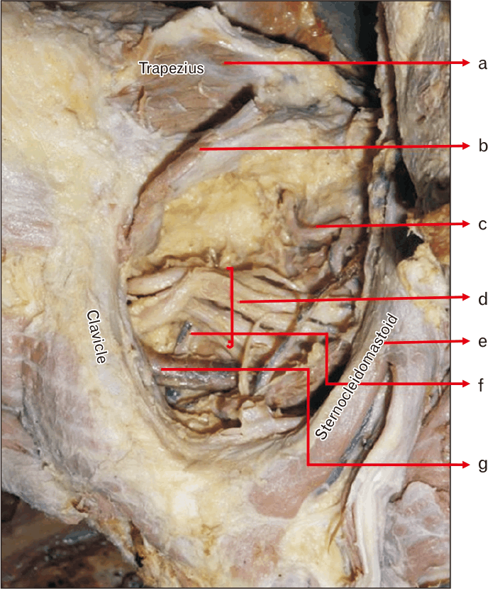
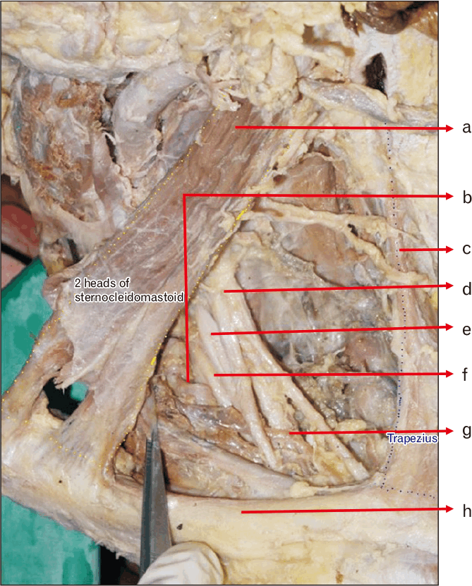
The cadaveric-based observation study involved 40 bilateral sides from 20 formalin-fixed human adult cadavers from Southern India, aged between 55 and 70 years. An equal number of male and female cadavers were dissected. The dominance of the hand was not considered due to a lack of history. The death was natural in all the cases, primarily due to cardiovascular causes. Only anatomically intact specimens were included in the study. The dissection was carried out in the neck region, tracing to the back, following the guidelines from the standard dissection manual. A curvilinear incision in the supraclavicular triangle was made. Incision was done lateral to the clavicular origin of the sternocleidomastoid muscle and extended approximately 4–5 cm parallel to the clavicle. The external jugular vein was identified and retracted medially. The internal jugular vein was also secured away to expose the scalenus anterior muscle and the SCA. All the branches of the SCA were identified. The origin of the DSA was noted, and the course was traced beyond till its termination. Simultaneously, its relation to the brachial plexus was observed. The observed values were calculated with descriptive statistical methods.
Results
Variations in the course of the artery and its relation to the brachial plexus
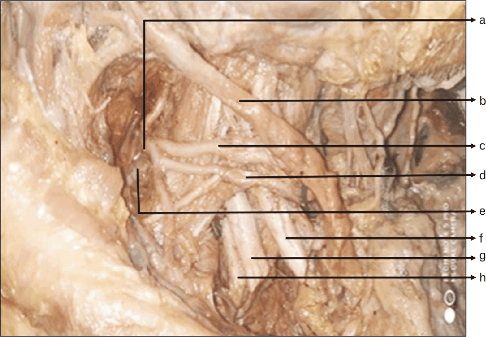
In the examined 40 specimens, 75% of them showed DSA coursing through the upper and middle trunks (Fig. 3) and 20% presented with the artery coursing between the middle and the lower trunks. While 5% of the specimens had the DSA running superior to the brachial plexus. All the specimens coursing superior to the brachial plexus were on the left side (Table 1).
Table 1 shows the variation in the DSA course among male and female specimens across three anatomical pathways. No significant sex-based difference was observed in the distribution of these variations.
Males and females displayed a similar pattern when analysed by sex, with 70% of the structures in males and 80% in females aligned with the upper/middle trunk. In the present study 10% of male specimens showed the course of the DSA superior to the upper trunk. In contrast, no such observations were made in female specimens. There is no significant statistical difference in the origin or course of the artery when considering laterality or sex differences in the specimens (Tables 2 and 3).
Table 2 compares right- and left-side variations of the DSA course in male and female cadavers. The distribution patterns were similar across sexes and sides, with no significant differences observed.
Table 3 summarises DSA course variations by side across all specimens, independent of sex. The overall pattern showed no significant difference between the right and left sides. Data were presented as frequency and percentage. The chi-square test was used to determine the association between the categorical variables. Significance was defined by P-values less than 0.05 using a two-tailed test. Data analysis was performed using IBM SPSS Statistics version 25.0 (IBM Co.).
Discussion
The DSA is under extensive investigation due to its importance to surgeons and anaesthetists. The detailed anatomy of its origin, course, and distribution makes it an interesting topic of debate. Global studies consistently corroborate the predominant origin of DSA from the second and third segments of the SCA [3, 8, 9, 13]. The studies encompassed both cadaveric and radiographic investigations, including sonographic and arteriographic studies [9, 14]. The DSA frequently originates from the TCA when it is not a direct branch of the SCA [6, 8, 13, 15]. Consistent with prior research, our study also confirmed the predominant origin from the SCA. This type of origin, arising as direct branches of the SCA or the TCA, corresponds to the Type II classification in our study [7]. Hence, the present study conducted on the South Indian population aligns with studies conducted on the European population [8]. Reports of the DSA arising primarily from the TCT are mentioned in studies on Thai, Japanese, and other Asian populations [7]. This observation expands upon the existing data and highlights the importance of considering ethnic variation when comparing the frequency of presentation of these variations. The DSA is also known to arise from the axillary artery, the SpsA, the cervico-dorsoscapular trunk, the costocervical trunk, the vertebral artery, the inferior thyroid artery, and the SbsA [2]. There could be more than one branch arising on the same side, a tendency in females. Ikka et al. [2] adding to this body of evidence, our analysis revealed bilaterality more frequently than previously reported, a finding of relevance for surgical planning.
The compression of the nerve by an artery is the most likely cause in patients with symptoms of compression of the lower trunk of the brachial plexus. Various studies have confirmed that the DSA is the artery most frequently involved in compression of the plexus, primarily when located between the middle and lower trunks, resulting in thoracic outlet syndrome [10]. The TCA that emerges from the proximal portion of the SCA tends to cross the lower part of the brachial plexus, which may also compress the brachial plexus sometimes [16].
Hong et al. [17] documented an unexpected visualization of the DSA during a pre-operative ultrasonographic assessment of a patient scheduled for a corner pocket approach, which is a common technique for achieving ulnar anaesthesia. In that case, the artery’s position directly in the needle’s path made the corner pocket technique impossible, necessitating the use of an alternative inter-truncal approach [9, 18]. This case highlighted the importance of conducting further research studies on the relationship between the DSA and the trunks of the brachial plexus. Motivated by this finding, the authors conducted a study in their South Indian population to provide more data on these anatomical variations.
The course of the DSA through the brachial plexus is as follows: the consistent location of the DSA is between the upper and the middle trunks in both sexes if it arises from the SCA. These findings collectively reinforce and add nuance to the anatomical descriptions of the DSA, emphasizing specific trends in our studied cohort that may impact preoperative planning. There is only a 10% chance of DSA passing through the trunks of the brachial plexus if it arises from the TCT, and a 3% chance if it arises from TCA, compared to an average of 70% chance if it arises from SCA. The data help us understand that the risk of compression of the brachial plexus by the artery is higher if its origin is from the SCA. Interestingly, the SpsA is found under the brachial plexus 100% of the time if it arises from the TCT or TCA. In line with the prior sonographic risk assessments, our data highlight a comparable risk profile, reinforcing the clinical importance of individualised vascular mapping. Based on the ultrasonographic studies, 90% of patients undergoing supraclavicular brachial plexus block are at risk of vessel rupture [9]. The artery lies in the path of the block needle if it is in the upper and middle zones. The data from the present study shows that the DSA coursed between the upper and the middle trunks in the majority of the population, showing that this could be the best anatomical course of the artery to avoid compression naturally, but simultaneously at a high risk for vessel rupture, as this is the most typical route for supraclavicular brachial plexus block.
The DSA can be conveniently used as the recipient pedicle for free tissue transfer due to its large calibre and favourable geometry for microvascular anastomosis. It can be identified in the deeper part of the posterior triangle after emerging through the brachial plexus. The mean calibre of the perforators was 0.8±0.2 mm and the length measured 8.1±2.8 cm according to Wang et al. [19]. The artery was traced from its origin to its termination below the rhomboids. The artery showed perforators, its calibre route; however, its calibre or number was not considered for statistical purposes. The average size of the cutaneous territory perfused by perforators from the superficial branch of the DSA was 7.8 cm×8.5 cm, as mentioned in another research by Kany et al. [20]. The extensive anastomosis between the DSA, the SpsA, and the circumflex scapular artery around the scapula provides an extensive vascularity to the muscles around the region, especially the trapezius [20]. Hence, the trapezius perforator flaps, and associated fascio-cutaneous flaps are used in the head and neck reconstructive surgeries [6, 15, 19, 21]. Furthermore, our findings regarding the DSA’s territory and anastomotic relationships corroborate its utility in various surgical contexts, as widely reported in the existing literature.
The DSA can replace the vascularity of the trapezius and supraspinatus in the absence of the SpsA [22]. The DSA supplying the subscapularis muscle was studied in detail. This knowledge can also be applied to various surgeries involving the back region and to procedures involving graft transfers [23]. Hematoma of DSA is implicated in thoracic cage injuries and may sometimes present as a pseudoaneurysm as well [24, 25]. The identification of DSA becomes essential, and procedures like 5 in 1 injection could be performed more precisely by injecting into the medial vicinity of DSA [26]. In summary, our findings not only corroborate many of the anatomical patterns described in the literature but also highlight distinct trends within our population, which hold implications for surgical and anaesthetic strategies in the region.
In conclusion, studies across the globe have confirmed that the origin of DSA is mainly from the SCA, either directly or indirectly through its branches, which coincides with the present study. An attempt was made to identify the origin and course of the artery in relation to sex, a factor not previously considered in studies. However, no statistically significant differences were noted in our study. Similarly, there were no significant differences in origin or course of the vessel between the right and left sides. It can also be concluded that the DSA coursed mostly between the upper and middle trunk. This knowledge is fundamental during the brachial plexus block. The artery was consistently seen coursing through either of the trunks of the brachial plexus, making it a potential culprit in cases of brachial plexus compression syndrome.
 XML Download