



 PDF
 Citation
 Print


Introduction
The sciatic nerve (SN) is the largest and longest nerve in the human body. It originates from the sacral plexus and extends down to the lower limbs. SN provides motor and sensory innervation to the lower extremities [1-3]. Typically, the SN divides into the tibial nerve (TN) and common fibular nerve (CFN) at the superior angle of the popliteal fossa [4]. However, cadaveric studies have shown that it can divide at various levels, including the pelvic region, gluteal region, upper or middle third of the thigh, or within the popliteal fossa.
Anatomical variation of the SN division has various clinical implications, potentially contributing to the development of piriformis syndrome and sciatica. The manifestations of these conditions can vary depending on the level at which the SN divides into its terminal branches [5-8]. In addition, variation of SN can affect intramuscular injections and surgical procedures [9, 10]. Complete SN palsy can lead to disabling outcomes, including flail feet and severe gait impairment [3, 11].
Although the variability of SN division is clinically important, the pooled prevalence and its patterns are not well documented in cadaver studies. Previous meta-analyses [12-16] examined SN variants related to the piriformis muscle, outcomes of injuries after acetabular fractures, and the effectiveness of combined sciatic and femoral nerve blocks in knee arthroplasty patients. Studies also explored outcomes in sciatic neuropathy and the relationship between anatomical variants on magnetic resonance imaging (MRI) and piriformis syndrome.
This study aimed to determine the pooled prevalence of variability in SN division in the cadaver studies and the associated clinical importance. This review examined the occurrence of SN division in various regions of the lower limb, including the pelvic region, the gluteal region, the upper third of the posterior thigh, the middle third of the thigh, and within the popliteal fossa.
Methodology
Search strategy
This review was carried out according to the guidelines of the Evidence-Based Anatomy Working Group [17] and PRISMA [18]. Studies were searched in PubMed, Google Scholar, ScienceDirect, and African Journals Online.
Keywords were combined using Boolean operators (OR and AND). We retrieved articles using the following key terms: (“anatomical variations” OR “topographic variations” OR “variant anatomy” OR “anatomic deviation” OR “anatomic differences” OR “anatomic disparities” OR “anatomic irregularities”) AND (“nervus ischiadicus” OR “ischiatic nerve” OR “lower limb major nerve”) AND (“division” OR “branching” OR “separation” OR “divergence” OR “splitting” OR “subdivision”) AND (“cadaver” OR “postmortem” OR “cadaverous” OR “cadaver dissection” OR “postmortem dissection” OR “cadaver examination” OR “human cadaver”) AND (“clinical importance” OR “clinical significance” OR “clinical relevance” OR “clinical implications” OR “medical importance”). All fields and MeSH terms were used to search in PubMed.
Unpublished studies were retrieved from Google, Google Scholar, and university institutional repositories. A manual search was performed in the major anatomical journals. Reference lists of the articles included in this study were examined. No date-range restrictions were employed. WAL and SA have searched independently.
Extraction
First, articles were sorted by titles and abstracts. Then, full-text articles were examined. Only studies conducted in English were included. Expert opinions, case reports, conference abstracts, and letters to the editor were also excluded. Articles that did not clearly state the anatomical variations of the SN division were excluded. Studies that contained damaged cadavers, unclear or incomplete data, patient cases, and imaging modalities (X-ray, CT, MRI) were excluded.
Data extraction was conducted using an Excel sheet. The extraction included the author’s name, publication year, number of lower limbs, country of origin, SN division in the pelvic region, gluteal region, upper third of the thigh, middle third of the thigh, and the popliteal fossa. W.A.L. and S.A. independently performed the data screening and data extraction process.
Quality assessment
Eligible studies were evaluated using the Anatomical Quality Assessment (AQUA) tool [19] (Supplementary Table 1). Five domains were evaluated: (1) objectives and subject characteristics, (2) study design, (3) methodology, (4) descriptive anatomy, and (5) reporting of results. We categorized each domain as either ‘low, high, or unclear’ risk of bias. The signaling questions “Yes,” “No,” and “Unclear” indicate low, high, and unclear risk of bias, respectively. A “low” risk of bias was made for all signalling questions answered with “yes” in the domain, and any signalling question answered with “no” in the domain was decided to have “high” potential for bias. The “Unclear” option was used for insufficient reported data. WAL and SA independently assessed the quality assessment of the studies. Differences in opinion between reviewers were discussed until a consensus was reached.
Data analysis
Data analysis was performed using Stata 14 software (Stata Corp.). A random-effects meta-analysis was employed to calculate the pooled prevalence [20]. Point prevalences with 95% confidence interval (CI) were presented in the forest plot [21]. Heterogeneity among the included studies was assessed using the Cochran’s Q test (with significance set at P<0.05) and quantified using the I2. Publication bias was evaluated using a funnel plot and Egger’s test. Subgroup analysis was carried out on the anatomic variation of SN at various levels of the lower extremities. A meta-regression was performed on geographic region, sample mean, publication year, Indian versus other, and methodological differences.
Outcome variables
The primary outcome of this study was the overall SN division in all cadavers. It was reported in the original paper as a percentage and/or as the number of variabilities within the SN division. Variabilities in the SN division refer to cases where the nerve divides outside the standard anatomical landmarks, specifically not within the superior angle of the popliteal fossa or the lower one-third of the thigh. Any deviation from this standard division site, either above or below the superior angle of the popliteal fossa, was considered an anatomical variation of the SN [1-3, 22].
Results
Characteristics of the studies
The characteristics of studies included for this review are shown in Table 2 [3, 6, 10, 22-38]. Twenty cadaver studies examined a total of 1,684 lower limbs. The sample size ranges from 24 to 338. Most of the studies (55%) were conducted on Indian cadavers, while the remaining were from Ethiopia, Kenya, South Africa, Turkey, Nepal, Malaysia, Poland, and Russia. The studies demonstrated great variability in SN division, ranging from 6.3% to 87.3%.
Meta-analysis
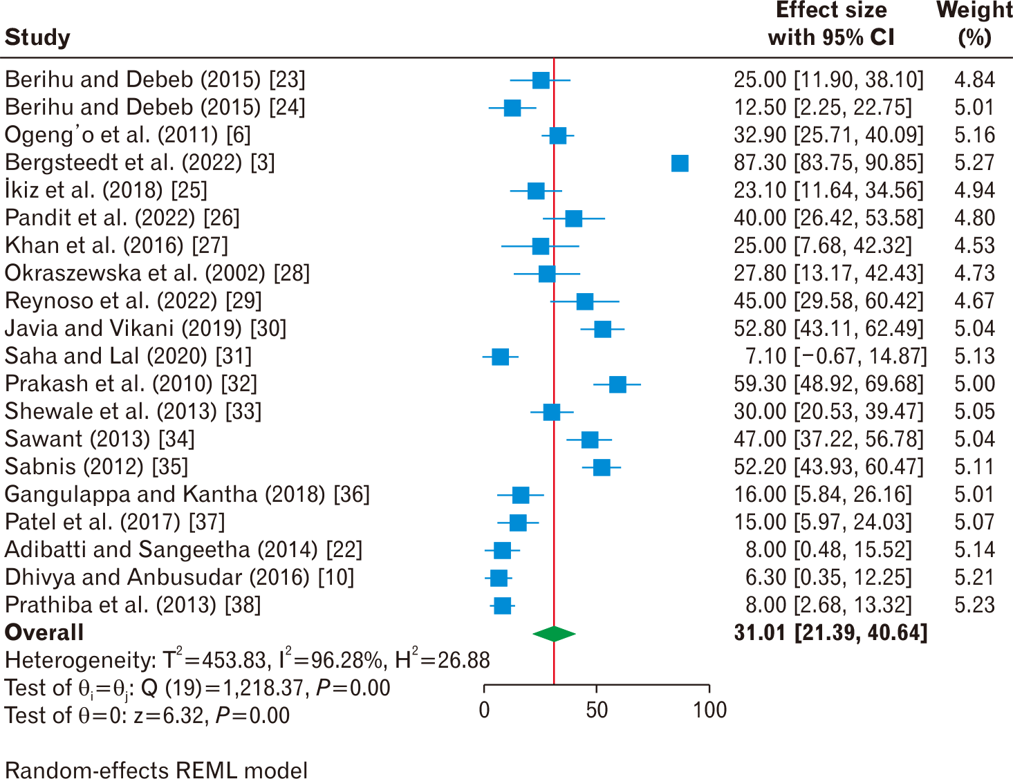
Fig. 1 [3, 6, 10, 22-38] presents a forest plot of a meta-analysis examining the anatomical variation of SN division among 20 studies. The pooled prevalence estimated was 31.01% (95% CI: 16.24–45.78; I2=98.4%), indicating that approximately one-third of the SN divides outside the typical pattern. The analysis showed statistical heterogeneity among the included studies (I2=98.4%; P<0.001), as visually evidenced by the wide scatter of individual study estimates (squares) and their CIs (horizontal lines) across the plot. Individual study estimates range from as low as 6.30% to as high as 87.30%.
Publication bias
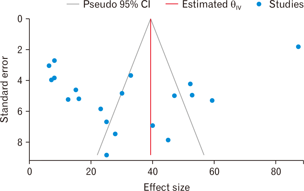
Visual inspection of the funnel plot showed an asymmetric distribution, suggesting potential bias or small-study effects (Fig. 2). Although visual inspection of the funnel plot suggested some degree of asymmetry, Egger’s regression test did not provide statistical evidence of publication bias (P=0.8437).
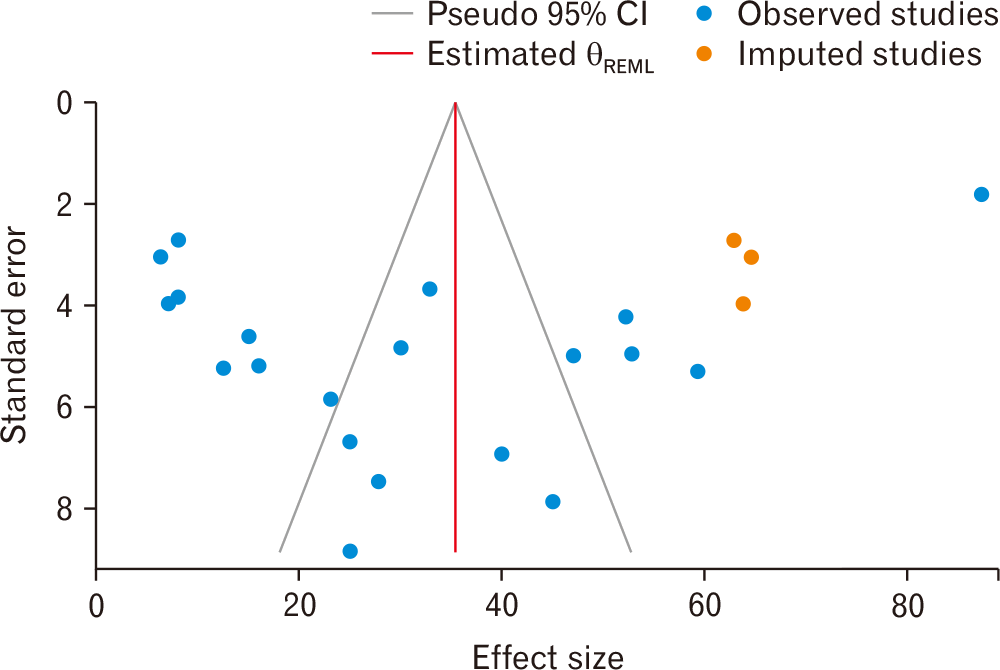
The trim-and-fill method imputed three studies on the right, resulting in an adjusted pooled prevalence of 35.38% (95% CI: 25.86–44.90), which is higher than the original estimate (31.01%; 95% CI: 16.24–45.78) (Fig. 3).
Subgroup analysis by anatomical region
Subgroup analysis of the SN division at various levels in the lower limb is presented in Table 4 [3, 6, 10, 22, 25-36]. The highest variability was observed in the popliteal fossa, with 21.17% (I2=98.2%), followed by the pelvic region, at 11.33% (I2=78.8%). Divisions in the gluteal region (4.86%) and the upper third of the thigh (4.74%) demonstrated the lowest prevalence and considerably lower heterogeneity (I2=37.4% and 33.5%, respectively). Detailed forest plots for each anatomical subgroup are provided in the corresponding (Supplementary Figs. 1–5).
Sources of heterogeneity
Given substantial heterogeneity in the overall analysis (I2=98.4%; P<0.001) and the popliteal fossa subgroup (I2=98.2%; P<0.001), we performed meta-regression and subgroup analyses based on geographic region, sample mean, publication year, Indian versus other, and methodological differences (Table 5) [3, 6, 10, 22-39]. Detailed forest plots for each subgroup analysis are provided in the corresponding (Supplementary Figs. 6–10).
Meta-regression identified sample mean and methodological differences as a significant source of heterogeneity (P<0.05). Subgroup analysis by geographic region (P=0.78), publication year (P=0.87), and studies conducted in India versus other areas (P=0.39) were not statistically significant. Subgroup analysis by sample mean demonstrated a high pooled estimate in studies with a sample size greater than the mean (≥84) (46.2%; 95% CI: 29.69, 62.71; I2=97.77%) compared to studies with with sample size less than mean (<84) (19.61%; 95% CI: 12.70, 26.53; I2=81.58%). Studies reporting of all five anatomical sites showed the highest prevalence (47.18%; 95% CI: 34.84, 59.52; I2=84.16%), followed by poplitel fossa and some others (47.75%; 95% CI: 22.25, 63.25; I2=96.56%), while studies with no popliteal fossa data demonstrated the lowest prevalence (16.88%; 95% CI: 9.35, 24.41; I2=87.83%).
Discussion
Prevalence of the anatomical variation of the sciatic nerve division
The anatomical variation of the SN division can be attributed to developmental and genetic factors. During the embryonic period, the lumbar plexus and sacral plexus extend into the limbs and differentiate into dorsal and ventral rami of the spinal nerve fibers [40]. The fibers of the sacral plexus converge and form the SN. The SN remains encased within a connective tissue sheath before bifurcating into the CFN and the TN [33, 40, 41]. The branching patterns of the SN are influenced by the location where this sheath ends, leading to variations in nerve separation levels in regions such as the pelvic area, the gluteal region, and the popliteal fossa.
Anatomical variations of the SN division can be further influenced by signaling molecules and factors during embryonic development [42-44]. These factors regulate axonal specification, migration, and projection [45-48]. The SN develops from axons that extend from the ventral rami of the lumbosacral plexus to innervate the lower extremities. Variations in SN branching may arise from differences in the expression or activity of these factors during axonal extension. For example, changes in the expression of genes such as retinoic acid receptors can alter guidance signals [49, 50], leading to different axonal growth patterns of the SN. Consequently, some individuals may have a single undivided SN, while others may exhibit multiple branches originating at various points along its course.
In the current study, atypical division of SN occurs in approximately 31.01% of individuals; thus, different branching patterns are present in approximately one in three nerves. The most common site for division was within the popliteal fossa (21.17%), followed by more proximal sites, the pelvic region (11.33%). This high prevalence challenges the traditional teachings in many anatomy curricula and the critical importance of this variation for clinicians across multiple specialties.
Meta-regression identified sample mean and methodological differences as a significant source of heterogeneity. The analysis showed a significantly higher pooled prevalence in studies with a sample size greater than the mean compared to studies with a sample size less than the mean.
Studies reporting all five anatomical sites showed the highest prevalence, followed by the popliteal fossa and related regions. Studies lacking a popliteal fossa reported the lowest prevalence. These patterns, a methodological framework, sample size, and anatomical sites act as a critical filter that systematically influences the observed result. Consequently, the extreme heterogeneity (I2=98.4%) is not only due to statistics but also driven by these fundamental differences in study sample size and comprehensive reporting of anatomical sites for the division of the SN, which can contribute disproportionately to higher and more variable estimates.
Clinical implications of sciatic nerve division
The division of the SN within the pelvic region, the gluteal region, and the upper third of the thigh is often classified as a high-level division. Divisions within the middle third of the thigh and the popliteal fossa are considered low-level divisions. High division alters the anatomic pattern of the SN, which affects the surgical approaches in the pelvic and gluteal regions. It increases the risk of SN injury during hip arthroplasty or trauma-related surgeries [51-55]. The surgeon can inadvertently cut, stretch, or compress the branches while accessing the hip joint or performing other surgical maneuvers. Tissue traction and manipulation may occur during surgery and may affect the SN. Anatomical variants of the SN can alter its susceptibility to tension, compression, or entrapment during these surgical maneuvers, potentially increasing the risk of nerve injury. Furthermore, these variations can contribute to injuries during surgeries performed in the gluteal region and the development of conditions such as piriformis syndrome, coccygodynia, muscle atrophy, and non-discogenic pain [56-58]. A higher division of the SN allows the TN and the CFN to follow different pathways, allowing one nerve to function independently if the other is affected by injury or torn [5, 37]. This pattern might help to preserve motor and sensory functions in the lower extremities. It can also lead to nerve compression and conditions such as sciatica, piriformis syndrome, and complications during surgical procedures.
A low division of the SN in the popliteal fossa, where the nerve splits at a lower level or even below the fossa, has significant implications for surgical procedures and popliteal blocks in the lower limbs [59-64]. If the surgeon is unaware of the low division, there is a risk of damaging or transecting nerve branches, leading to possible sensory disturbances, motor deficits, and chronic pain in the lower leg and foot.
The prevalence estimates for the SN division at various levels quantified in this study allow for an evidence-based approach to clinical risk assessment and procedural planning. The following integrates our subgroup point estimates and their heterogeneity with the specific clinical implications.
(1) Popliteal fossa (21.17%; 95% CI: 9.45–32.89; I2=98.2%): this is the most common site of division, affecting approximately one in five individuals. However, the high heterogeneity (I2=98.2%) indicates that this figure is not uniform across populations; the true prevalence is context-dependent and could range from under 9.5% to 32.9%. This uncertainty has direct clinical consequences. For surgeons operating in the popliteal fossa, this variability mandates a “look-before-you-leap” approach; they cannot rely on anatomical assumptions and must perform meticulous dissection to identify and protect the often-already-divided nerve components to avoid iatrogenic injury [60]. The high prevalence also explains why single-injection popliteal SN blocks have a known failure rate [65]; our data suggests that in some populations, up to a third of patients may have a division high enough to necessitate a dual-block technique for complete success.
(2) Pelvic region (11.33%; 95% CI: 6.45–16.21; I2=78.8%): this finding indicates that over 1 in 10 individuals have SN division within the pelvis, which directly quantifies the risk for procedures in this area. The high heterogeneity (I2=78.8%) indicates this risk may vary between populations, but the point estimate suggests it is never negligible. Pre-operative imaging or the use of nerve monitoring are important for complex pelvic surgeries. For hip arthroplasty and pelvic tumor resection, surgeons may anticipate a substantial risk of injuring either the TN or CFN as separate structures [26, 66]. For anesthesia, the 11% prevalence provides an evidence-based rationale for the systematic use of dual-injection techniques for SN blocks [22, 39, 67-69].
(3) Gluteal region (4.86%; 95% CI: 3.12–6.60; I2=37.4%): while less common (affecting ~1 in 20 individuals), a division in the gluteal region carries severe consequences, as this is the most critical finding for gluteal intramuscular injections. Low heterogeneity (I2=37.4%) may give clinicians confidence in this 5% risk estimate. The 5% probability that the CFN is a separate, superficial structure translates directly to a 5% baseline risk of iatrogenic injury and subsequent foot drop [22, 39, 67-69]. Due to the potential for surgically altered nerve pathways and the variable anatomy of the superior gluteal neurovascular bundle at the intramuscular junction, extreme care must be exercised during both gluteal surgical procedures and routine intramuscular injections [56-58].
(4) Upper third of thigh (4.74%; 95% CI: 2.98–6.50; I2=33.5%): with a prevalence similar to the gluteal region and low heterogeneity, this finding is relatively reliable. In cases of femoral shaft fractures or thigh compartment syndrome, clinicians should be aware that nearly 5% of patients are at risk of distinct and potentially isolated TN or CFN deficits. This knowledge is crucial for accurate neurological assessment and surgical planning in vascular and trauma surgery.
(5) Middle third of thigh (7.11%; 95% CI: 4.55–9.67; I2=17.9%): this subgroup has the most reliable estimate, with very low heterogeneity (I2=17.9%). The 7% prevalence indicates a tangible risk for procedures in the mid-thigh. During biopsies, fracture fixation, or trauma exploration, there is a well-defined risk of causing an isolated common peroneal nerve injury and foot drop. This specific, quantifiable risk should be part of the preoperative consent process and guide surgical dissection in this region.
Recommendations
(1) Pre-operative imaging/planning: for pelvic and high-gluteal surgeries, the 11% risk of pelvic division and 5% risk of gluteal division justify considering advanced imaging or neurophysiological planning to avoid iatrogenic injury.
(2) Anesthesia protocol: the combined 21% (popliteal) and 11% (pelvic) divisions proximal to the traditional block site strongly support adopting dual-injection or proximal catheter techniques for comprehensive SN blocks to minimize failure.
(3) Injection safety: the 5% prevalence of gluteal division provides a powerful, quantitative argument for mandating the ventrogluteal site for intramuscular injections to prevent foot drop.
(4) Surgical vigilance: surgeons operating in the popliteal fossa must be prepared for anatomical variation in nearly one-quarter of cases, while those operating in the mid-thigh should be vigilant for separate nerves in 7% of cases.
(5) Informed consent: these prevalence rates provide specific, evidence-based numbers that can be used to inform patients about the risks of nerve injury during relevant surgical procedures.
Limitations
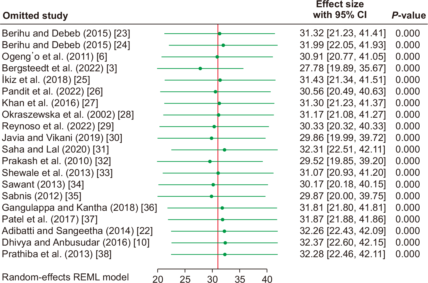
This study provided scientific evidence on the anatomical variation of the SN division and its clinical importance in cadaver studies. However, the following limitations might be taken into consideration. First, the exclusion of non-English publications may introduce selection bias and limit the global representativeness of the finding, as limiting the search to English-language studies may have led to the omission of region-specific data and consequently reduced the global generalizability of the results. Second, the high statistical heterogeneity and the presence of wide CIs in some analyses (including those with negative lower bounds) reflect substantial statistical uncertainty. This is attributable to small sample sizes in some studies and the high methodological diversity across the included cadaveric studies. Third, while meta-regression and subgroup analysis identified sample size and methodological framework as significant sources of heterogeneity, residual heterogeneity remains high. Our analysis was constrained to study-level variables, and we lacked data on finer methodological details or cadaver demographics, which are likely additional, unmeasured sources of variation. Finally, the geographic distribution of the available literature is imbalanced, with a predominant number of studies from India. Although a sensitivity analysis confirmed the stability of our main pooled estimate, data from other regions remain scarce. An assessment of publication bias was inconclusive, as funnel plot asymmetry is likely a product of both high clinical heterogeneity and potential publication bias.
Future directions
Based on the gaps and heterogeneities identified in this review, we suggest several key directions for future research. First, imaging-based validation studies, such as MRI or ultrasound, are necessary to correlate our cadaveric findings with in vivo anatomy. Second, genetic investigations are also vital to explore potential developmental and hereditary factors underlying these anatomical differences. Furthermore, combining anatomical data with clinical studies would help clarify the clinical importance of these variations and guide safer, more personalized procedural planning.
Conclusion
In this study, the variability in SN division was high, and one in three SNs divides outside the typical pattern. Along the path of the lower extremity, five types of SN variability were identified: pelvic region (11.33%), gluteal region (4.86%), upper third of the thigh (4.74%), middle third of the thigh (7.11%), and popliteal fossa (21.17%). The unusual patterns of SN division can affect surgical approaches and increase the risk of nerve injury during hip arthroplasty or trauma-related surgeries. These anatomical variations complicate anesthetic administration. Therefore, it is important to consider variations in SN anatomy during surgical and anesthetic procedures for the lower limb.
 XML Download