Abstract
The cerebral aqueduct (CA), or aqueduct of Sylvius, is the narrowest portion of the ventricular system, connecting the third and fourth ventricles and providing an essential conduit for cerebrospinal fluid (CSF) circulation. Despite its small caliber, the CA transmits over 500 ml of CSF daily, and its vulnerability to obstruction makes it central to the pathophysiology of hydrocephalus. This review synthesizes current knowledge of the CA across developmental, anatomical, and clinical dimensions. We examine its embryological formation and the contribution of structures such as the subcommissural organ and Reissner’s fiber, highlighting comparative features across species, and evaluate the dynamic physiology of CSF flow in relation to age and disease states. Advances in imaging modalities, including high-resolution magnetic resonance imaging and fetal ultrasound, are summarized in the contexts of both research and clinical practice. Pathological considerations are addressed, ranging from congenital anomalies (e.g., aqueductal webs, Chiari malformations, cavernomas, and forking) to acquired lesions (e.g., gliosis, neoplasia, and trauma-induced stenosis). The genetic underpinnings of aqueductal stenosis are reviewed, emphasizing L1CAM mutations and broader molecular pathways implicated in congenital hydrocephalus. Surgical strategies, including endoscopic third ventriculostomy, aqueductoplasty, and transaqueductal stenting, are critically appraised in terms of efficacy, complication profiles, and long-term outcomes. Overall, this review underscores the CA as a nexus of neurodevelopment, neuropathology, and neurosurgical innovation, and identifies gaps in knowledge that merit future investigation.
The cerebral aqueduct (CA), also known as the aqueduct of Sylvius, is a narrow channel within the midbrain that connects the third and fourth ventricles and plays a critical role in cerebrospinal fluid (CSF) circulation. Despite its small caliber, it serves as a key conduit through which a substantial volume of CSF passes daily, rendering it particularly susceptible to obstruction and pathological alteration.
Given its clinical importance and the evolving understanding of its anatomy, physiology, and pathology, a comprehensive synthesis is warranted. This review aims to provide an integrated overview of the CA, emphasizing its anatomy, development, function, imaging characteristics, and clinical relevance.
The protocol of the study did not require approval by the ethical committees or informed consent. The study followed the Declaration of Helsinki (64th WMA General Assembly, Fortaleza, Brazil, October 2013).
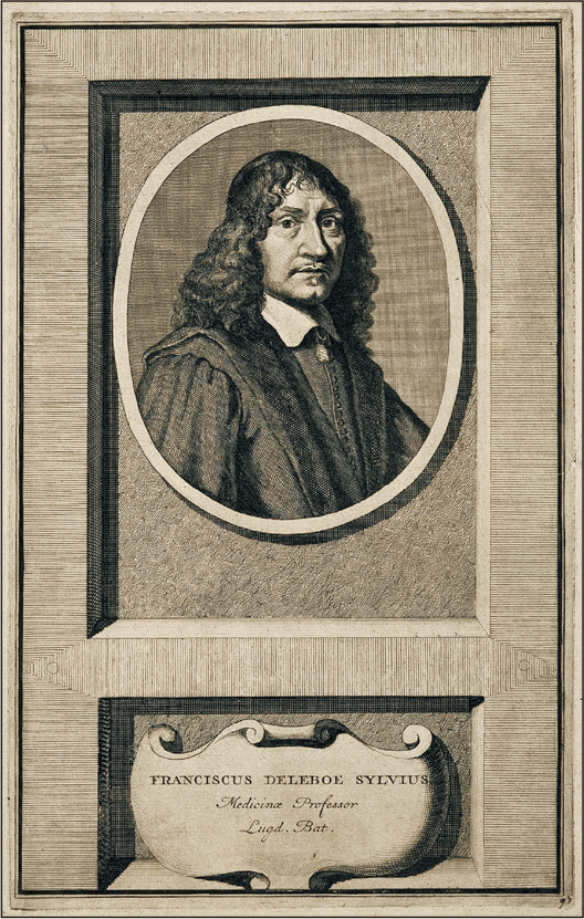
The CA (midbrain aqueduct, aqueduct of Sylvius) unites the third and fourth ventricles and was described in 1650 by the anatomist Franciscus de la Boe Sylvius (Fig. 1) [1, 2]. It is still remembered eponymously after Sylvius, though it had first been described by Jacopo Berengario da Carpi 25 years earlier.
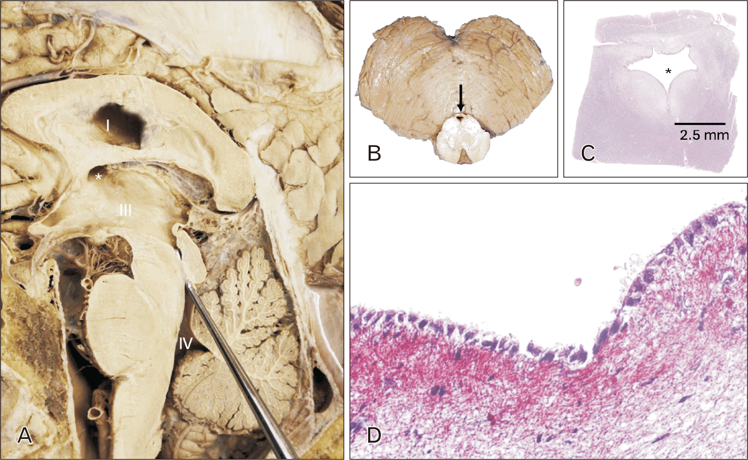
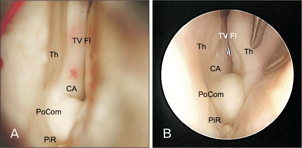
The CA is in the dorsal midbrain, surrounded by the periaqueductal gray matter [3]. The third ventricle connects superiorly to it, while the fourth ventricle connects inferiorly to it (Fig. 2) [4]. The posterior commissure (Fig. 3) and lamina tecti lie posterior to the CA, while the medial longitudinal fasciculus, red nucleus, and CN III/IV nuclei lie anterior to it [3]. The CA has a cross-sectional area of approximately 0.5 mm2 in children and 0.8 mm2 in adults. Although its small size makes it the narrowest part of the CSF path, more than 500 ml of CSF pass through the channel per day in the human brain [4, 5].
The CA is formed by foldings of the ventricular wall comprising small longitudinal striae that run parallel to the median sulci [6]. Histologically, the CA and the central canal of the spinal cord are lined by a single layer of ependymal cells (Fig. 2) [7]. These cells have apical kinocilia and are firmly attached to neighboring ependymal cells [7]. They often show variant anatomy, such as a lining thicker than a single cell layer or small diverticula [3]. The subependymal glial plate can also present with many variations, sometimes in only one part of the CA, or surrounding the CA, or being indiscernible from the ependymal layer of cells, or containing ependymal cells [3, 6]. If the subependymal glial plate is indiscernible, then the CA is bordered by glial cells of the brain stem. If these glial cells proliferate, the glia can enter the canal and obstruct the flow of CSF (gliosis) (see section on Acquired Malformations) [3, 4].
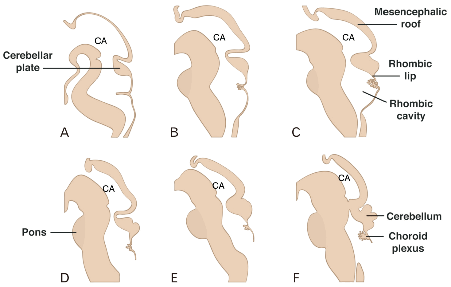
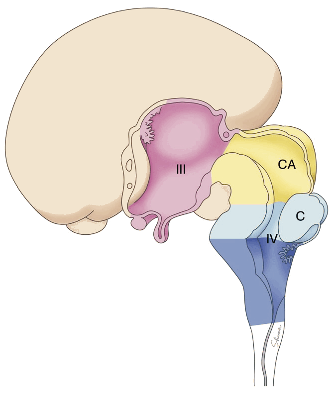
During the 3rd and 4th weeks of gestation, primary neurulation begins through a complex signaling and regulatory process that starts with an open neural plate and culminates in sections that proliferate, fold, and pinch off to form the neural tube [8, 9]. Following neural tube closure, three dilations develop on its cranial end. These embryonic brain vesicles become the prosencephalon (which later becomes the telencephalon and diencephalon), mesencephalon, and rhombencephalon (which later becomes the metencephalon and the myelencephalon). Cavities inside the vesicles later develop into the ventricles, producing CSF and facilitating its transport. The cavity of the rhombencephalon forms the fourth ventricle; the cavity of the diencephalon becomes the third ventricle; the cavities within the telencephalon develop into the lateral ventricles. The lateral and third ventricles communicate via the interventricular foramina of Monro, while the third and fourth ventricles communicate via the CA (Figs. 4, 5) [10, 11]. The ventricles reach adult size before the brain does, so the ventricular configuration is altered as the brain continues to develop [12]. During infancy, the length of the CA rapidly increases from 12.8 mm at birth to 18 mm after one year postpartum [3]. Both the lumen and the length of the CA vary among individuals.
Located at the entrance of the CA is the subcommissural organ (SCO). This structure is thought to contribute to the formation of the Reissner’s fiber (RF), a fibrous aggregation consisting mostly of SCO-secreted glycoproteins of a type of called SCO-spondins [13]. This enigmatic structure has been thought essential for proper formation of a straight body axis in vertebrates and to be significant in maintaining normal CSF circulation, because its absence or injury results in collapse of the CA leading to hydrocephalus [14-19]. However, while it is clear that the RF is important in the embryological development of the CA, more research is needed to elucidate its function and mechanism precisely (Table 1).
This table summarizes the main developmental milestones of the CA, from neural tube formation in early gestation to postnatal growth and the contribution of the SCO and RF to aqueductal patency.
Usually, CSF is produced primarily in the lateral ventricles by the choroid plexus, a collection of specialized ependymal cells and capillaries [20-22]. It flows unidirectionally from the lateral ventricles through the interventricular foramina to reach the third ventricle and then travels through the CA from the third to the fourth ventricles [23]. CSF in the fourth ventricle then flows through the foramina of Magendie and Luschka into the subarachnoid space [23, 24].
CSF flow is pulsatile and is highly dependent on the cardiac cycle. It begins approximately 200 ms after the early phase of ventricular depolarization and lasts for approximately 60% of the cardiac cycle [25]. This is followed by a small amount of backflow from the fourth to the third ventricle when the brain and intracranial vasculature contract during diastole [25, 26]. CSF flow is influenced by both respiration and cardiac pulsation, and can be characterized by measurements such as pulsatility, velocity, and amplitude [27]. Many studies have shown that CSF flow through the CA is largely driven by arterial pulsation, but the peak flow velocity varies with age [28-30]. One study showed that peak CSF flow velocity ranged from 3.8 to 24.7 mm/s (mean=11.3±5.8 mm/s) in the elderly, while another study found the amplitude of peak velocity in healthy adolescent controls to be 6.6±2.3 cm/s (mean=2.6±0.9 cm/s) [28, 30]. The difference highlights the decrease in flow rate in elderly populations. Similarly, a study comparing stroke volume (the total volume of CSF displaced throughout the cardiac cycle) between younger and older patients revealed a significantly greater stroke volume in younger patients, suggesting a loss of compliance with increasing age [31].
The CSF pulse in the CA can be used to evaluate intraparenchymal structural changes and as a marker for different pathologies, increased pulsatility being linked to conditions such as normal pressure hydrocephalus and multiple sclerosis [32]. Additionally, it has been theorized that a change in pulsatile flow dynamics within the CA contributes to the development of ventricular enlargement in patients with communicating hydrocephalus [33].
Vertebrates have a highly conserved ventricular system [34]. Animals such as sharks, frogs, iguanas, rabbits, cats, dogs, and primates all have a CA that connects CSF flow between the third and fourth ventricles [35]. The CA connects to the optic ventricles in non-mammalian animals such as sharks, frogs, and iguanas with a prominent optic lobe [35]. Besides anatomical and spatial differences in the CA across species, its size differs as expected. For instance, in the adult rat brain, the cone-shaped CA is approximately 3.5 mm long, while in humans, the tube-shaped channel is approximately 15 mm [22, 29].
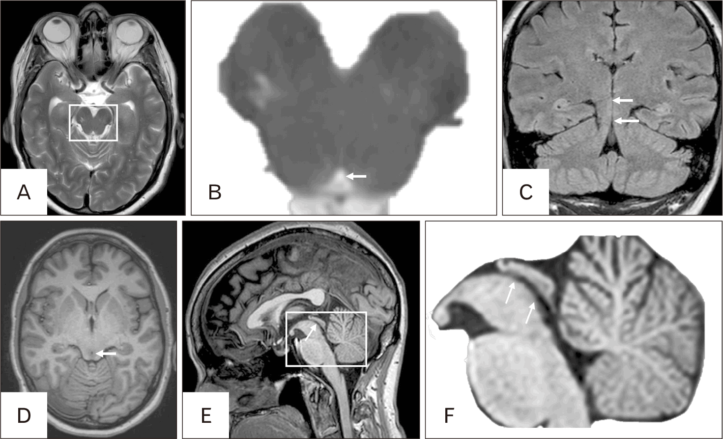
Imaging of the CA is best accomplished using magnetic resonance imaging (MRI) (Fig. 6). Conventional 1.5 T T1- and T2-weighted MRI provides excellent anatomical detail of aqueductal configuration and can identify structural stenosis or congenital anomalies [36]. Beyond static morphology, phase-contrast MRI has been particularly valuable for evaluating CSF dynamics, enabling quantification of velocity and pulsatile flow through the aqueduct to be quantified [37]. These measurements allow detection of subtle flow abnormalities to be detected that are not always evident on structural sequences alone, and have proven useful in distinguishing obstructive from communicating forms of hydrocephalus [37].
The aqueduct can also be assessed during early development with fetal ultrasound, where it serves as an easily identifiable landmark in the first trimester owing to its echogenic “box-like” appearance [38]. This characteristic appearance permits evaluation of midline integrity and ventricular connectivity to be evaluated, and measuring the distance between the CA to the anterior border of the occiput has been used to detect midbrain displacement in fetuses with spina bifida [38]. Such ultrasonographic findings can therefore act as an early marker of neural tube defects, supporting both diagnosis and prognostic evaluation.
Together, these imaging modalities not only define the structural anatomy of the CA but also provide insight into its functional physiology, establishing a critical role for MRI and ultrasound in both clinical practice and research.
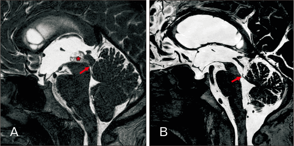
Aqueductal stenosis (AS) is a partial or complete blockage of the CA, which can lead to hydrocephalus (Fig. 7) [39]. A cross-sectional area as narrow as 0.1 mm2 has been reported in non-hydrocephalic patients, but how narrow the channel must be to result in hydrocephalus is unknown [3]. AS is thought to be responsible for up to 10% of adult hydrocephalus cases [40]. In AS, the third ventricle and the lateral ventricles enlarge, while the fourth ventricle typically remains normally sized [41]. The blockage can be congenital or acquired via such factors as infection, hemorrhage, or tumor (Fig. 7) [39]. Symptoms that have been described in AS include headache, diplopia, gait impairment, urinary incontinence, and mental deterioration [42]. However, symptoms and their severity vary widely among patients with hydrocephalus, including but not limited to headaches, vision problems, loss of urinary bladder continence, loss of coordination, seizures, lethargy, irritability, and muscle weakness [43].
Sylvian aqueduct syndrome is characterized by pupillary anomalies, impairment of conjugate upward gaze, nystagmus, and palsies of the extraocular muscles caused by a transtentorial pressure gradient through the CA [44, 45]. This unique constellation of symptoms indicates a periaqueductal location and can have a variety of causes, including encephalitis, trauma, multiple sclerosis, and congenital disease [46].
Many congenital malformations result in pathology concerning the CA, including but not limited to Chiari malformations, aqueductal webs, periaqueductal cysts, tectal plate tumors, Sylvian aqueduct syndrome, and aberrant vasculature nearby.
Congenital narrowing of the aqueduct can be due to intrinsic obstruction or extrinsic compression. One example of an intrinsic obstruction is an aqueductal web, a membrane composed of ependymal cells and fibrillary neuroglia between the third and fourth ventricles, which blocks the flow of CSF (Fig. 7) [40]. If this obstruction severely restricts the flow of CSF, procedures such as an endoscopic third ventriculostomy, endoscopic aqueductoplasty, or ventricular shunting can be performed (see Surgery section) [47].
Another rare vascular malformation that can result in obstruction of the CA is a cavernoma, a cluster of capillaries that form lesions in the central nervous system (CNS) [48, 49]. This pathology is relatively rare, occurring in approximately 0.4% of people; intraventricular cavernomas constitute approximately 2.5%–10.8% of all cerebral cavernomas [48, 49]. While an aqueductal location is extremely rare, there have been cases of cavernous angiomas appearing in the CA and causing hydrocephalus [48, 50]. In contrast to this are pathologies that typically cause increased pressure on the CA, such as periaqueductal cysts and developmental venous anomalies located adjacent to the CA. Because those structures are close to the CA, these conditions have been documented to cause non-communicating hydrocephalus [51, 52]. The channel can also become kinked and compressed owing to structural abnormalities or brain masses such as tumors [53].
Another anatomical variation that can cause partial obstruction is forking of the CA. Incomplete merging of the median fissure causes a septum to form, resulting in two separate canals (forking) [22, 54]. This septum is typically located in the inferior part of the CA and is composed of fibrillary neuroglia with clumps of ependymal cells and ependymal canaliculi [3]. The canals thus formed can either lead to the fourth ventricle or be blind pouches, which can result in obstruction [22]. In patients with hydrocephalus associated with a myelomeningocele, the CA can also undergo structural changes that shorten it and lead to forking at its dorsal part [22].
While forking can arise pathologically over time, a diagnosis of developmental-based forking requires histopathological evidence of ependymal-lined channels [54]. Fetal infections and teratogens can cause pathophysiological changes in the ciliated ependymal cells, leading to such conditions as atresia and obstructed CSF flow, and forking of the CA is thought to arise from a similar developmental anomaly [4, 54].
In addition to obstruction/compression of the CA, another pathological congenital condition that can lead to AS is a Chiari malformation. There are two types of Chiari malformation, Chiari I and Chiari II. A Chiari I malformation includes downward displacement of the cerebellar tonsils through the foramen magnum. A Chiari II malformation is characterized by displacement of the brainstem, cerebellar vermis, and fourth ventricle caudally [55, 56]. In a Chiari II malformation, both the fourth and third ventricles appear small, while the lateral ventricles often appear asymmetrically dilated [57]. Vimentin, an intermediate filament crucial for coordinating signaling pathways, is frequently overexpressed in ependymal cells in patients with Chiari II malformations and has been implicated in the pathogenesis of both Chiari types, though the exact mechanism remains unclear [58, 59]. Both types of Chiari malformation are associated with AS and can be a congenital cause of hydrocephalus [3]. AS can also be associated with other embryological conditions such as Dandy–Walker malformation (cerebellar vermian hypoplasia) and agenesis of the corpus callosum [41, 60].
Congenital AS can be sporadic or hereditary. Interestingly, this condition affects almost twice as many males as females, and an estimated 40% of patients have a genetic etiology, with relevant genes listed in Table 2 [61-63].
These genes have been implicated in congenital AS, but many more gene pathways can also be identified in congenital hydrocephalus [62, 63]. Notably, not only have mutations in L1CAM been known to cause X-linked stenosis of the CA with resultant hydrocephalus, but they can also manifest in a wide range of pathological conditions. This syndrome, dubbed L1 syndrome, can present with isolated agenesis of the corpus callosum, corticospinal tract hypoplasia, anterior cerebellar vermian hypoplasia, thalamic fusion, adducted thumbs, and spastic paraplegia [63]. A duplication on the short arm of chromosome X in the 22.33 region, deletions in the long arm of chromosome 9, and deletions in the long arm of chromosome 12 in the 22–23.1 region have also been identified [62].
Acquired malformations are generally thought to be a result from another process, such as trauma or illness. Gliosis is one such reactive processes in which glial cells (particularly astrocytes) proliferate and hypertrophy in response to a brain insult or trauma [64]. Histology of such gliosis can reveal disruption of the ependymal or subependymal layer. It may also be accompanied by an inflammatory process, leading to signs and symptoms of increased intracranial pressure [64-67]. In neonates, intraventricular hemorrhage and subsequent scarring is a major cause of posthemorrhagic AS. Reactive gliosis following perinatal hemorrhage has been described as a key mechanism, with astrocytic proliferation and disruption of the ependymal or subependymal layers leading to narrowing of the aqueduct and impaired CSF circulation [68]. Similarly, meningitis and other CNS infections can provoke an inflammatory reaction in ependymal lining. This inflammation often results in gliotic changes and structural alterations that compromise aqueductal patency, contributing to secondary hydrocephalus.
Neoplasias such as tectal gliomas have also been documented as an etiology for acquired AS, producing obstruction either by direct infiltration or through a mass effect on the aqueduct [61, 69]. Collectively, hemorrhage, infection, gliosis, trauma, and tumor growth are the principal acquired contributors to AS and highlight the clinical importance of early recognition and management of those risk factors.
For patients who present with symptomatic hydrocephalus, various procedures can be performed, such as a ventricular shunt, e.g., a ventriculoperitoneal shunt [70]. One such procedure, endoscopic cerebral aqueductoplasty, is used to widen the CA to facilitate CSF flow. This procedure can correct pathologies such as AS or a trapped fourth ventricle. There are many ways to perform this procedure, including the traditional approach beginning at the coronal suture and continuing through the lateral ventricle into the third ventricle to reach the CA, and a newer foramen magnum trans-fourth ventricle approach [71]. Irrespective of approach, this is considered a highly successful procedure, with postoperative cine MRI demonstrating normal CSF flow rates and resolution of symptoms [71, 72]. Stent placement is also recommended to prevent postoperative occlusion of the CA from scarring and reclosure, which could necessitate reoperation [73, 74]. This procedure is thought to restore CSF flow more physiologically than others and to entail less risk to major arteries in the brain, such as the basilar artery and its branches [73]. However, it also carries a risk to surrounding midbrain structures, which can result in conditions such as Parinaud syndrome and various nerve palsies (CNs III and IV) [73].
A related procedure is endoscopic transaqueductal stenting, in which a stent is placed in the CA without necessarily widening it. However, like all procedures in which a foreign body is introduced, there is a chance of infection or malfunction, causing most surgeons to opt for alternatives [75].
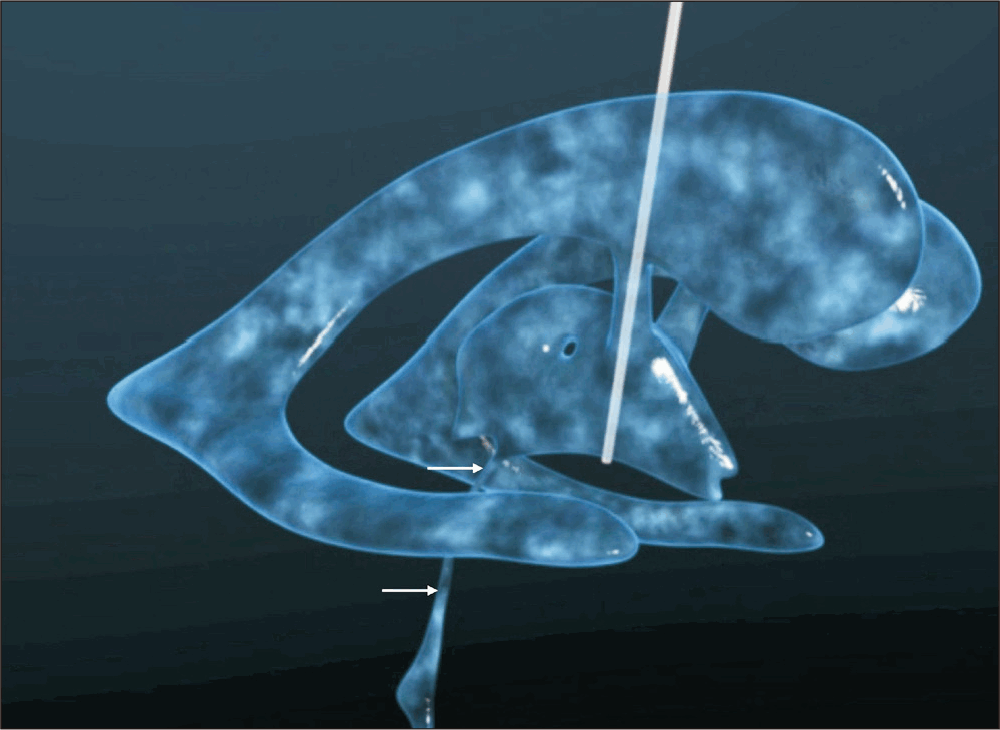
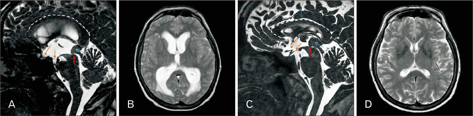
One such alternative is endoscopic third ventriculostomy, in which surgeons create a hole in the floor of the third ventricle using an endoscope (Figs. 8, 9) [76]. This procedure is performed by creating a burr hole around the coronal suture on a line that extends from the foramen of Monro to the interpeduncular cistern [77]. Once an adequate trajectory has been analyzed and the endoscope inserted, advancement through the third ventricle can be achieved. Risks for this procedure include hemorrhage (0.21% rate of basilar rupture), hematomas, infections, CSF leaks, and subdural hygromas [78, 79]. Neurological conditions such as gaze palsy, hemiparesis, and altered consciousness have also been documented postoperatively. This procedure is not an option for children under six months of age, as there has been a high documented rate of failure. One study reported an overall complication rate of 8.5% and a lower infection rate than with to shunt placement [78, 80]. It has been well documented as a successful treatment for obstructive hydrocephalus secondary to conditions such as AS, with a success rate of 80% at two-year follow-up [79].
The CA occupies a uniquely vulnerable and clinically consequential position within the ventricular system. Its minute caliber, variable embryological development, and surrounding neuroanatomical relationships make it highly susceptible to congenital and acquired pathologies. AS remains a major contributor to obstructive hydrocephalus, with genetic, structural, and inflammatory mechanisms often intersecting. Advances in imaging have improved diagnostic precision, making earlier detection of aqueductal abnormalities and a more nuanced assessment of CSF dynamics possible. Modern surgical interventions, particularly endoscopic techniques, have shifted the therapeutic landscape by offering more physiological and durable alternatives to shunting, though risks to adjacent midbrain structures remain significant.
Looking forward, further elucidation of genetic and molecular pathways, coupled with innovations in minimally invasive imaging and operative technologies, could enhance prevention, diagnosis, and treatment. A comprehensive understanding of the CA is indispensable not only for clinicians managing hydrocephalus but also for neuroscientists investigating the fundamental biology of CSF flow and brain development.
Notes
Author Contributions
Conceptualization: JI, CJB, JL, ASD, RST, GD, NK, NEO, NV, CAD, FB, AF. Data acquisition: CAD, JI, CJB, JL, ASD, RST. Data analysis or interpretation: JI, RST. Drafting of the manuscript: GD, JL. Critical revision of the manuscript: JI, CJB, ASD, RST. Approval of the final version of the manuscript: all authors.
References
1. García-González O, Mireles-Cano JN, Silva-Cerecedo P, Rueda-Franco F. 2014; The aqueduct. Childs Nerv Syst. 30:1147–9. DOI: 10.1007/s00381-014-2431-6. PMID: 24866226.
2. https://wellcomecollection.org/works/rv4evxja.
3. Spennato P, Cinalli G, Cianciulli E, d'Armiento M. Aqueductal stenosis and hydrocephalus. In: Cinalli G, Ozek M, Sainte-Rose C, editors. Pediatric Hydrocephalus. Springer; 2019. p.1-30.
10.1007/978-3-319-31889-9_19-1
4. Scott DE. Ventricular system. In: Ramachandran VS, editor. Encyclopedia of the Human Brain. Academic Press; 2002. p.661-85.
10.1016/B0-12-227210-2/00356-3
5. Zarei F, Zeinali-Rafsanjani B, Iranpour P, Sefidbakht S. 2022; A case of aqueductal obstruction by web with no sign except a headache. Radiol Case Rep. 17:3767–9. DOI: 10.1016/j.radcr.2022.07.036. PMID: 35965940. PMCID: PMC9364052.
6. Alvarez LA, Kato T, Llena JF, Hirano A. 1987; Ependymal foldings and other related ependymal structures in the cerebral aqueduct and fourth ventricle of man. Acta Anat (Basel). 129:305–9. DOI: 10.1159/000146420. PMID: 3630618.
7. Stanković G, Nikolić V, Puskas L, Filipović B, Stojsić-Dzunja L, Krivokuća D. 2005; A histological study of cerebral aqueduct. Med Pregl. 58:534–40. DOI: 10.2298/MPNS0512534S. PMID: 16673854.
8. Gilbert SF. Formation of the neural tube. In: Gilbert SF, editor. Developmental Biology. 6th ed. Sunderland; 2000.
9. https://www.ncbi.nlm.nih.gov/books/NBK542285/.
10. Sadler TW. Langman's medical embryology. 12th ed. Lippincott Williams & Wilkins; 2012.
11. Hochstetter F. [Beiträge zur Entwicklungsgeschichte des menschlichen Gehirns]. Deuticke, 1929.
12. Mortazavi MM, Adeeb N, Griessenauer CJ, Sheikh H, Shahidi S, Tubbs RI, Tubbs RS. 2014; The ventricular system of the brain: a comprehensive review of its history, anatomy, histology, embryology, and surgical considerations. Childs Nerv Syst. 30:19–35. DOI: 10.1007/s00381-013-2321-3. PMID: 24240520.
13. Zhang T, Ai D, Wei P, Xu Y, Bi Z, Ma F, Li F, Chen XJ, Zhang Z, Zou X, Guo Z, Zhao Y, Li JL, Ye M, Feng Z, Zhang X, Zheng L, Yu J, Li C, Tu T, Zeng H, Lei J, Zhang H, Hong T, Zhang L, Luo B, Li Z, Xing C, Jia C, Li L, Sun W, Ge WP. 2024; The subcommissural organ regulates brain development via secreted peptides. Nat Neurosci. 27:1103–15. DOI: 10.1038/s41593-024-01639-x. PMID: 38741020.
14. Aboitiz F, Montiel JF. 2021; The enigmatic Reissner's fiber and the origin of chordates. Front Neuroanat. 15:703835. DOI: 10.3389/fnana.2021.703835. PMID: 34248511. PMCID: PMC8261243.
15. Bagnat M, Gray RS. 2020; Development of a straight vertebrate body axis. Development. 147:dev175794. DOI: 10.1242/dev.175794. PMID: 33023886. PMCID: PMC7561478.
16. Driever W. 2018; Developmental biology: Reissner's fiber and straightening of the body axis. Curr Biol. 28:R833–5. DOI: 10.1016/j.cub.2018.05.080. PMID: 30086316.
17. Grondona JM, Hoyo-Becerra C, Visser R, Fernández-Llebrez P, López-Ávalos MD. 2012; The subcommissural organ and the development of the posterior commissure. Int Rev Cell Mol Biol. 296:63–137. DOI: 10.1016/B978-0-12-394307-1.00002-3. PMID: 22559938.
18. Pérez-Fígares JM, Jimenez AJ, Rodríguez EM. 2001; Subcommissural organ, cerebrospinal fluid circulation, and hydrocephalus. Microsc Res Tech. 52:591–607. DOI: 10.1002/1097-0029(20010301)52:5<591::AID-JEMT1043>3.0.CO;2-7. PMID: 11241868.
19. Vio K, Rodríguez S, Yulis CR, Oliver C, Rodríguez EM. 2008; The subcommissural organ of the rat secretes Reissner's fiber glycoproteins and CSF-soluble proteins reaching the internal and external CSF compartments. Cerebrospinal Fluid Res. 5:3. DOI: 10.1186/1743-8454-5-3. PMID: 18218138. PMCID: PMC2265671.
20. Daggubati LC, Akbari SHA. Aqueductal stenosis. In: Tubbs RS, Iwanaga J, Rizk EB, D'Antoni AV, Dumont AS, editors. Cerebrospinal Fluid and Subarachnoid Space: Pathology and Disorders: Volume 2. Elsevier; 2022. p.401-14.
10.1016/B978-0-12-819507-9.00022-3
21. https://www.ncbi.nlm.nih.gov/books/NBK538156/.
22. https://www.ncbi.nlm.nih.gov/books/NBK540988/.
23. https://www.ncbi.nlm.nih.gov/books/NBK519007/.
24. Wright BL, Lai JT, Sinclair AJ. 2012; Cerebrospinal fluid and lumbar puncture: a practical review. J Neurol. 259:1530–45. DOI: 10.1007/s00415-012-6413-x. PMID: 22278331.
25. Schroth G, Klose U. 1992; Cerebrospinal fluid flow. I. Physiology of cardiac-related pulsation. Neuroradiology. 35:1–9. DOI: 10.1007/BF00588270. PMID: 1289731.
26. Bradley WG. 2015; CSF flow in the brain in the context of normal pressure hydrocephalus. AJNR Am J Neuroradiol. 36:831–8. DOI: 10.3174/ajnr.A4124. PMID: 25355813. PMCID: PMC7990574.
27. Yamada S, Miyazaki M, Yamashita Y, Ouyang C, Yui M, Nakahashi M, Shimizu S, Aoki I, Morohoshi Y, McComb JG. 2013; Influence of respiration on cerebrospinal fluid movement using magnetic resonance spin labeling. Fluids Barriers CNS. 10:36. DOI: 10.1186/2045-8118-10-36. PMID: 24373186. PMCID: PMC3895787.
28. Bapuraj JR, Londy FJ, Delavari N, Maher CO, Garton HJ, Martin BA, Muraszko KM, Ibrahim el-SH, Quint DJ. 2016; Cerebrospinal fluid velocity amplitudes within the cerebral aqueduct in healthy children and patients with Chiari I malformation. J Magn Reson Imaging. 44:463–70. DOI: 10.1002/jmri.25160. PMID: 26788935.
29. Ding G, Li L, Chopp M, Zhang L, Li Q, Luo H, Wei M, Zhang J, Boyd E, Zhang Z, Jiang Q. 2024; Velocity of cerebrospinal fluid in the aqueduct measured by phase-contrast MRI in rat. NMR Biomed. 37:e5233. DOI: 10.1002/nbm.5233. PMID: 39104053.
30. Sharma AK, Gaikwad S, Gupta V, Garg A, Mishra NK. 2008; Measurement of peak CSF flow velocity at cerebral aqueduct, before and after lumbar CSF drainage, by use of phase-contrast MRI: utility in the management of idiopathic normal pressure hydrocephalus. Clin Neurol Neurosurg. 110:363–8. DOI: 10.1016/j.clineuro.2007.12.021. PMID: 18282655.
31. Stoquart-ElSankari S, Balédent O, Gondry-Jouet C, Makki M, Godefroy O, Meyer ME. 2007; Aging effects on cerebral blood and cerebrospinal fluid flows. J Cereb Blood Flow Metab. 27:1563–72. DOI: 10.1038/sj.jcbfm.9600462. PMID: 17311079.
32. Beggs CB, Shepherd SJ, Cecconi P, Lagana MM. 2019; Predicting the aqueductal cerebrospinal fluid pulse: a statistical approach. Appl Sci. 9:2131. DOI: 10.3390/app9102131.
33. Holmlund P, Qvarlander S, Malm J, Eklund A. 2019; Can pulsatile CSF flow across the cerebral aqueduct cause ventriculomegaly? A prospective study of patients with communicating hydrocephalus. Fluids Barriers CNS. 16:40. DOI: 10.1186/s12987-019-0159-0. PMID: 31865917. PMCID: PMC6927212.
34. Lowery LA, Sive H. 2009; Totally tubular: the mystery behind function and origin of the brain ventricular system. Bioessays. 31:446–58. DOI: 10.1002/bies.200800207. PMID: 19274662. PMCID: PMC3003255.
35. Kier EL, Kalra VB, Conlogue GJ, Filippi CG, Saluja S. 2022; Comparative anatomy of dissected optic lobes, optic ventricles, midbrain tectum, collicular ventricles, and aqueduct: evolutionary modifications as potential explanation for non-tumoral aqueductal anomalies in humans. Childs Nerv Syst. 38:287–94. DOI: 10.1007/s00381-021-05408-0. PMID: 34812897. PMCID: PMC8789712.
36. Kemp SS, Zimmerman RA, Bilaniuk LT, Hackney DB, Goldberg HI, Grossman RI. 1987; Magnetic resonance imaging of the cerebral aqueduct. Neuroradiology. 29:430–6. DOI: 10.1007/BF00341738. PMID: 3683831.
37. Stoquart-El Sankari S, Lehmann P, Gondry-Jouet C, Fichten A, Godefroy O, Meyer ME, Baledent O. 2009; Phase-contrast MR imaging support for the diagnosis of aqueductal stenosis. AJNR Am J Neuroradiol. 30:209–14. DOI: 10.3174/ajnr.A1308. PMID: 18832663. PMCID: PMC7051720.
38. Finn M, Sutton D, Atkinson S, Ransome K, Sujenthiran P, Ditcham V, Wakefield P, Meagher S. 2011; The aqueduct of Sylvius: a sonographic landmark for neural tube defects in the first trimester. Ultrasound Obstet Gynecol. 38:640–5. DOI: 10.1002/uog.10088. PMID: 22045650.
39. Moini J, Piran P. Brainstem. In: Moini J, Piran P, editors. Functional and Clinical Neuroanatomy. Academic Press; 2020. p.293-317.
10.1016/B978-0-12-817424-1.00009-4
40. Yilmaz TF, Sari L, Toprak H. 2024; Advanced cerebrospinal fluid flow MRI findings of aqueductal stenosis caused by web. J Clin Ultrasound. 52:201–7. DOI: 10.1002/jcu.23606. PMID: 38013602.
41. Shekdar K, Feygin T. 2011; Fetal neuroimaging. Neuroimaging Clin N Am. 21:677–703. ixDOI: 10.1016/j.nic.2011.05.010. PMID: 21807318.
42. Tisell M, Tullberg M, Hellström P, Blomsterwall E, Wikkelsø C. 2003; Neurological symptoms and signs in adult aqueductal stenosis. Acta Neurol Scand. 107:311–7. DOI: 10.1034/j.1600-0404.2003.00124.x. PMID: 12713521.
43. https://www.mayoclinic.org/diseases-conditions/hydrocephalus/symptoms-causes/syc-20373604.
44. Park JJ, Park BH, Lee HS, Lee JS. 2006; A Parkinsonism as a component of Sylvian aqueduct syndrome: effect of floating cranioplasty and distal catheter elongation. J Korean Neurosurg Soc. 39:438–42.
45. Swash M. 1974; Periaqueductal dysfunction (the Sylvian aqueduct syndrome): a sign of hydrocephalus? J Neurol Neurosurg Psychiatry. 37:21–6. DOI: 10.1136/jnnp.37.1.21. PMID: 4544322. PMCID: PMC494558.
46. Chattha AS, Delong GR. 1975; Sylvian aqueduct syndrome as a sign of acute obstructive hydrocephalus in children. J Neurol Neurosurg Psychiatry. 38:288–96. DOI: 10.1136/jnnp.38.3.288. PMID: 1151409. PMCID: PMC491910.
47. Masarwy A, Watterson C, Boyke A, Bonda D, Danielpour M. 2024; Obstructive hydrocephalus of uncommon etiology: case report and neurosurgical management of aqueductal web presenting in adolescence. Childs Nerv Syst. 40:4389–92. DOI: 10.1007/s00381-024-06645-9. PMID: 39482561. PMCID: PMC11579050.
48. Feletti A, Dimitriadis S, Pavesi G. 2017; Cavernous angioma of the cerebral aqueduct. World Neurosurg. 98:876.e15–876.e22. DOI: 10.1016/j.wneu.2016.11.096. PMID: 27890756.
49. https://www.uclahealth.org/medical-services/neurosurgery/conditions-treated/cavernous-angioma.
50. Ortega-Porcayo LA, Perdomo-Pantoja A, Palacios-Ortíz IJ, Cohen SC, González-Mosqueda JP, Gómez-Amador JL. 2017; Endoscopic management of a cavernous malformation on the floor of third ventricle and aqueduct of Sylvius: technical case report and review of the literature. Surg Neurol Int. 8:237. DOI: 10.4103/sni.sni_165_17. PMID: 29026673. PMCID: PMC5629841.
51. Hayashi T, Uchino A, Tokushige K, Baba Y. 2024; Aqueductal developmental venous anomaly causing obstructive hydrocephalus: a case report and review of the literature. Radiol Case Rep. 19:2024–30. DOI: 10.1016/j.radcr.2024.02.023. PMID: 38449483. PMCID: PMC10917638.
52. Roberti F, Magram G. 2005; Fiberscopic fenestration and prophylactic stenting of periaqueductal cysts: technical report on three cases. Minim Invasive Neurosurg. 48:202–6. DOI: 10.1055/s-2005-870929. PMID: 16172964.
53. Ohaegbulam C, Black P. Hydrocephalus. In: Ramachandran VS, editor. Encyclopedia of the Human Brain. Academic Press; 2002. p.517-24.
10.1016/B0-12-227210-2/00170-9
54. Feletti A, Fiorindi A, Longatti P. 2016; Split cerebral aqueduct: a neuroendoscopic illustration. Childs Nerv Syst. 32:199–203. DOI: 10.1007/s00381-015-2827-y. PMID: 26231565.
55. https://www.ncbi.nlm.nih.gov/books/NBK557498/.
56. https://www.ninds.nih.gov/health-information/disorders/chiari-malformations.
57. Naidich TP, Pudlowski RM, Naidich JB. 1980; Computed tomographic signs of the Chiari II malformation. III: Ventricles and cisterns. Radiology. 134:657–63. DOI: 10.1148/radiology.134.3.6965544. PMID: 6965544.
58. Ridge KM, Eriksson JE, Pekny M, Goldman RD. 2022; Roles of vimentin in health and disease. Genes Dev. 36:391–407. DOI: 10.1101/gad.349358.122. PMID: 35487686. PMCID: PMC9067405.
59. Sarnat HB. 2004; Regional ependymal upregulation of vimentin in Chiari II malformation, aqueductal stenosis, and hydromyelia. Pediatr Dev Pathol. 7:48–60. DOI: 10.1007/s10024-003-2127-5. PMID: 15255035.
60. https://www.ncbi.nlm.nih.gov/books/NBK538197/.
61. Shankar P, Zamora C, Castillo M. 2016; Congenital malformations of the brain and spine. Handb Clin Neurol. 136:1121–37. DOI: 10.1016/B978-0-444-53486-6.00058-2. PMID: 27430461.
62. Hale AT, Boudreau H, Devulapalli R, Duy PQ, Atchley TJ, Dewan MC, Goolam M, Fieggen G, Spader HL, Smith AA, Blount JP, Johnston JM, Rocque BG, Rozzelle CJ, Chong Z, Strahle JM, Schiff SJ, Kahle KT. 2024; The genetic basis of hydrocephalus: genes, pathways, mechanisms, and global impact. Fluids Barriers CNS. 21:24. DOI: 10.1186/s12987-024-00513-z. PMID: 38439105. PMCID: PMC10913327.
63. Kousi M, Katsanis N. 2016; The genetic basis of hydrocephalus. Annu Rev Neurosci. 39:409–35. DOI: 10.1146/annurev-neuro-070815-014023. PMID: 27145913.
64. Messam CA, Hou J, Janabi N, Monaco MC, Gravell M, Major EO. Glial cell types. In: Ramachandran VS, editor. Encyclopedia of the Human Brain. Academic Press; 2002. p.369-87.
10.1016/B0-12-227210-2/00152-7
65. Arienta C, Cappricci E, Baiguini M. 1986; Non tumoral aqueduct stenosis. Multiple microscopic subependymal glial protuberances demonstrated at histological examination. J Neurosurg Sci. 30:67–70.
66. O'Callaghan WJ, Robertson DM. 1966; Gliosis of the aqueduct. Can Med Assoc J. 95:1260–3.
67. Russell DS. Observations on the pathology of hydrocephalus. Her Majesty's Stationery Office; 1949.
68. Scavarda D, Bednarek N, Litre F, Koch C, Lena G, Morville P, Rousseaux P. 2003; Acquired aqueductal stenosis in preterm infants: an indication for neuroendoscopic third ventriculostomy. Childs Nerv Syst. 19:756–9. DOI: 10.1007/s00381-003-0805-2. PMID: 12908116.
69. Jandhyala NR, Negash B, Garcia MR, Allen J, Wisoff JH, Segal D. 2025; Tectal gliomas as a rare finding in presumed idiopathic congenital aqueductal stenosis: patient series. J Neurosurg Case Lessons. 10:CASE24695. DOI: 10.3171/CASE24695. PMID: 40720906. PMCID: PMC12305356.
70. https://www.ncbi.nlm.nih.gov/books/NBK459351/.
71. Sansone JM, Iskandar BJ. 2005; Endoscopic cerebral aqueductoplasty: a trans-fourth ventricle approach. J Neurosurg. 103:388–92. DOI: 10.3171/ped.2005.103.5.0388. PMID: 16302609.
72. Erşahin Y. 2007; Endoscopic aqueductoplasty. Childs Nerv Syst. 23:143–50. DOI: 10.1007/s00381-006-0227-z. PMID: 17053938.
73. Fleck S, Schroeder HWS. Aqueductoplasty and aqueductal stenting. In: Cinalli G, Özek M, Sainte-Rose C, editors. Pediatric Hydrocephalus. Springer; 2019. p.1483-504.
10.1007/978-3-319-27250-4_80
74. Fritsch MJ, Schroeder HW. 2013; Endoscopic aqueductoplasty and stenting. World Neurosurg. 79:S20.e15–8. DOI: 10.1016/j.wneu.2012.02.013. PMID: 22381819.
75. Prajsnar-Borak A, Schroeder HWS, Oertel J. 2024; Endoscopic transaqueductal stent placement for tumor-related aqueductal compression in pediatric patients: surgical consideration, technique, and results. Childs Nerv Syst. 40:395–405. DOI: 10.1007/s00381-023-06171-0. PMID: 37823956. PMCID: PMC10837227.
76. Demerdash A, Rocque BG, Johnston J, Rozzelle CJ, Yalcin B, Oskouian R, Delashaw J, Tubbs RS. 2017; Endoscopic third ventriculostomy: a historical review. Br J Neurosurg. 31:28–32. DOI: 10.1080/02688697.2016.1245848. PMID: 27774823. PMCID: PMC5922250.
77. Yadav YR, Parihar V, Pande S, Namdev H, Agarwal M. 2012; Endoscopic third ventriculostomy. J Neurosci Rural Pract. 3:163–73. DOI: 10.4103/0976-3147.98222. PMID: 22865970. PMCID: PMC3409989.
78. Bouras T, Sgouros S. 2011; Complications of endoscopic third ventriculostomy. J Neurosurg Pediatr. 7:643–9. DOI: 10.3171/2011.4.PEDS10503. PMID: 21631203.
79. Rahman MM, Khan SIMKN, Khan RA, Islam R, Sarker MH. 2021; Endoscopic third ventriculostomy in children: problems and surgical outcome: analysis of 34 cases. Chin Neurosurg J. 7:3. DOI: 10.1186/s41016-020-00228-8. PMID: 33407946. PMCID: PMC7786960.
80. Lu L, Chen H, Weng S, Xu Y. 2019; Endoscopic Third Ventriculostomy versus Ventriculoperitoneal Shunt in Patients with Obstructive Hydrocephalus: Meta-Analysis of Randomized Controlled Trials. World Neurosurg. 129:334–40. DOI: 10.1016/j.wneu.2019.04.255. PMID: 31136836.
81. Iwanaga J, Kim HJ, Akita K, Logan BM, Hutchings RT, Ottone N, Nonaka Y, Anand M, Burns D, Singh V, Peris-Celda M, Martinez-Soriano F, Apaydin N, Hanna A, Yoshioka N, Fernandez-Miranda J, Hur MS, Shoja MM, Saremi F, Reina F, Tabira Y, Carrera A, Spratt JD, Ho SY, Mori S, Komune N, Watanabe K, Prats-Galino A, De Andrés J, Reina MA, Abrahams PH, Anderson RH, Ibaragi S, Loukas M, Tubbs RS. 2025; Ethical Use of Cadaveric Images in Anatomical Textbooks, Atlases, and Journals: a consensus response from authors and editors. Clin Anat. 38:222–5. DOI: 10.1002/ca.24258. PMID: 39754475.
82. Iwanaga J, Obata K, Kato T, Samrid R, Lesser ER, Cardona JJ, Kikuchi K, Kim CY, Ono K, D'Antoni A, Komune N, Tabira Y, Hur MS, Kitagawa N, Kim HJ, Loukas M, Watanabe K, Tubbs RS. 2026; What is the appropriate sample size in human cadaveric studies? A quantitative review of 770 articles. Clin Anat. 39:14–9. DOI: 10.1002/ca.70007. PMID: 40692167. PMCID: PMC12747640.
83. Iwanaga J, Takeda S, Walocha JA, Ribatti D, Del Sol M, Ravi KS, Moryś J, Paulsen F, Singh V, Apaydin N, Kim HJ, Adds P, Duparc F, Loukas M, Tubbs RS. 2025; Guidelines Against Discrimination and Bias in Anatomical Research Papers (GDBARP): recommendations from anatomical journal editors. Clin Anat. 38:613–8. DOI: 10.1002/ca.24293. PMID: 40384060.
84. Iwanaga J, Kikuchi K, Tabuchi K, Dave M, Anbalagan M, Fukino K, Kitagawa N, Reina MA, Reina F, Carrera A, Nonaka T, Rajaram-Gilkes M, Khalil MK, Matsushita Y, Tubbs RS. 2024; A histology guide for performing human cadaveric studies: SQIP 2024 what to look for with light microscopy. Clin Anat. 37:555–62. DOI: 10.1002/ca.24156. PMID: 38469731.
Fig. 2
Gross anatomical and histological views of the cerebral aqueduct. (A) Sagittal section of the human brain showing the cerebral aqueduct (probe). (B) Axial cut through the midbrain illustrating the cerebral aqueduct (arrow). (C) Histological section (low power, H&E) through the aqueduct (asterisk). (D) Histological section of the lining of the aqueduct (100×, Masson’s trichrome). I, right lateral ventricle; asterisk, foramen of Monro; III, third ventricle; IV, fourth ventricle.
Fig. 3
Microscopic (A) and endoscopic (B) views of the floor of the third ventricle and the proximal opening of the cerebral aqueduct. TV Fl, third ventricular floor; Th, thalamus; CA, cerebral aqueduct; PoCom, posterior commissure; PiR, pineal recess.
Fig. 4
Sequential sagittal drawings of the brainstem and developing cerebral aqueduct (CA) in the fetus. Schematic drawing of the CA in the entire brain of developing embryo (after Hochstetter) [11]. (A) The cerebral aqueduct is large and the cerebellar plate small. (B) The brainstem thickens as well as the dorsal wall of the cerebral aqueduct. (C) The rhombic cavity is well formed and the cerebellar plate enlarges. (D) The cerebral aqueduct begins to decrease in size. (E) The cerebellar plate enlarges and the rhombic cavity is now fully open to the subarachnoid space. (F) The early cerebellum is clear, including choroid plexus of the fourth ventricle. The cerebral aqueduct continues to narrow in size.
Fig. 5
The diencephalon is shown in pink, the telencephalon in tan, the midbrain in yellow, the metencephalon in light blue, the myelencephalon in dark blue, and the spinal cord in white. CA, cerebral aqueduct; III, third ventricle; IV, fourth ventricle; C, cerebellum. ©2025 IT Clinical Anatomy Research Lab. Used with permission.
Fig. 6
MRI of the cerebral aqueduct. (A) T2-weighted axial. (B) Cerebral aqueduct (arrow) in rectangular area of (A). (C) Cerebral aqueduct (arrows) in T1-weighted coronal. (D) Cerebral aqueduct (arrow) in T1-weighted axial. (E) Cerebral aqueduct (arrow) in T1-weighted sagittal. (F) Cerebral aqueduct (arrows) in rectangular area of (E).
Fig. 7
Aqueductal stenosis (arrows) in sagittal T2-weighted image resulting from (A) a third-ventricle lesion (asterisk) and (B) an arachnoidal web.
Fig. 8
Drawing of the ventricular system undergoing a third ventriculostomy. The cerebral aqueduct is seen at the arrows.
Fig. 9
Pineal region lesion (blue arrows) causing aqueductal stenosis (red arrows). Preoperative sagittal T2-weighted MRI images (A, sagittal; B, axial) show triventricular hydrocephalus with upward displacement and thinning of the corpus callosum (dashed white line), and ballooning of the supraoptic and infundibular recesses of the third ventricle (orange arrows). Postoperative T2-weighted MRI images following an endoscopic third ventriculostomy show resolution of triventricular hydrocephalus, and normalization of both corpus callosum(dashed white line) and supraoptic and infundibular recesses of the third ventricle (orange arrows) (C, sagittal; D, axial).
Table 1
Chronological sequence of embryonic and early postnatal development of the cerebral aqueduct


PDF
Citation
Print


XML Download