



 PDF
 Citation
 Print


Introduction
Acute-on-chronic liver failure (ACLF) refers to a life-threatening syndrome that occurs on the basis of chronic liver disease and is characterized by acute decompensation of liver function and extrahepatic organ failure (1). While aetiologies vary globally, hepatitis B virus (HBV) infection remains the predominant trigger in the East, accounting for 82% of cases in China (2). ACLF patients commonly exhibit multiorgan dysfunction, including liver, kidneys, and respiratory system, with the 90-day mortality rate reaching 58% (3). Currently, owing to the lack of specific drugs, existing treatments for ACLF are limited to organ support and complication management (4). Although liver transplantation represents an effective treatment, its widespread application is severely limited by donor shortages (5). Consequently, mesenchymal stem cell (MSC)-based therapy has emerged as a promising alternative strategy because of its potential for tissue regeneration and immunomodulation (6-9).
MSCs are multipotent stem cells characterized by self-renewal capacity, robust proliferative potential, and multilineage differentiation ability. They exhibit a broad spectrum of biological functions, including anti-inflammatory effects, anti-apoptotic effects, immune modulation, growth factor secretion, and tissue repair (10). In recent years, MSC-based therapy for ACLF has been extensively investigated in both experimental models and clinical trials, yielding encouraging preliminary results (6, 7). However, bone marrow MSCs (BM-MSCs) derived from autologous chronic hepatitis B patients often display impaired proliferation capacity and decreased expression of growth factor receptors (11). Thus, umbilical cord MSCs (UC-MSCs) and allogeneic BM-MSCs are currently the main sources of MSCs in clinical trials (8, 9).
Despite the transition to healthier allogeneic cell sources, the clinical efficacy of MSCs transplantation remains inconsistent. The translation of MSC-based therapies from bench to bedside has been substantially hindered by two core obstacles: low targeting efficiency and uncertain long-term outcomes (12). Currently, the majority of research efforts focus on the mechanisms by which MSCs exert their therapeutic effect and how to enhance their homing and targeting efficiency. In contrast, the critical factors that dictate long-term therapeutic efficacy are often overlooked. A major barrier to sustainable clinical efficacy is the low survival rate of MSCs post-transplantation (13). This phenomenon is at least partially attributable to the pro-inflammatory and pro-oxidative host microenvironment that predominates at injured hepatic sites, which may render the targeted cells dysfunctional or senescent. In autoimmune diseases such as systemic lupus erythematosus (SLE), patient-derived serum can directly induce MSCs senescence, thereby accelerating disease progression (14). Similarly, serum from ACLF patients has been shown to impair the function of MSCs by altering cytokine secretion. High levels of interleukin-1β in ACLF patient serum can upregulate interleukin-8 expression and ultimately reduce the therapeutic efficacy of hUC-MSCs transplantation (15).
Oxidative stress plays a pivotal role in the pathophysiology of ACLF and acts as a primary driver of microenvironmental deterioration (16). It has been reported to trigger cell apoptosis and tissue damage through intrinsic pathways (17). Such a persistent oxidative state can lead to the accumulation of advanced oxidation protein products (AOPPs). AOPPs are dityrosine-containing and cross-linking protein products that are formed after the reaction of albumin with chlorinated oxidants during oxidative stress. They are involved in monocyte and neutrophil activation and induces respiratory bursts. AOPPs are important biomarkers of protein damage and can reflect the oxidative stress levels of patients with ACLF and serve as independent risk factors for HBV-related ACLF prognosis (18). A previous study demonstrated that AOPPs can inhibit the osteogenic differentiation and proliferation capacity of MSCs in a dose- and time-dependent manner in vitro (19). However, whether AOPPs serve as the key target in compromising MSCs therapeutic efficacy within the complex ACLF microenvironment remains unknown. Therefore, the present study aimed to investigate the biological alterations of MSCs exposed to ACLF patient serum. We evaluated changes in cell morphology, proliferation, migration, energy metabolism, and senescence, as well as how AOPPs affect these changes, providing a theoretical basis for the selection of suitable patients to optimize MSC-based therapy. These results help clarify the pivotal role of AOPPs in mediating MSCs senescence and functional impairment, and highlight that monitoring serum AOPPs levels may be a promising biomarker for determining the optimal time window for MSCs transplantation in patients with ACLF.
Materials and Methods
Ethical statement
This study was conducted in accordance with the Declaration of Helsinki. The approval was granted by the Ethics Committee of China-Japan Union Hospital of Jilin University (20221020027) and the Ethics Committee of Jilin Hepatobiliary Disease Hospital (2023-Reaearch Ethics Review-004). Written informed consent was obtained from all participants prior to inclusion in the study.
Human subjects and serum collection
To investigate the pathological effects of ACLF patient serum on MSCs, serum samples were harvested from 20 ACLF patients (mean age: 48.64±10.57 years; 18 males) and 20 healthy donors (mean age: 45.06±8.34 years; 14 males). Detailed clinical and biological profiles of the participants are summarized in Table 1. The inclusion and exclusion criteria for patients with ACLF were defined according to the Asian Pacific Association for the Study of the Liver (2). The ACLF grade was based on the criteria established by the Chinese Group on the Study of Severe Hepatitis B-ACLF (20, 21). Peripheral blood samples were collected in sterile vacuum tubes without anticoagulants. To obtain serum, the blood samples were centrifuged at 2,000 ×g for 15 minutes at 4℃. The supernatant was collected, heat-inactivated at 56℃ for 30 minutes to deactivate complement, and subsequently stored at −80℃ for further use.
Mesenchymal stem cell (MSCs) culture and pretreatment
Primary human bone marrow-derived MSCs were purchased from Hefei Wanwu Biotechnology Co., Ltd. (Cat. No. Delf-10702). The complete culture medium for the MSCs was formulated as Dulbecco’s modified Eagle’s medium (DMEM) (Cat. No. 11885084) supplemented with 10% foetal bovine serum (FBS) (Cat. No. a5256701) and 1% penicillin‒streptomycin (Cat. No. C0222; Beyotime). The cells were cultured in a humidified incubator maintained at 37℃ with 5% CO2, and the culture medium was changed every 3 to 4 days. To ensure cellular homogeneity and functional stability, MSCs at passages 3∼5 were selected for all subsequent assays.
Before the MSCs were cultured with serum from ACLF patients and healthy donors, the serum samples stored in a −80℃ freezer were thawed at room temperature and subjected to sterile filtration using a 0.22-μm membrane filter. When MSCs were grown to 60%∼70% confluence in 60-mm culture dishes, the medium was replaced with 5 mL of fresh culture medium containing 10% (v/v) ACLF patient or healthy donor serum, followed by 72 hours of continuous culture. A parallel negative control (NC) group in which MSCs were cultured in complete growth medium without additional serum supplementation was established. The three experimental cohorts were designated as the ACLF patient (AP) group, healthy control (HC) group, and NC group.
To quantitatively assess morphological changes, the cell aspect ratio and cell area were measured using Image J (NIH). For each group, at least 50 cells were randomly selected.
Hematoxylin and eosin staining
To visually assess the morphological transformation of MSCs, hematoxylin and eosin (HE) staining was performed. Briefly, MSCs were seeded onto sterile glass coverslips placed in 6-well plates and subjected to the indicated serum treatments for 72 hours. Prior to staining, the cells were washed with phosphate-buffered saline (PBS) and fixed with 4% paraformaldehyde (Cat. No. P0099-3 L; Beyotime) for 20 minutes at room temperature. The fixed cells were then stained with hematoxylin solution for 5 minutes to label the nuclei, followed by a brief rinse and counterstaining with eosin solution for 2 minutes to visualize the cytoplasm. After a final wash and dehydration, the cellular morphology was observed and photographed using a light microscope (Olympus).
Proliferation assay
To assess the impact of ACLF patient serum on MSCs proliferation, the Cell Counting Kit-8 (CCK-8) assay was performed according to the manufacturer’s instructions (Cat. No. C0037; Beyotime). Briefly, MSCs were seeded into 96-well plates (Corning) at a density of 5×10³ cells/well. Cell viability was measured at 1-, 3-, 5-, and 7-days post-seeding. At each time point, 10 μL of CCK-8 reagent was added to each well, and the plates were incubated at 37℃ for 2 hours. The optical density at 450 nm (OD450) was measured using a Multiskan microplate reader (Thermo Fisher Scientific). The proliferation inhibition rate was calculated as follows: Inhibition rate (%)=(ODNC−ODAP/HC)/ODNC)×100%.
Flow cytometric analysis cell surface phenotype
To characterize the surface marker profile of MSCs under different culture conditions, flow cytometric analysis was performed. Cells (5×10⁵ per sample) were harvested, washed with PBS, and incubated with the following fluorochrome-conjugated mouse anti-human monoclonal antibodies: anti-CD44-FITC (Cat. No. 103006; BioLegend), anti-CD105-FITC (Cat. No. 323208; BioLegend), anti-CD29-PE (Cat. No. 303003; BioLegend), and anti-CD34-PE (Cat. No. 343606; BioLegend). Isotype-matched immunoglobulins were used as negative controls. Data were acquired with a flow cytometer (FACS Calibur; BD Biosciences), and subsequently analysed to confirm the positive expression of mesenchymal markers and the negative expression of haematopoietic markers.
Apoptosis assay
To determine whether serum from patients with ACLF could induce apoptosis in MSCs, an Annexin V-FITC Apoptosis Kit (Cat. No. 556547; BD Biosciences) was used. After 72 hours of serum treatment, the MSCs were harvested, washed with cold PBS, and resuspended in binding buffer. The cells were stained with Annexin V-FITC and PI for 30 minutes at room temperature in the dark according to the manufacturer’s protocol. Apoptotic rates were analysed by flow cytometry immediately after staining.
Cell cycle analysis
To investigate cell cycle distribution, a cell cycle and apoptosis analysis kit (Cat. No. C1052; Beyotime) was used. After treatment, the MSCs (2×10⁵ cells/well) were harvested and fixed in 70% ice-cold ethanol at −20℃ overnight. The fixed cells were washed with PBS and incubated with PI staining solution containing RNase A for 30 minutes at room temperature in the dark. DNA content was measured by flow cytometry to determine the percentage of cells in G0/G1, S, and G2/M phases.
Scratch assay
To evaluate cell migration ability, a scratch assay was performed according to previously reported methods (22). MSCs treated with different media were seeded into 6-well plates and cultured for 24 hours. A linear scratch was created in the monolayer using a sterile 200-μL pipette tip. Detached cells were removed by washing with PBS, and the cells were cultured in medium supplemented with 1% serum to minimize proliferation effects. Images of the scratch area were captured at 0 hour and 24 hours using an inverted microscope. The wound closure area was quantified using Image J and the wound closure rate was calculated according to the following formula: Wound closure rate (%)=(S0−St)/S0×100%. where S0 denotes the initial scratch area at 0 hours and St represents the residual scratch area at 24 hours post-scratching.
Transwell assay
To further validate the migration potential, Transwell assays were conducted using an 8-μm pores-sized transwell chamber in 24-well plates (Corning) as previously reported (23). Treated MSCs (5×10⁴ cells) were resuspended in serum-free medium and seeded into the upper chamber. The lower chamber was filled with DMEM containing 20% FBS as a chemoattractant. After 24 hours of incubation, the cells were fixed with 4% paraformaldehyde for 15 minutes, after which nonmigrating cells on the upper surface were gently removed with a cotton swab. The migrated cells were then stained with 0.1% crystal violet (Cat. No. C0121; Beyotime). Migrated cells were imaged under a light microscope, and the number of cells in each field was then manually counted.
Seahorse assay
Mitochondrial respiration and glycolysis were assessed using a Seahorse XF24 Analyser (Seahorse Bioscience). The oxygen consumption rate (OCR) and extracellular acidification rate (ECAR) were measured using the Seahorse XF Cell Mito Stress Test Kit (Cat. No. 103015-100; Seahorse Bioscience) and the Glycolysis Stress Test Kit (Cat. No. 102194-100; Seahorse Bioscience), respectively, for the evaluation of oxidative phosphorylation (OXPHOS) activity and glycolytic function. Treated MSCs were seeded into XF24 cell culture microplates at a density of 1×10⁴ cells/well and incubated overnight at 37℃ with 5% CO2 to allow cell adhesion. To measure the OCR, the cells were incubated in XF Base Medium supplemented with glucose, pyruvate, and glutamine. The following inhibitors were sequentially injected to probe mitochondrial function: (1) oligomycin (1 μM) to inhibit adenosine triphosphate (ATP) synthase and measure ATP-linked respiration; (2) Carbonyl cyanide-4-(trifluoromethoxy)phenylhydrazone (FCCP, 1 μM) to uncouple the electron transport chain and measure maximal respiratory capacity; (3) rotenone/antimycin A (0.5 μM) to shut down mitochondrial respiration and determine nonmitochondrial oxygen consumption. To measure the ECAR, cells were incubated in glucose-free medium. The following reagents were sequentially injected: (1) glucose (10 mM) to induce glycolysis; (2) oligomycin (1 μM) to inhibit mitochondrial ATP production and drive maximal glycolysis; and (3) 2-deoxy-D-glucose (50 mM) to inhibit glycolysis and measure non-glycolytic acidification. All data were normalized according to cell number (1×10⁴ cells per cell).
Senescence-associated β-dalactosidase assay
To detect cellular senescence, senescence-associated β-dalactosidase (SA-β-gal) activity was measured using a staining kit (Cat. No. C0602; Beyotime) according to the manufacturer’s instructions. Pretreated MSCs were fixed with the provided fixative solution for 30 minutes at room temperature and then incubated with the freshly prepared staining solution overnight at 37℃ in a CO2-free incubator. Senescent cells, identified by the development of a blue colour, were visualized under a light microscope. The percentage of SA-β-gal-positive cells was calculated by dividing the number of blue-stained cells by the total number of cells in each field of view.
Western blot analysis
To quantify the expression of senescence-related proteins, Western blot analysis was performed following established protocols (14). Cells were harvested and lysed in radio immunoprecipitation assay lysis buffer (Cat. No. P0013B; Beyotime) supplemented with protease (Cat. No. P1005; Beyotime) and phosphatase inhibitors (Cat. No. P1005; Beyotime) on ice for 30 minutes. Protein concentration was determined using a BCA protein assay kit (Cat. No. P0012; Beyotime). Subsequently, equal amounts of proteins were separatedby 12.5% sodium dodecyl sulfate-polyacrylamide gel electrophoresis and transferred onto polyvinylidene fluoride membranes. The membranes were blocked with 5% non-fat milk for 1 hour at room temperature and subsequently incubated overnight at 4℃ with the following primary antibodies: anti-p53 (1:1,000; Cat. No. 2524S; Cell Signaling Technology), anti-p21 (1:1,000; Cat. No. SC-6246; Santa Cruz Biotechnology), anti-p16 (1:1,000; Cat. No. 80772S; Cell Signaling Technology), and anti-β-actin (1:5,000; Cat. No. 20536-1-AP; Proteintech). After being washed, the membranes were incubated with horseradish peroxidase-conjugated secondary antibodies (1:5,000; Cat. No. ZB-2301; ZSGB-BIO) for 1 hour at room temperature. Protein bands were visualized using an electrochemiluminescence detection kit (Cat. No. 180-5001; Tanon) and quantified using Image J software. β-actin was used as an internal reference to normalize protein expression levels.
Intracellular reactive oxygen species detection
Intracellular reactive oxygen species (ROS) levels were quantified using a reactive oxygen species assay kit (Cat. No. S0033S) with 2',7'-dichlorodihydrofluorescein diacetate (DCFH-DA) as the fluorescent probe. Following treatment, MSCs were incubated with 10 μM DCFH-DA in serum-free medium at 37℃ for 20 minutes in the dark. Cells were subsequently washed three times with PBS to remove extracellular probe, and the fluorescence intensity of 2',7'-dichlorofluorescein (DCF), which is correlated with intracellular ROS levels, was measured by flow cytometry with an excitation wavelength of 488 nm and an emission wavelength of 525 nm. ROS levels were expressed as the mean fluorescence intensity (MFI) of DCF.
Detection of advanced oxidation protein products
To evaluate serum oxidative stress levels, AOPPs concentrations were measured using an AOPPs Assay Kit (Cat. No. K7811; Immundiagnostik AG) according to the manufacturer’s instructions. Briefly, 125 μL of the serum samples were incubated with 25 μL of delipidation reagent at room temperature for 10 minutes to minimize interference. After centrifugation, 100 μL of delipidated serum was mixed with 400 μL of assay buffer. A total of 200 μL of standards, controls, or diluted samples was then added to the corresponding wells. The absorbance was measured at 340 nm using a microplate reader. AOPPs concentrations were calculated using a standard curve and are expressed as μmol/L.
Statistical analysis
Statistical analyses were performed using SPSS 22.0 software (IBM Corp.) and GraphPad Prism 8.0 (GraphPad Software). All experiments were performed independently at least three times (n=3), and the data are presented as mean±standard deviation. The normality of the data distribution was assessed using the Shapiro-Wilk test. For normally distributed data, comparisons between two groups were performed using the two-tailed Student’s t-test, and comparisons among multiple groups were performed using one-way analysis of variance (ANOVA) followed by Tukey’s post hoc test. For nonnormally distributed data, comparisons were performed using the Mann-Whitney U-test (for two groups) or the Kruskal-Wallis test followed by Dunn’s post hoc test (for multiple groups). Values were considered significantly different at p<0.05.
Results
ACLF patient serum microenvironment altered MSCs morphology without affecting phenotype
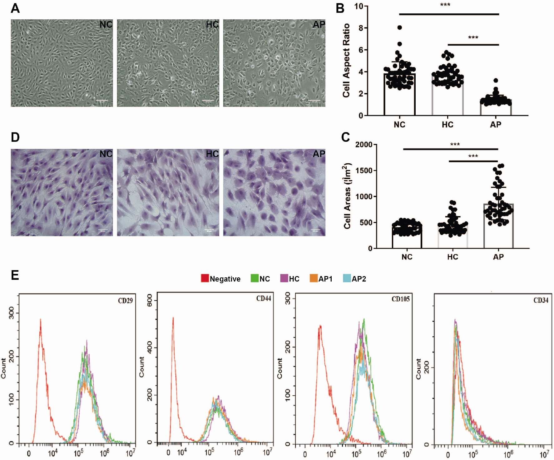
First, we examined the morphological changes in MSCs following 72 hours of pretreatment with different types of serum. Microscopic analysis revealed that MSCs in the AP group no longer exhibited a typical spindle-like morphology, as they did in the NC and HC groups. Instead, many cells showed a polygonal, elongated and flattened morphology, with increased intercellular gaps and the appearance of many coarse particles around their nuclei. In contrast, the MSCs in the HC group changed slightly, maintaining their characteristic spindle shape, smooth cell surfaces, and regular growth pattern (Fig. 1A). To validate the morphological changes quantitatively, we analysed the cell aspect ratio and the cell area of MSCs from the original micrographs (n=50 cells per group) using ImageJ software. Compared with those in the NC and HC groups, the aspect ratio and area of the MSCs in the AP group were significantly lower and greater, respectively (Fig. 1B, 1C). Additionally, HE staining confirmed the similar transformation of MSCs from spindle-shaped to star-shaped in the AP group (Fig. 1D). Second, we analysed the phenotype of MSCs treated with various serum samples and found no significant differences among the groups. The MSCs in all the groups were positive for CD29, CD44, and CD105 but negative for CD34 (Fig. 1E). These results suggested that the pathological microenvironment of ACLF patient serum might adversely impact MSCs morphology, while their phenotypes remain preserved.
ACLF patient serum microenvironment impaired the proliferation capacity of MSCs
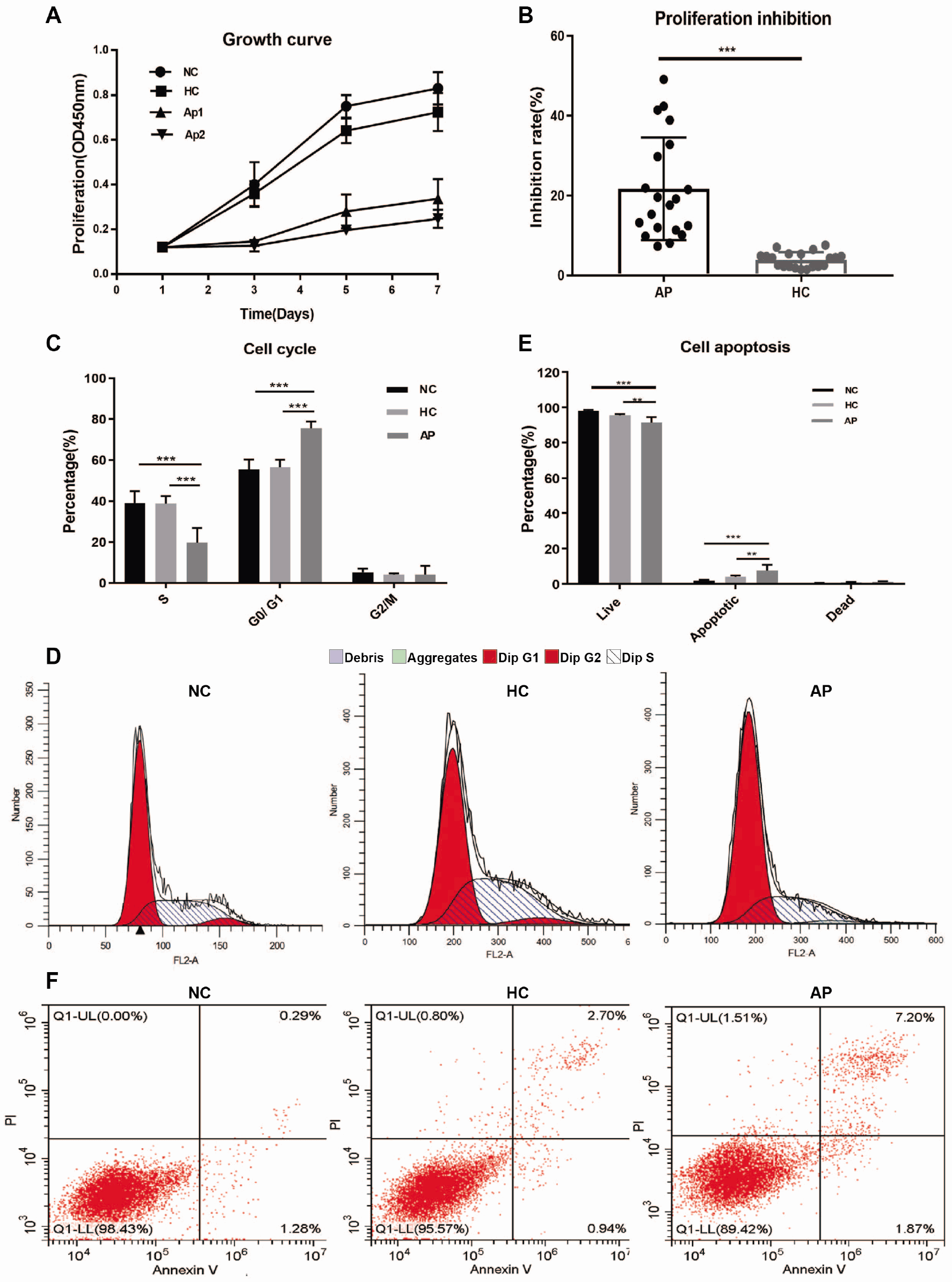
To determine whether the serum microenvironment of ACLF patients affected the proliferation, cell cycle progression, and apoptosis of MSCs, we first cultured MSCs for 7 days and measured their growth curves and proliferation inhibition rates after 72 hours of serum treatment. Compared with those in the NC and HC groups, the proliferation rate of MSCs in the AP group markedly decreased (Fig. 2A). The proliferation inhibition rates in the AP and HC groups were 21.70%±12.87% and 3.94%±1.90%, respectively (p<0.001) (Fig. 2B). The proliferation inhibition rates in the AP group was greater than those in the HC group, with the NC group serving as the negative control. These findings clearly revealed that the proliferation ability of MSCs was markedly inhibited under the ACLF patient serum microenvironment.
On the basis of these results, we further explored the dynamics of cell cycle progression. The percentage of cells in the G0/G1 phase was higher in the AP group (75.71%±3.23%) than in the NC group (55.89%±5.53%, p<0.001) and HC group (56.71%±3.53%, p<0.001). Correspondingly, the proportion of cells in the S phase was significantly lower in the AP group (19.90%±7.07%) compared with NC group (39.07%±5.85%, p<0.001) and HC group (28.29%±3.72%, p<0.001). In addition, the distribution of cells across the G0/G1 and S phases did not significantly differ between the HC and NC group (Fig. 2C, 2D). These results suggested that the impaired proliferation of MSCs in the AP group was likely attributed to cell cycle arrest in G0/G1 phase.
Finally, we evaluated the cell viability of MSCs and found that the percentage of apoptotic cells in the AP group (7.63%±3.11%) slightly increased compared with that in NC group (1.81%±0.45%, p<0.001) and HC group (3.89%±0.72%, p=0.008). Moreover, compared with NC group, the proportion of dead cells increased in both HC group (0.73%±0.25%, p<0.001) and AP group (0.99%±0.35%, p=0.008) (Fig. 2E, 2F).
ACLF patient serum microenvironment impaired the migration ability of MSCs
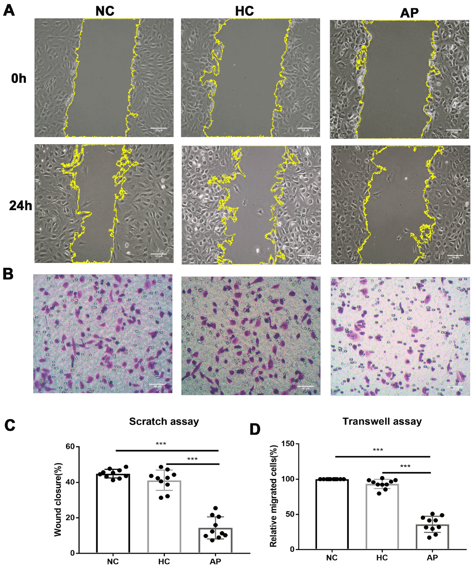
The clinical efficacy of MSC-based treatment is closely dependent on the ability of MSCs to migrate and integrate into injured tissues (24). To investigate the migration ability of MSCs pretreated with different types of serum for 72 hours, we performed scratch wound assay. The results showed that the wound closure rate in AP group (14.40%±6.17%) was significantly lower than that in the NC group (44.89%±2.41%, p<0.001) and HC group (42.00%±4.81%, p<0.001), while no significant difference was observed between the NC group and HC group (p=0.184) (Fig. 3A, 3C). Given the proliferation inhibition observed in Fig. 2, this delayed closure likely reflects a combined impairment of both migration and proliferation. To specifically isolate the migratory potential from proliferation effects, we further performed the Transwell assay. Consistently, the results of the transwell assay indicated that the percentage of migrated cells in the AP group (35.97%±11.37%) was significantly decreased compared with that in HC group (93.86%±6.68%, p<0.001) (Fig. 3B, 3D).
ACLF patient serum microenvironment impaired OXPHOS and glycolysis in MSCs
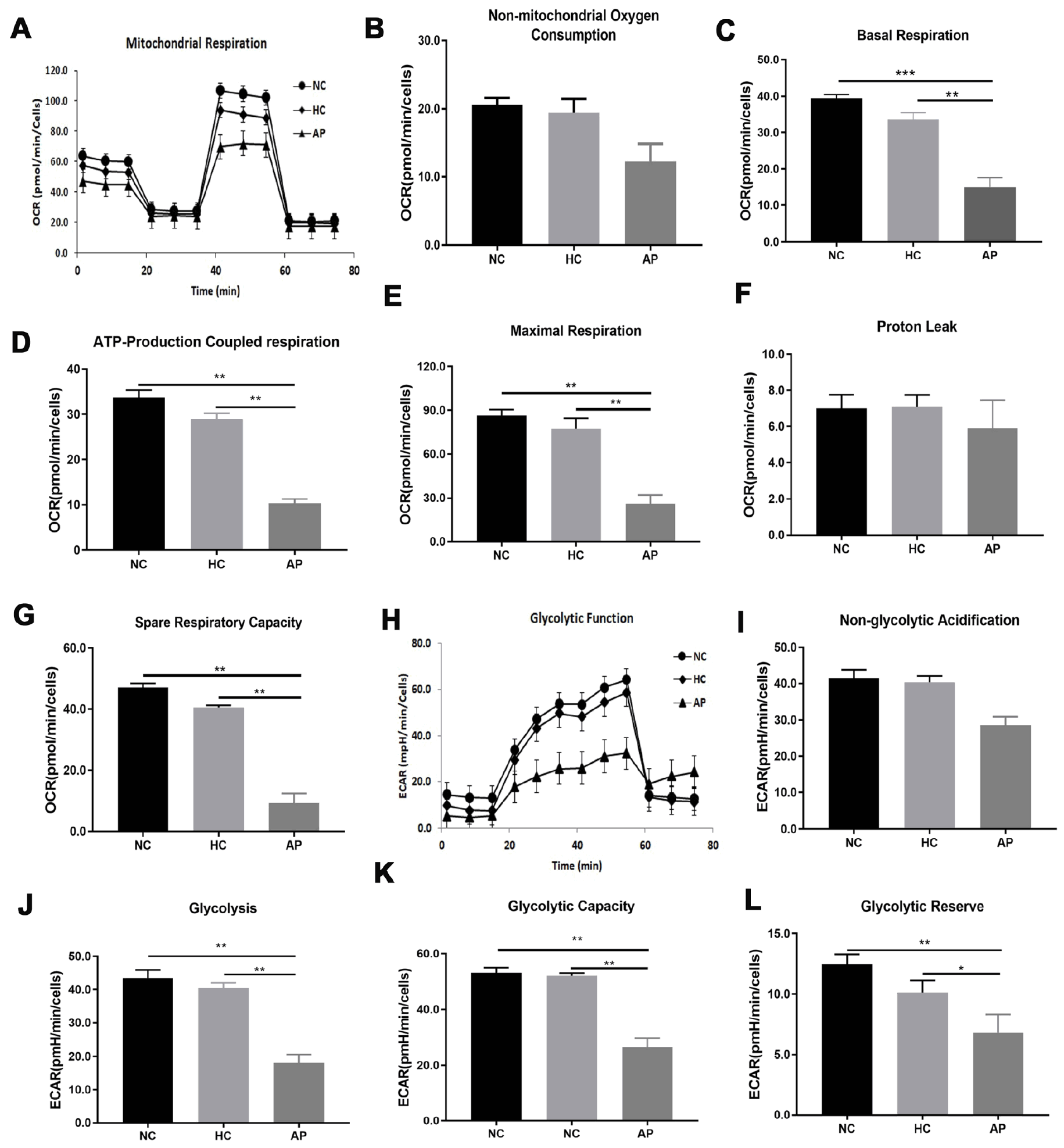
We next detected whether the energy metabolism of MSCs was affected in the ACLF patient serum microenvironment. Both OCR, a key indicator of mitochondrial OXPHOS function, and ECAR, which reflects cellular glycolytic activity, were measured under baseline conditions and various stress states designed to maximize energy production (Fig. 4A, 4H). Basal respiration level in the AP group were significantly lower than those in the NC and HC groups (Fig. 4C). Following the addition of the ATP synthase inhibitor oligomycin, ATP-production coupled respiration was markedly reduced (Fig. 4D). Mitochondrial spare respiratory capacity can be reflected by the increase in oxygen consumption induce by the uncoupling agent FCCP. The results showed that the maximal respiration of AP group was significantly lower than that in the NC and HC groups (Fig. 4E). Furthermore, the spare respiratory capacity, assessed after rotenone addition, was also significantly decreased in the AP group (Fig. 4G). In addition, although the difference was not statistically significant, non-mitochondrial oxygen consumption (Fig. 4B) and proton leak (Fig. 4F) were lower in the AP group than in the NC and HC groups.
To further clarify the metabolic changes in MSCs, we evaluated the ECAR across different groups. Our data revealed that the level of non-glycolytic acidification in the AP group was slightly lower than that in the NC and HC groups, although the difference was not significant (Fig. 4I). However, compared with those in the NC and HC groups, the overall glycolysis of MSCs in the AP group was significantly reduced (Fig. 4J). To assess their maximum glycolytic potential, oligomycin was added to inhibit mitochondrial ATP production and force reliance on glycolysis. Both the glycolytic capacity and the glycolytic reserve were lower in the AP group than in the NC and HC groups (Fig. 4K, 4L). In conclusion, these findings indicated that the ACLF patient serum microenvironment may impair the energy metabolic activity of MSCs.
ACLF patient serum microenvironment induced MSCs senescence
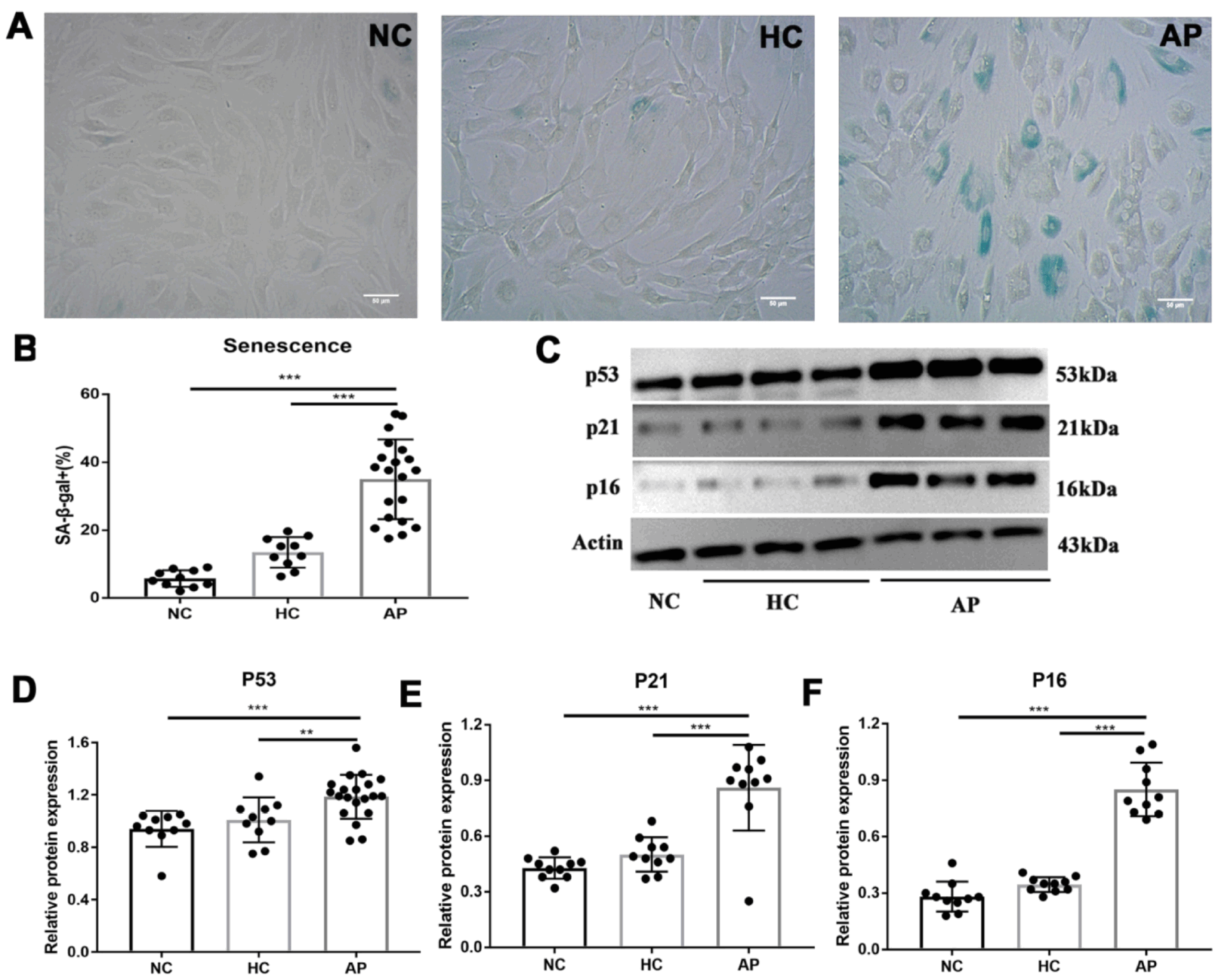
To determine the effect of ACLF serum microenvironment on MSCs senescence, we performed SA-β-gal staining. The results revealed that the proportion of SA-β-gal-positive cells in the AP group was markedly higher (34.98%±11.73%) than that in the NC group (5.73%±2.44%, p<0.001) and the HC group (13.44%±4.52%, p<0.001) (Fig. 5A, 5B). In addition, we examined the expression of the cell cycle arrest-related protein (p53, p21 and p16) using western blot analysis. The expression levels of all three proteins were significantly upregulated in the AP group compared with both the HC group (p<0.001) and NC group (p<0.001) (Fig. 5C-5E). Collectively, these findings demonstrated that the ACLF patient serum microenvironment dramatically accelerated MSCs senescence.
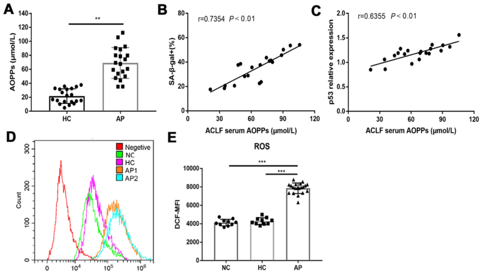
Oxidative stress plays a pivotal role in the pathophysiology of ACLF. AOPPs are generated during oxidative stress and are recognized as reliable biomarkers for evaluating the severity of oxidative stress in ACLF patients. Elevated serum AOPPs levels in ACLF patients have been documented in previous studies (25). Accumulating evidence also indicated that elevated AOPPs level mediated the senescence-promoting effect of serum derived from patients with ankylosing spondylitis (AS) (26). In the present study, we measured the serum AOPPs concentration in ACLF patients, and the results revealed that the AOPPs concentration was significantly greater in the AP group (69.09±11.73 μmol/L) than in the HC group (21.65±10.45 μmol/L, p=0.004) (Fig. 6A). Furthermore, correlation analyses were performed to explore the relationship between serum AOPPs levels and two senescence-related indicators, namely the percentage of SA-β-gal-positive cells and the expression level of p53 protein. The results demonstrated a strong positive correlation between serum AOPPs levels and both senescence markers (Fig. 6B, 6C). Collectively, these results indicated that elevated serum AOPPs may serve as a key mediator of the senescence-inducing effect of ACLF patient serum.
As previously reported, AOPPs can induce ROS production and play a vital role in triggering cellular senescence (27). Therefore, we detected the intracellular ROS levels in MSCs after 72 hours of pretreatment with different types of serum using the probe DCFH-DA by flow cytometry. The results revealed that MSCs exposed to ACLF patient serum exhibited significantly higher ROS levels, as evidenced by a higher MFI of DCF (Fig. 6D, 6E). Taken together, these findings support the hypothesis that serum AOPPs promote MSCs senescence by inducing excessive ROS accumulation.
Discussion
At present, liver transplantation remains the most effective treatment for liver failure. However, its widespread clinical application is severely limited by three key constraints: a critical shortage of donor organs, prohibitive treatment costs, and severe post-transplantation immune rejection. MSCs transplantation has been regarded as a potential alternative strategy, which can significantly improve the 24-week survival rate of patients with ACLF by enhancing liver function and reducing the incidence of infection (28). Nevertheless, accumulating evidence suggests that a subset of patients exhibit a suboptimal response to MSCs therapy (29). Notably, previous studies on MSCs transplantation have predominantly focused on its therapeutic efficacy and underlying mechanism, while systematic investigations into the causes of treatment failure or non-response remain scarce.
Oxidative stress serves as a cornerstone in the development and progression of ACLF. When the production of ROS overwhelms the antioxidant defence system, excessive ROS trigger oxidative stress-related pathological injury, including cellular apoptosis, tissue destruction, and deterioration of organ function (30-32). Given that MSCs are transplanted into this highly hostile oxidative environment, their therapeutic potential may be significantly impaired. To date, the specific impact of the ACLF-related oxidative stress environment on MSCs function has not been fully elucidated. In the present study, we investigated the underlying mechanisms by which serum derived from ACLF patients modulates MSCs biological activity, and provided empirical evidence to facilitate the optimization of clinical efficacy of MSCs transplantation for the treatment of ACLF patients.
To determine the impact of ACLF serum environment on the biological activity of MSCs in vitro, we conducted a series of experiments. Compared with the standard culture conditions, ACLF patient serum treatment significantly inhibited MSCs proliferation. However, despite this suppression, the MSCs phenotypes remained preserved. Subsequent analysis of cell cycle distribution and apoptotic status revealed marked G0/G1 phase arrest and increased apoptosis in the AP group. Taken together, these results demonstrate that the compromised proliferative capacity of MSCs after ACLF patient serum exposure is a consequence of cell cycle arrest and apoptosis.
Although MSCs are known to promote liver repair through mechanisms including hepatogenic differentiation, immunomodulation, and paracrine signalling, their therapeutic effect is often hindered by poor in vivo engraftment (33, 34). Our findings demonstrated that the ACLF patient serum microenvironment severely compromised the migratory capacity of MSCs. However, considering that this functional decline occurred alongside G0/G1 phase arrest and increased apoptosis, the extent to which cellular stresses independently contribute to impaired migratory capacity requires further clarification. To bridge this gap, future research should focus on whether ACLF patient serum microenvironment directly impairs MSCs migration and how it leads to impaired binding to injured hepatic sites in vivo. Collectively, we propose that the proinflammatory and oxidative microenvironment of ACLF impairs the fundamental biological activities of MSCs, thereby providing a mechanistic explanation for the suboptimal clinical responses observed in a subset of patients.
Although MSCs rely mainly on glycolysis to produce ATP, they also possess abundant, fully functional mitochondria, and their functions are orchestrated by the integration of both mitochondrial OXPHOS and glycolysis (35). The metabolic balance between OXPHOS and glycolysis determines MSCs fate; specifically, inhibiting OXPHOS has been shown to enhance stemness, whereas suppressing glycolysis drives the transition towards differentiation (36, 37). Our results demonstrated that ACLF patient serum microenvironment induced both mitochondrial OXPHOS and glycolytic dysfunction in MSCs. This mitochondrial impairment leads to a deficit in cellular ATP, which is known to trigger p53 activation and subsequent cell cycle arrest (38). Consistently, we observed significant upregulation of the cell cycle arrest-related proteins p53, p21, and p16 in the AP group. These molecular changes are consistent with the results of our functional cell cycle assays, collectively suggesting that energy deficiency is a key driver of MSCs dysfunction in the ACLF patient serum microenvironment.
MSCs senescence is generally classified into 4 types: replicative senescence, oncogene-induced senescence, stress-induced premature senescence (SIPS), and developmental senescence (39, 40). Given that elevated oxidative stress is a key driver of ACLF pathogenesis, we hypothesized that the ACLF patient serum microenvironment triggers SIPS in transplanted MSCs (41). Similar phenomena have been observed in other chronic inflammatory conditions, such as AS, SLE, and systemic sclerosis, where the pro-oxidative serum microenvironment significantly aggravates MSCs senescence. In line with these findings, our study demonstrated that MSCs in the AP groups exhibited a premature senescence phenotype. This phenotype was characterized by a significant increase in the proportion of SA-β-gal-positive cells and the marked upregulation of key cell cycle arrest proteins, including p53, p21, and p16. These results confirmed that ACLF patient serum could induce MSCs senescence. Furthermore, senescent MSCs exhibit a senescence-associated secretory phenotype and secrete proinflammatory cytokines and degradative enzymes (42). These features not only impair the single-cell function but also promote inflammation and senescence within the injured liver tissue, ultimately compromising the overall therapeutic efficacy of MSC-based interventions in ACLF patients.
To identify the specific factors driving this senescence, we focused on AOPPs. As novel protein markers of oxidative stress, AOPPs are generated under conditions of severe oxidative damage and are implicated in multiple oxidation-associated diseases, including chronic hepatitis and liver failure (3). Previous studies have reported significantly elevated AOPPs levels in the serum of ACLF patients, which are positively correlated with total bilirubin levels, Child–Pugh scores, and Model for End-Stage Liver Disease scores, and serve as independent risk factors for poor prognosis (43). Consistent with these findings, we confirmed that AOPPs levels in ACLF patient serum increase parallel with disease severity. Collectively, given this strong clinical correlation, it is reasonable to hypothesize that high concentrations of AOPPs in the ACLF patient serum microenvironment may contribute to the functional impairment of MSCs.
Evidence from other disease models supports this hypothesis, confirming that AOPPs exert multifaceted regulatory effects across diverse cell types. For example, AOPPs can suppress MSCs proliferation and osteogenic differentiation, induce endothelial cell senescence and apoptosis evasion, promote dose- and time-dependent intracellular ROS production and trigger ROS-dependent S-phase arrest in hepatocytes (44). Notably, research on AS has demonstrated that serum AOPPs levels are positively correlated with both the proportion of SA-β-gal-positive cells and the expression of p53, p21, and p16, suggesting that AOPPs serve as key mediators of serum-induced MSCs senescence in patients with AS (45). Similarly, our study revealed that AOPPs levels in ACLF patient serum were positively correlated with the percentage of SA-β-gal-positive cells and p53 expression levels in the AP group. These correlations strongly suggest that AOPPs are primary drivers of MSCs senescence in the ACLF patient serum microenvironment. Mechanistically, AOPPs facilitate cellular senescence by triggering nicotinamide adenine dinucleotide phosphate oxidase-dependent ROS generation (46, 47). To validate this in our model, we detected intracellular ROS levels using the probe DCFH-DA via flow cytometry. As expected, MSCs treated with ACLF serum exhibited significantly elevated ROS production, providing a mechanistic link between AOPPs accumulation and the observed senescent phenotype. Taken together, our findings demonstrated that AOPPs in ACLF patient serum not only acted as active pathogenic factors driving MSCs towards premature senescence via ROS induction but also hold potential as a promising biomarker for identifying the optimal time window for MSCs transplantation.
Accumulating evidence indicates that enhancing the antioxidant capacity of transplanted stem cells can improve their therapeutic effect in a variety of diseases (48). Thus, increasing the endogenous antioxidant capacity of MSCs is a promising strategy for optimizing transplantation outcomes in the pro-oxidative microenvironment of ACLF patients (49, 50). To date, several methods have been attempted. Small-molecule antioxidants and ROS scavengers (e.g., edaravone and N-acetyl-L-cysteine) have been investigated for their ability to enhance stress resistance in MSCs (51, 52). In addition, genetic engineering has provided a more targeted strategy. A transient plasmid-mediated pretreatment strategy targeting nuclear factor erythroid 2-related factor 2 and Dickkopf-1 has been developed to increase MSCs stress tolerance and enhance the regulatory effects of paracrine signalling on injured hepatocytes (53). Herein, our study provides a new perspective for protecting MSCs and helping them act properly by directly intervening in the pathological microenvironment. As reported by a recent network meta-analysis, the combination treatment of plasma exchange (PE) and MSCs transplantation was the most effective therapy for improving the prognosis of patients with HBV-ACLF (54). This phenomenon is likely attributed to the clearance of systemic toxins and oxidative products. Specifically, it has been confirmed that PE can significantly reduce AOPPs levels (44). Considering the pivotal role of AOPPs in mediating MSCs senescence and functional impairment, monitoring serum AOPPs levels may be a promising biomarker for evaluating the optimal time window for MSCs transplantation in patients with ACLF.
Despite these promising findings, our study has several limitations that should be acknowledged. First, our study provides a preliminary observation of how ACLF patient serum affects the core biological functions of MSCs; however, the precise molecular signalling pathways downstream of AOPP-induced oxidative stress remain to be fully elucidated. In addition, the current findings are limited by the relatively small sample size. Therefore, well-designed prospective studies involving larger, well-characterized cohorts are warranted to validate the impact of serum AOPPs levels on the therapeutic efficacy of MSCs transplantation. Such research will be essential for establishing AOPPs as robust biomarkers for patient stratification and treatment optimization in clinical practice for patients with ACLF.
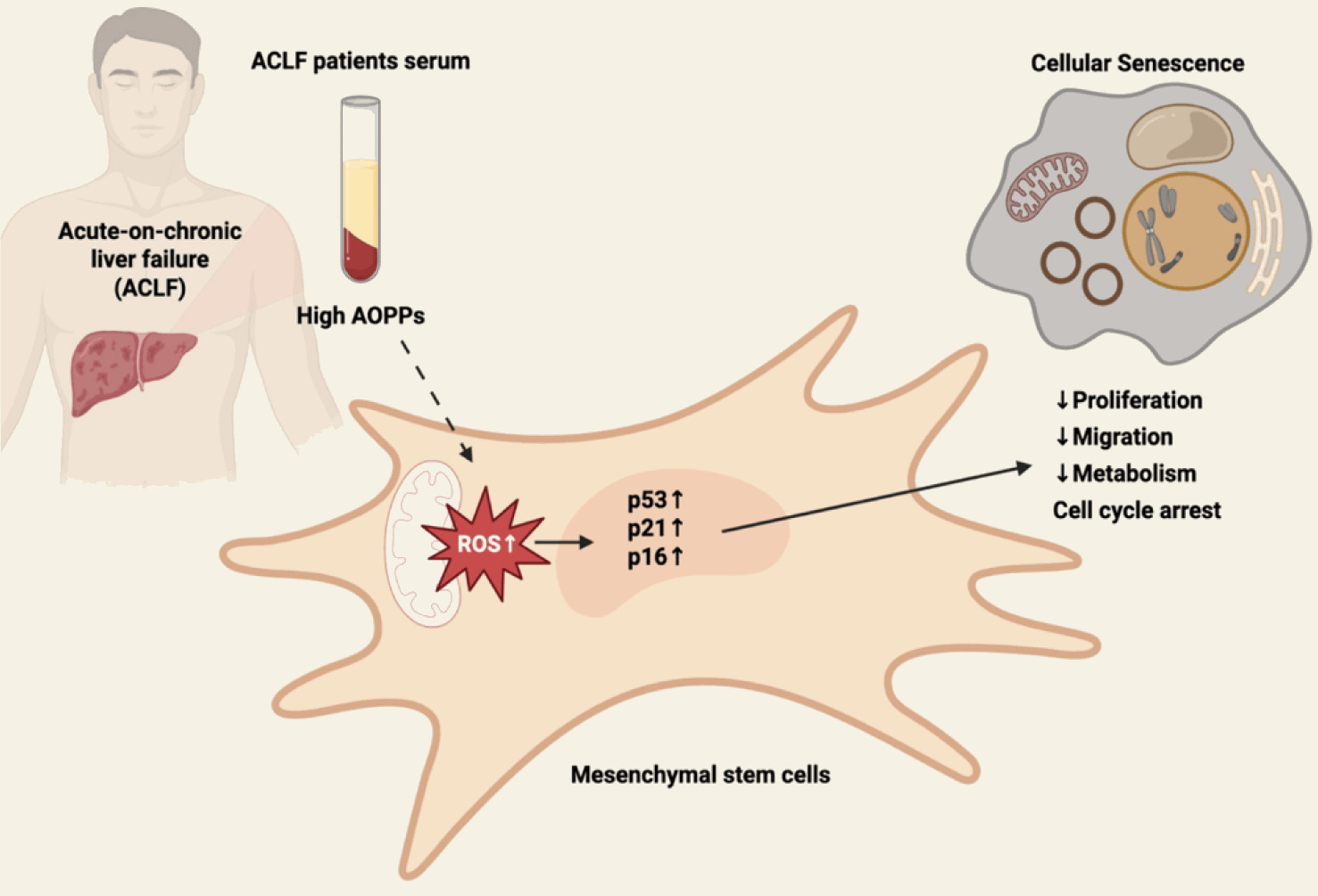
In summary, our results demonstrate that the ACLF serum microenvironment impairs the proliferative and migratory potential of MSCs, disrupts energy metabolism, and accelerates cellular senescence. These findings may explain why a subset of ACLF patients respond poorly to MSC-based therapy. As summarized in Fig. 7, we propose a working model in which elevated levels of AOPPs in ACLF serum may trigger mitochondrial dysfunction and ROS accumulation. This oxidative stress coincides with the activation of p53/p21/p16 signalling pathways, likely driving cell cycle arrest and senescence. Consequently, this cascade potentially impairs key MSCs functions, including proliferation, migration, and metabolism. Since stress-induced senescence severely limits the clinical translation of MSC-based therapy, further investigations are needed to evaluate serum AOPPs levels as a predictive biomarker for MSC-based therapy in ACLF patients.
 XML Download