



 PDF
 Citation
 Print


Introduction
Preterm premature rupture of membranes (PPROM) refers to the rupture of the fetal membranes and leakage of amniotic fluid prior to the onset of labor before 37 weeks of gestation (1). PPROM can lead to serious complications, including preterm delivery, intrauterine infections, and umbilical cord prolapse—each posing significant risks to fetal and neonatal health (2). Despite its clinical significance, the exact etiology of PPROM remains poorly understood, which hinders the development of effective preventive and therapeutic interventions. For PPROM occurring before 37 weeks of gestation, the standard clinical approach is expectant management aimed at prolonging pregnancy latency (3). Latency—defined as the interval between membrane rupture and delivery—tends to be longer when rupture occurs at earlier gestational ages. Prolonged latency is positively associated with improved neonatal outcomes, as gestational age at delivery is a key determinant of neonatal health (4).
Pre-viable PPROM, usually defined as PPROM before 24 weeks of gestation, occurs in less than 1% of pregnancies (5). For these patients, current management options include termination of pregnancy, expectant management, or aggressive intervention with prophylactic antibiotics, antenatal corticosteroids, tocolytic therapy, and/or amnioinfusion (1, 6). Several studies have been investigated in an effort to heal the ruptured fetal membranes, including cervical adapter, transcervical use of fibrin glue, collagen slurry under endoscopic guidance, and intra-amniotic injection of autologous platelets and cryoprecipitate (amniopatch technique) (7-10). However, none of these treatments are promising in terms of success rate, and therefore they are not yet included in standard management of pre-viable PPROM (11).
To extend latency and improve pregnancy outcomes, regenerative medicine, particularly stem cell-based therapy, has emerged as a promising alternative. Mesenchymal stem cells (MSCs) have been widely investigated for their regenerative, anti-inflammatory, and immunomodulatory properties. Recent advances in stem cell research have demonstrated therapeutic potential across a wide range of diseases, including neurodegenerative disorders (12), skeletal injuries (13), cardiovascular diseases (14), and diabetes (15). MSC-based therapies are also being evaluated for reproductive conditions such as ovarian dysfunction and uterine scarring, both of which are contributors to infertility (16-18). MSCs can be isolated from various tissues, including bone marrow, adipose tissue, umbilical cord, and placenta. Their multifunctional roles—immunomodulation, angiogenesis, inflammation regulation, and tissue regeneration—make them key players in regenerative medicine (19).
Furthermore, MSCs possess the capacity to differentiate into various cell types, including osteoblasts, adipocytes, chondrocytes, and stromal cells, depending on environmental cues in vivo or in vitro. Their innate ability to migrate to injury sites and modulate local immune responses has been explored in applications such as nerve regeneration (20) and wound healing. Importantly, much of their therapeutic benefit is attributed to paracrine signaling, which orchestrates the repair process by regulating the local microenvironment (21).
Given these regenerative capabilities, MSCs are considered a promising therapeutic option for PPROM, specifically the healing of fetal membranes injury. The present study aimed to identify the potential therapeutic agents for PPROM.
Materials and Methods
Ethical statement
Fetal membranes were collected from women after elective cesarean section at term gestation (n=30), with informed consent, and transported aseptically to the laboratory. An umbilical cord, used as the source of Wharton’s jelly-derived mesenchymal stem cells (WJ-MSCs), was similarly obtained with maternal consent. All procedures were conducted in accordance with protocols approved by the Institutional Review Board (IRB) of Samsung Medical Center. All participants provided written informed consent. Ethical approvals were granted by the IRB for both fetal membranes collection (Approval No. 2021-07-055) and WJ-MSCs isolation (Approval No. 2016-07-102-037).
Wharton’s jelly-derived mesenchymal stem cells (WJ-MSCs)
WJ-MSCs used in this study were manufactured under Good Manufacturing Practice (GMP)-compliant protocol by ENCell Co., Ltd. and were approved by the Korean Ministry of Food and Drug Safety as an investigational cell therapy product, ensuring clinical-grade quality and safety. The cells met the minimal criteria for MSCs as defined by the International Society for Cellular Therapy (22), exhibiting positive expression of CD44, CD73, CD90, CD105, and CD166 (≥95%) and negative expression of CD11b, CD14, CD19, CD34, CD45, and HLA-DR (≤0.5%). These immunophenotypic characteristics were verified by flow-cytometric analysis as part of GMP quality-control testing.
WJ-MSCs were isolated from a human umbilical cord from one donor and cultured as previously described (23). Briefly, mononuclear cells were isolated from Wharton’s jelly, part of the umbilical cord tissue, seeded in culture flasks, and expanded for approximately 20 days with three serial passages to obtain a sufficient cell population. WJ-MSCs at passage 3 was cryopreserved in liquid nitrogen until use. Before each experiment, frozen cells were rapidly thawed in a 37℃ water bath for 3∼5 minutes, washed, and resuspended at the desired cell concentration in the appropriate culture medium.
Cell culture and wound healing assay
Human amniotic epithelial cells (hAEpi; ScienCell-#7110) were cultured in flasks coated with poly-L-lysine (2 μg/cm2) using Epithelial Cell Medium (EpiCGS, Cat. #4101) supplemented with 2% fetal bovine serum (EpiCGS, Cat. #0010) and 1% Epithelial Cell Growth Supplement (EpiCGS, Cat. #4152) at 37℃ in 5% CO2, following the manufacturer’s instruction.
The effect of WJ-MSCs on the proliferation of amniotic epithelial cells was evaluated using a wound healing assay. hAEpi were seeded at a density of 100,000 cells/well in 6-well plates and cultured in a CO2 incubator. Once the cells reached 90% confluence, a linear scratch was made at the center of each well using a sterile pipette tip. After washing with Dulbecco’s phosphate buffered saline (DPBS; Biowest), the following conditions were tested: (i) +WJ-MSC, where WJ-MSCs were seeded at 100,000 cells/well in Transwell inserts (0.4 μm pore size; BD Biosciences) and co-cultured with hAEpi, and (ii) CM, in which conditioned medium collected from WJ-MSCs was treated to hAEpi. Cultures were maintained for up to 24 hours, and wound closure was assessed microscopically at 0, 6, 12, and 24 hours. The extent of wound healing was quantified by measuring the repopulated wound area using Image J (Version 1.53e; NIH).
RNA isolation and reverse transcription-quantitative PCR
Total RNA was isolated from hAEpi cells using the AccuPrepⓇ Universal RNA Extraction Kit (Bioneer, Cat. #K-3140) according to the manufacturer’s instructions. cDNA was synthesized from 1 μg of RNA using the ABScript Neo RT Master Mix for qPCR with gDNA Remover (ABclonal, Cat. #RK20433-100). Quantitative PCR was performed with SYBR Green Master Mix (Applied Biosystems; Thermo Fisher Scientific, Inc.) on the QuantStudioTM 6 Pro Real-time PCR System (Applied Biosystems; Thermo Fisher Scientific, Inc.). The thermal cycling steps were as follows: pre-denaturation at 50℃ for 2 minutes and 95℃ for 10 minutes; followed by 40 amplification cycles of 95℃ for 30 seconds, 58℃ for 30 seconds, and 72℃ for 30 seconds; and a melting curve stage at 95℃ for 15 seconds, 60℃ for 1 minute, and 95℃ for 15 seconds. GAPDH was used as the internal control, and relative expression levels were calculated using the 2−ΔΔCT method. The primer sequences used were as follows: GAPDH: F, 5'-GAAGGTGAAGGTCGGAGT-3'; R, 5'-TGGCAACAATATCC ACTTTACCA-3', Ki67: F, 5'-CGCAGTTGACAAGCACATGT-3'; R, 5'-GTGGGGAGCAGAGG TTCTTC-3', MMP1: F, 5'-ATCGGGGCTTTGATGTACCC-3'; R, 5'-GGCTGGACAGGATTTTGGG A-3'.
Ex vivo model of preterm premature rupture of membranes
Fetal membranes were washed several times with DPBS (Biowest) until all visible blood clots were removed. Residual clots and blood vessels were manually excised. The membranes were then trimmed to a cross-sectional area not exceeding 25 cm2, and an incision measuring approximately 6∼7 mm in length was made using a surgical scalpel (Paragon) to simulate the membrane rupture observed in PPROM. This incision range was selected based on preliminary experiments, which confirmed that it consistently induced measurable leakage suitable for evaluating the therapeutic effect of WJ-MSCs. The injured membranes were transferred to 60 mm culture dishes containing 5 mL of medium, and WJ-MSCs were directly applied to the incision site at three doses (2.5×10⁵, 5.0×10⁵, and 1.0×10⁶ cells). The tissues were then incubated at 37℃ in 5% CO2 for 24 hours before subsequent analysis.
Leak test for evaluating WJ-MSCs’ effectiveness
Sixty milliliters of 0.01% Trypan blue solution were added to a Square PETG Bottle (60 mL, Nalgene). Instead of sealing the bottle with a cap, the opening was covered with fetal membrane tissue that had been cultured for 24 hours, and the membrane was secured tightly using a rubber band.
To evaluate leakage, the prepared container was gently inverted onto an absorbent pad, ensuring direct contact between the fetal membrane and the pad. This allowed the Trypan blue solution to pass through any damaged regions and be absorbed by the pad. After 1 minute, the container was removed, and an image of the stained area was captured using a camera positioned at a fixed height. The extent of the leakage was quantified by measuring the stained area on the absorbent pad using Image J software (Version 1.53e, NIH).
Histological analysis
Fetal membrane tissue samples were fixed overnight at 4℃ in 4% paraformaldehyde (Biosesang), then dehydrated and embedded in paraffin. The paraffin-embedded tissue blocks were sectioned into 4 μm-thick sections using a microtome. Sections were deparaffinized using Histoclear (National Diagnostics) for a total of 9 minutes (3 minutes×3) with a Leica ST5010 Autostainer XL (Leica Biosystems), followed by rehydration through graded ethanol (100% and 95%).
The sections were stained with hematoxylin for 8 minutes, rinsed with PBS or tap water, and briefly immersed in 0.1% hydrochloric acid for 1 second. After additional washing, eosin staining was performed for 40 seconds at room temperature (25℃). Slides were then dehydrated using 95% and 100% ethanol and mounted. Following air-drying in a fume hood, stained slides were scanned using a high-resolution digital slide scanner (ScanScope Aperio AT2; Leica Biosystems).
Statistical analysis
All experimental data were analyzed using paired t-tests, one-way or two-way ANOVA, followed by post hoc multiple comparison tests (Dunn’s or Šídák’s, respectively), using GraphPad Prism software (version 8.0.1; GraphPad). Statistical significance was indicated as follows: p<0.05, p<0.01, and p<0.001. Data are presented as the mean±standard deviation.
Results
WJ-MSCs promoted the proliferation of amniotic epithelial cells
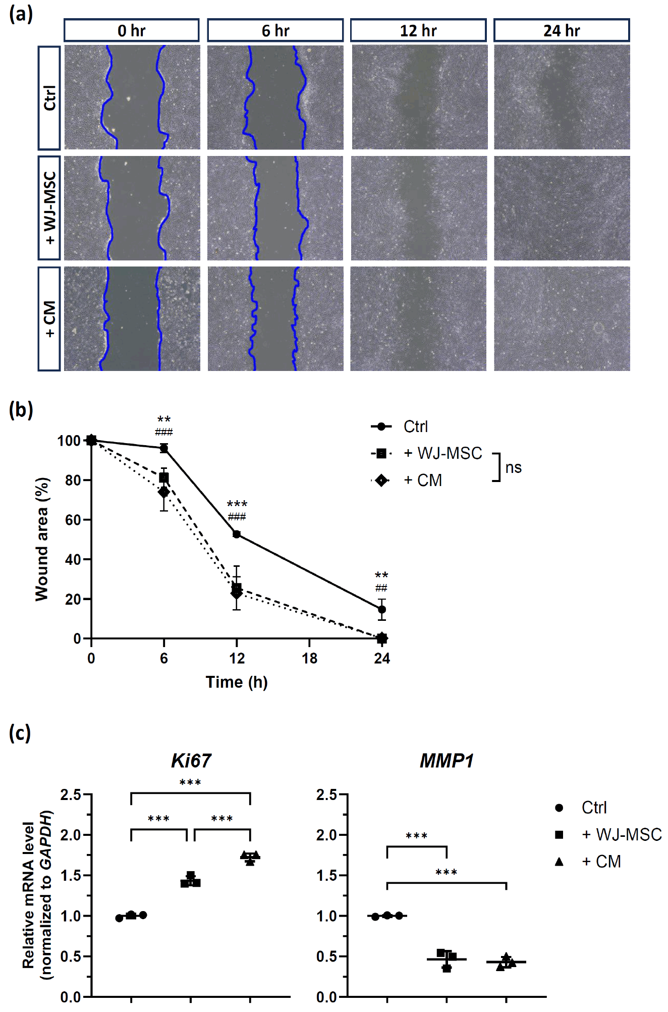
To investigate the regenerative potential of WJ-MSCs on injured hAEpi, a wound healing assay was performed (Fig. 1). After inducing a linear scratch on the hAEpi monolayer to simulate fetal membrane injury (an in vitro PPROM model), wound closure was assessed under three conditions (Fig. 1a, Supplementary Fig. S1): in vitro PPROM model (Ctrl), in vitro PPROM model co-cultured with WJ-MSCs (+WJ-MSC), and in vitro PPROM model treated with conditioned medium derived from WJ-MSCs (+CM).
We quantitatively measured the wound closure area (Fig. 1b). At the initial time point (0 hour), all groups started with the same wound area (100.0%±0.0%). At 6 hours, wound closure was modest in the control group (96.2%±2.3%), whereas both treatment groups showed accelerated closure. Compared to control, the wound area of +WJ-MSCs group increased to 81.2%±4.8% (p=0.006), and the +CM group exhibited an even greater reduction (74.1%±9.5%, p<0.001). At 12 hours, both treatment groups demonstrated greater improvements (+WJ-MSC: 25.6%±11.1%, p<0.001; +CM: 22.8%±8.4%, p<0.001) compared with the control group (52.7%±1.3%). At 24 hours, nearly complete closure was observed in +WJ-MSCs group (0.0%±0.0%, p=0.007) and +CM groups (0.2%±0.4%, p=0.008), while the control group still displayed a residual wound area (14.7%±5.3%). Collectively, these results demonstrate that WJ-MSCs significantly accelerate wound closure in hAEpi monolayers. Moreover, the comparable efficacy between WJ-MSCs and conditioned medium treatment suggests that the repair-promoting effect is largely mediated by soluble paracrine factors secreted by WJ-MSCs.
Next, the expressions of Ki67 and MMP1 were measured to assess transcriptomic changes within epithelial cells, followed by WJ-MSCs and their paracrine factors. Ki67 expression was significantly upregulated in both +WJ-MSCs and +CM groups compared with control (p<0.001 for both), with significant difference between the two treatment groups (p<0.001). In contrast, MMP1 expression was significantly downregulated in both treatment groups relative to control (p<0.001 for both), with no significant difference between the two treatment groups.
WJ-MSCs regenerated the damaged site of human fetal membranes
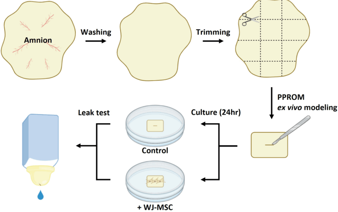
The wound healing assay demonstrated the recovery potential of WJ-MSCs on amniotic injury through paracrine effects. Next, we developed an ex vivo PPROM model to further evaluate the recovery efficacy of WJ-MSCs (Fig. 2). Fetal membranes from several donors (n=18) were incised, and WJ-MSCs were directly applied to the lesion sites, and the tissues were cultured for 24 hours. To assess membrane integrity and the therapeutic effects of WJ-MSCs, the extents of leakage were assessed (Fig. 3).
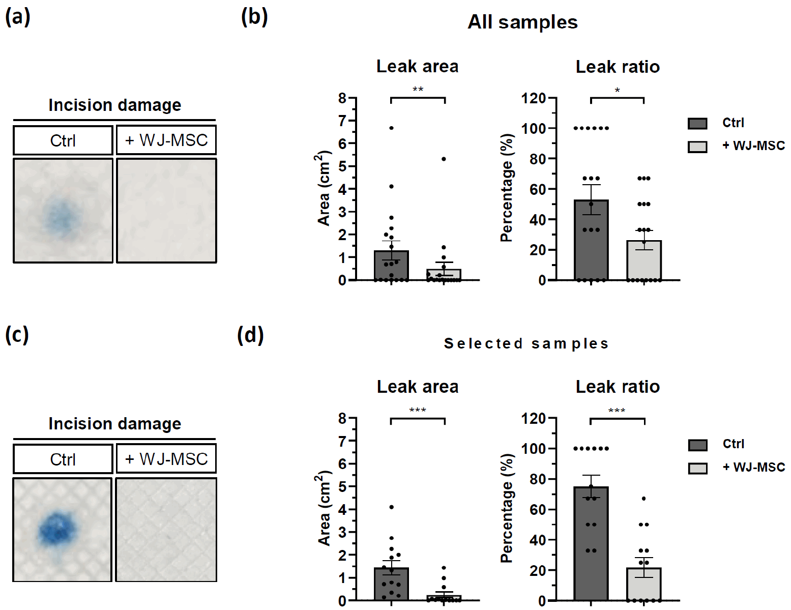
Representative images show demonstrated leakage reduction following WJ-MSC treatment compared with controls (Fig. 3a, 3b). As quantitative analysis, WJ-MSC treatment significantly reduced both the leak area (Ctrl: 1.307±1.789 cm2, +WJ-MSC: 0.434±1.266 cm2, p=0.004) and the leak ratio (Ctrl: 52.78%±41.37%, +WJ-MSC: 26.39%±26.93%, p=0.03). In particular, samples with insufficient baseline injury in the control group were excluded and a subset of selected samples (n=13) was reanalyzed (Fig. 3c, 3d). In this sub-analysis, WJ-MSC treatment produced a pronounced reduction in leak area (Ctrl: 1.433±1.15 cm2, +WJ-MSC: 0.244±0.466 cm2, p=0.001) and leak ratio (Ctrl: 75.00%±26.86%, +WJ-MSC: 21.77%±23.73%, p<0.001). These results indicate that WJ-MSCs can rapidly reinforce fetal membrane barrier function by decreasing both the extent and frequency of leakage in this ex vivo model.
The optimal dose of WJ-MSCs for PPROM treatment was determined at 5.0×10⁵ cells
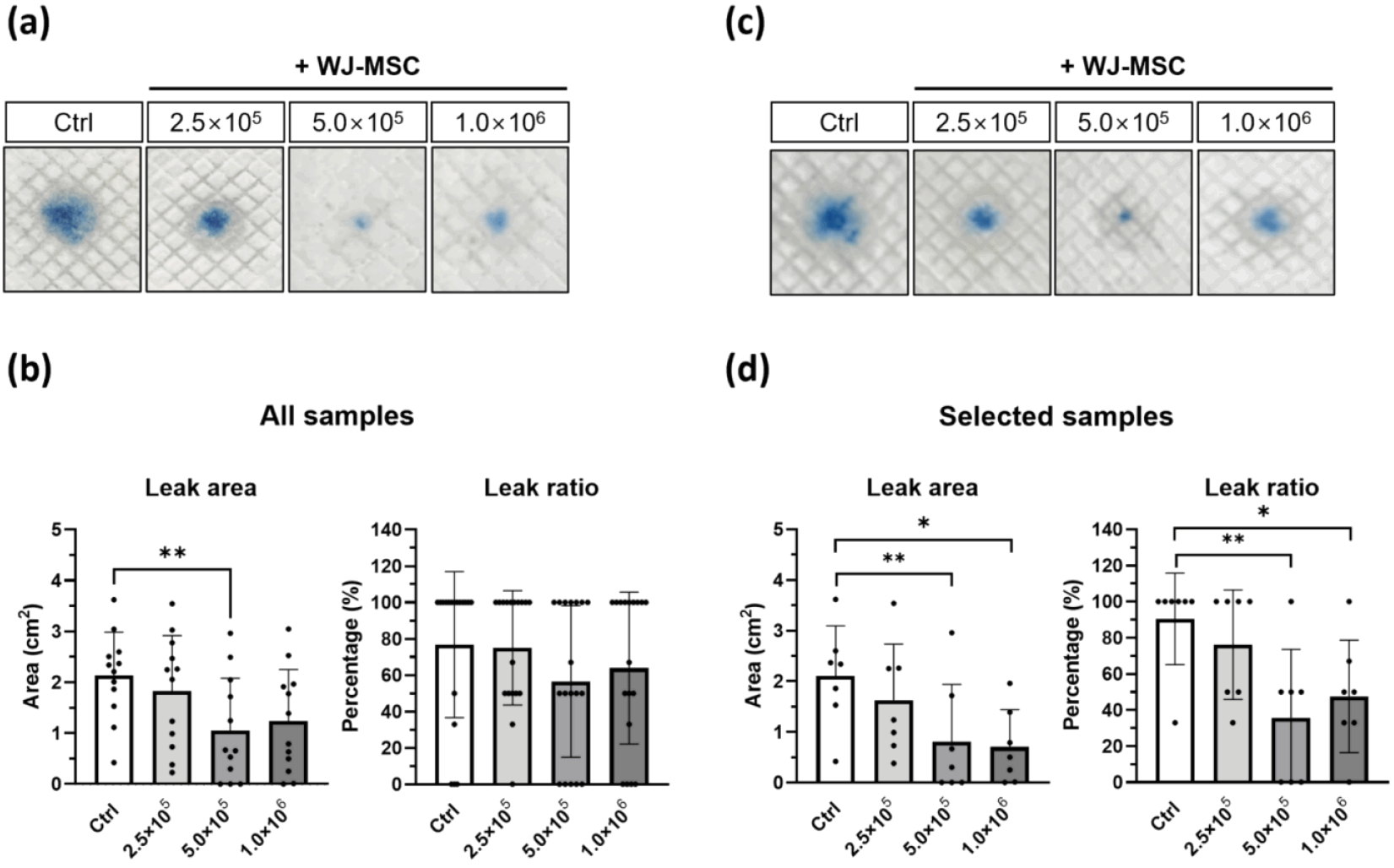
Next, we compared the therapeutic efficacy of varying doses of WJ-MSCs treatment to injured fetal membranes (Fig. 4): low-dose (2.5×10⁵ cells), medium-dose (5.0×10⁵ cells), and high-dose (1.0×10⁶ cells). After 24 hours of treatment, the leakage area and ratio were assessed to evaluate dose-dependent effects (n=12).
In the control group, the average leak area was 2.136±0.849 cm2. Treatment with medium-dose of WJ-MSCs (5.0×10⁵ cells) resulted in a significant decrease in leak area to 1.052±1.030 cm2 (p=0.001, Fig. 4a, 4b). Although the low-dose (2.5×10⁵ cells) and high-dose (1.0×10⁶ cells) WJ-MSCs treatment also decreased the leak area by 1.828±1.088 cm2 and 1.234±1.016 cm2, respectively, however, the differences were not statistically significant. A similar trend was observed for the leak ratio, with the medium-dose treatment group showing the greatest reduction, though this change did not reach significance.
In the sub-analysis of selected samples (n=7), the medium-dose group demonstrated a significant decrease in leak area to 0.807±1.133 cm2 (p=0.001), while the high-dose group also showed a reduction to 0.700±0.740 cm2 (p=0.03), compared to the control (2.106±0.991 cm2) (Fig. 4c, 4d). Furthermore, the leak ratio significantly decreased in both groups: 35.714%±37.796% in the medium-dose (p=0.009) and 47.571%±31.160% in the high-dose group (p=0.05).
Histological analysis revealed that WJ-MSCs regenerate the chorion of the fetal membrane
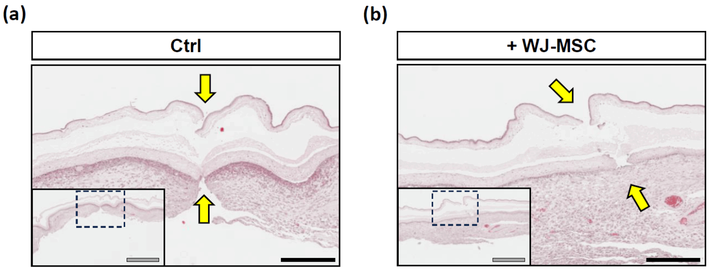
Among the fetal membranes evaluated, a representative sample demonstrating preliminary therapeutic efficacy was selected for histological analysis. Hematoxylin and eosin staining was performed to compare tissue morphology between the control group and WJ-MSCs treatment group. In the control group, the incision extending from the amnion to the chorion remained visible, indicating a complete rupture of the membrane. In contrast, WJ-MSCs-treated membrane exhibited partial tissue regeneration, particularly in the chorion layer, while the amniotic layer remained disrupted.
Discussion
PPROM is a major contributor to preterm birth and is associated with significant neonatal morbidity and mortality. Although its pathogenesis of PPROM is multifactorial and not yet fully understood, contributing factors include intrauterine infection (24, 25), inflammation (26), and altered enzymatic activity (27). Despite decades of research, no effective therapeutic approach currently exists to restore the integrity of ruptured fetal membranes or prevent associated complications. Clinical management remains largely supportive, relying on hospitalization, antibiotics, and corticosteroids to prolong pregnancy and reduce infection risk. However, these interventions do not address the underlying membrane damage, underscoring the urgent need for regenerative interventions.
In this study, wound healing assays demonstrated that WJ-MSCs significantly enhanced epithelial defect closure in hAEpi monolayers (Fig. 1). Both co-culture with WJ-MSCs and treatment with WJ-MSC–conditioned medium produced comparable wound-closure kinetics, supporting the interpretation that the pro-repair activity of WJ-MSCs is largely mediated by soluble paracrine factors rather than direct cell–cell contact alone. Supporting this interpretation, Ki67 expression was upregulated in WJ-MSC–treated conditions, indicating increased proliferative activity during the repair process, while MMP1 expression was downregulated, suggesting that WJ-MSCs concurrently suppress excessive extracellular matrix (ECM) degradation that could otherwise compromise barrier integrity (Fig. 1c). Taken together, these findings imply that WJ-MSCs promote epithelial repair through a coordinated enhancement of migration and proliferation, accompanied by ECM-stabilizing effects. These findings align with previous studies highlighting the wound-healing capabilities of MSCs in various tissues, including skin (28) and trachea (29).
We also established and validated a novel ex vivo screening platform to evaluate the regenerative potential of WJ-MSCs for the treatment of PPROM (Fig. 2). It is known that the physiological pressure naturally acting on the fetal membranes during pregnancy ranges from approximately 20 to 30 mmHg (30). Considering the potential for preterm labor, the intra-amniotic pressure may increase to around 40 mmHg. Based on this estimation, we established experimental conditions in which a pressure of approximately 40 mmHg could be applied to the fetal membranes secured at the opening of a container during a functional (leak) test. As a result, and taking into account the diameter of the container’s opening, we determined that filling the container with 60 mL of saline would generate the desired pressure. The amount of saline can be adjusted according to the desired pressure and the size of the container used. To quantify the amount of saline that leaked through the membranes onto an absorbent pad, we used Trypan blue diluted to a concentration of 0.01%, a non-cytotoxic dye suitable for this application. Through this setup, we established an ex vivo screening platform to functionally assess the recovery of fetal membranes damaged via incision. Rather than directly measuring the injured site, this platform offers an indirect evaluation of membrane integrity by assessing the membrane’s ability to prevent fluid leakage under physiologically relevant pressure conditions. This approach provides a practical means of evaluating functional efficacy in a manner that closely simulates clinical scenarios.
Consistent with the in vitro results, WJ-MSC treatment significantly reduced both leakage area and leakage ratio in the ex vivo PPROM model (Fig. 3). In this study, fetal membrane defects of approximately 6∼7 mm in length were used as a single incision model, based on preliminary experiments showing that lesions within this range produced consistent and reproducible leakage suitable for functional evaluation. This design ensured a standardized yet clinically relevant degree of injury while avoiding over-interpretation of minor differences in incision length. The size and morphology of membrane defects in PPROM are highly variable in clinical practice and play a critical role in healing outcomes. For example, spontaneous PPROM is often associated with larger, irregularly shaped defects, whereas iatrogenic PPROM (e.g., after amniocentesis or fetoscopic procedures) typically involves smaller and more defined injuries. In our previous study, the success rate of amniopatch was lower in patients with spontaneous PPROM (11.8%) than in patients with iatrogenic PPROM (36.4%), although the difference was not statistically significant (p=0.174) (31). Further studies are needed to evaluate the efficacy of WJ-MSCs on fetal membranes healing in larger and irregular defects.
MSCs have emerged as promising candidates for tissue repair and recovery in various clinical contexts. Particularly, it seems that MSC therapy would be the most effective in the treatment of PPROM. Endogenous MSCs are already present in the fetal membrane and play a role in their development. Amnion-derived mesenchymal stem cells (AMSCs), which reside in the mesenchymal (fibroblast) layer beneath the amniotic epithelium, contribute to the structural integrity and repair capacity of the fetal membranes (32, 33). AMSCs produce collagen type I, key structural proteins of the amnion and chorion (34). Since collagen synthesis is critical for membrane repair in PPROM, the administration of allogenic (exogenous) WJ-MSCs may help restore membrane integrity by supplementing collagen production. This hypothesis is supported by our histological findings showing recovery of the chorion layer in WJ-MSC-treated sample (Fig. 5).
Beyond structural support, membrane rupture in PPROM is driven by a complex interplay of pro-inflammatory cytokines, inflammasome activation, and matrix metalloproteinases (MMPs) (24, 35). Complementary in vitro findings (upregulation of Ki67 and downregulation of MMP1; Fig. 1c) suggest that WJ-MSCs stimulate endogenous epithelial cell activation during repair. However, definitive confirmation of this mechanism will require in vivo lineage-tracing studies. MSC are known to mitigate this process by inhibiting MMP-1 and MMP-2 activity while upregulating TIMP-3, preventing excessive collagen degradation (36, 37). They also suppress inflammatory cytokines (IL-1β, IL-6, IL-8, and TNF-α), which are elevated in amniotic fluid, maternal serum, and cervicovaginal fluid of PPROM patients (38, 39). Moreover, MSC could upregulate anti-inflammatory cytokines such as IL-10 and TGF-beta (40). These immunomodulatory effects further support the therapeutic basis of MSC for PPROM.
Dose-response analysis demonstrated that the medium-dose group (5.0×10⁵ cells) achieved the greatest therapeutic effect, while the low- and high-dose groups did not yield statistically significant results (Fig. 4). This highlights the importance of optimizing stem cell dosage, as excessive cell numbers may induce aggregation of administered stem cells, which could inhibit the paracrine actions or reduce the survival of stem cells, counteracting repair mechanisms (41), while insufficient numbers may fail to reach therapeutic thresholds. Collectively, our findings demonstrated that WJ-MSCs significantly accelerated the wound healing of amniotic epithelial cells and reduced leakage in a fetal membrane, suggesting their potential as a recovery therapeutic candidate for PPROM, but the exact mechanism of action is unproven in this study.
Taken together, MSCs represent a particularly attractive therapeutic option for PPROM. However, several limitations must be acknowledged. First, this study was conducted under static culture conditions and did not incorporate mechanical forces or cytokine environments that mimic the intrauterine milieu. Second, additional in vivo studies are required to evaluate the therapeutic efficacy of WJ-MSCs and assess various safety concerns, including reproductive toxicity. As an in vivo approach, it is necessary to establish a PPROM animal model to confirm the therapeutic potential of WJ-MSCs and to conduct comprehensive safety evaluations. Given that PPROM is a serious condition that can threaten fetal survival, it is also crucial to consider all potential risks to the fetus, possibly caused by therapeutic strategies. Therefore, WJ-MSC therapy must be accompanied by rigorous safety studies that remain open to all possible long-term effects on surviving offspring.
In conclusion, this study provides experimental evidence demonstrating the regenerative efficacy of WJ-MSCs in both in vitro and ex vivo models of PPROM and highlights their promise as an MSC-based therapeutic strategy. Future studies will focus on validating these findings in clinically relevant animal models and identifying the specific paracrine factors responsible for the observed therapeutic effects.
Supplementary Materials
Supplementary data including one figure can be found with this article online at https://doi.org/10.15283/ijsc25048
 XML Download