



 PDF
 Citation
 Print


Introduction
Vocal fold atrophy is characterized by the progressive degeneration of the thyroarytenoid (TA) muscle, leading to incomplete glottic closure and impaired phonation. This condition often results in persistent hoarseness and limits functional recovery even after treatment, because the atrophic TA muscle markedly reduces phonatory efficiency (1, 2). Vocal fold atrophy may arise from phono-trauma, laryngitis, systemic disease, surgical interventions, or simply aging. The risk increases with age, and the prevalence of vocal fold atrophy has been reported to be 5.84 per 100,000 in men and 0.65 per 100,000 in women (3).
Vocal fold injection augmentation and surgical reconstruction followed by voice therapy are common treatments for glottic insufficiency. Injection augmentation is performed by delivering biomaterials into paralyzed or atrophic vocal folds to enhance glottic closure and improve voice function. This minimal invasive procedure can be performed under simple local anesthesia in an outpatient setting. However, clinical outcomes vary widely depending on the injectable material used, as differences in durability, tissue integration, and viscoelastic properties can affect voice quality (4). For example, hyaluronic acid (HA) (TissuefillⓇ) is widely used due to its biocompatibility and viscoelastic properties similar to that of the native vocal fold, but its effects only last for 6∼12 months, often necessitating repeated injections (5-7). Collagen offers good tissue compatibility but carries a risk of allergic reactions, particularly with xenogeneic sources, and is rapidly resorbed (8). Autologous fat avoids immune rejection but exhibits unpredictable resorption rates, leading to variable outcomes (9). Calcium hydroxylapatite (CaHA) products (RadiesseⓇ) provide relatively long-lasting augmentation but may induce foreign body reactions, stiffness, or granuloma formation. Short-term agents such as carboxymethylcellulose are easy to inject but are rapidly absorbed, limiting their durability (10, 11). Collectively, while current injectable materials can transiently improve vocal fold function, their effects are short-lived, typically lasting less than a year, thus requiring repeated interventions (7, 12, 13). To overcome these limitations, stem cell–based therapies have been explored. Bone marrow– and adipose-derived mesenchymal stem cells (MSCs) have shown potential but remain limited by low engraftment rate, donor-site morbidity, and inconsistent long-term efficacy (12, 13). Recent strategies combining MSCs with biomaterial carriers have demonstrated enhanced extracellular matrix (ECM) restoration and improved phonatory outcomes in animal models (14, 15). Nevertheless, the optimal stem cell source for clinical translation remains unclear.
Tonsil-derived mesenchymal stem cells (TMSCs) have emerged as a particularly promising candidate. Readily obtainable in large quantities from routine tonsillectomy specimens without ethical concern, TMSCs exhibit high proliferative capacity as well as potent immunomodulatory and anti-fibrotic properties (16). These unique advantages suggest that TMSCs may overcome the limitations of current injectable materials and other MSC-based therapies, offering durable regeneration and functional recovery in vocal fold atrophy. TMSCs also possess multilineage differentiation potential across mesodermal, ectodermal, and endodermal lineages (17, 18). Particularly, several studies reported their robust myogenic potential, where their engraftment restored damaged muscle tissues from Duchenne muscular dystrophy in mice model (19). Furthermore, muscle cell transplantation proved to be an effective strategy for promoting myofiber genesis and functional tissue regeneration within a mouse injury model (20). This myogenic potential highlights their relevance for laryngeal disorders, where restoration of intrinsic muscle function is critical.
Therefore, in the present study, we evaluated the therapeutic potential of TMSCs suspended in HA for the regeneration of atrophied vocal fold muscles. By combining the viscoelastic support of HA with the regenerative capacity of TMSCs in a rabbit model of recurrent laryngeal nerve (RLN) injury–induced vocal fold atrophy, we aimed to establish a novel injection-based strategy for vocal fold regeneration.
Materials and Methods
Isolation of TMSCs
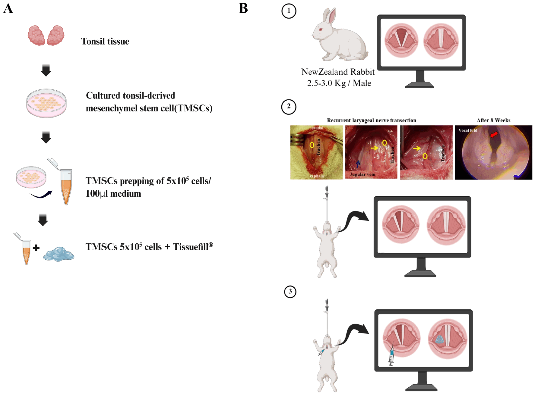
Human palatine tonsillar tissues were obtained from patients undergoing tonsillectomy after obtaining written informed consent, with approval from the Institutional Review Board of Ewha Womans University Medical Center (Approval No. ECT-11-58-37) (Fig. 1A, first and second steps). Fresh tissues were minced under sterile conditions and enzymatically digested in Dulbecco’s modified Eagle’s medium-high glucose (DMEM-HG; Welgene) containing 210 U/mL collagenase type I (Invitrogen) and 10 μg/mL DNase I (Sigma-Aldrich) at 37℃ for 30 minutes in a shaking incubator (200 rpm). The digested suspension was filtered through a sterile wire mesh and centrifuged at 1,300 rpm for 5 minutes at room temperature (Combi-514R, Hanil Science Medical). The cell pellet was washed with DMEM-HG containing 20% fetal bovine serum (FBS; Corning) and once with DMEM-HG containing 10% FBS. Mononuclear cells were obtained by Ficoll-Paque (density 1.077 g/mL, GE Healthcare) density gradient centrifugation. The collected mononuclear cells were washed with phosphate-buffered saline (PBS) twice and subsequently seeded into 100-mm culture dishes (TPP) with DMEM-HG supplemented with 10% FBS and 1% antibiotic-antimycotic solution (Thermo). The cells were incubated for 48 hours, and non-adherent cells were removed by replacing the medium.
Fluorescence-activated cell sorting analyses of MSC makers
To characterize the immunophenotype of TMSCs, flow cytometry was performed to assess the expression of surface markers associated with MSC phenotypes. TMSCs at passage 5 were harvested with 0.25% trypsin-EDTA (Gibco), washed twice with Dulbecco’s phosphate-buffered saline (DPBS; Welgene Inc.), and resuspended in staining buffer (BD Biosciences). A total of 1.0×10⁵ cells were incubated with fluorochrome-conjugated monoclonal antibodies against human CD73, CD90, and CD105 (positive MSC markers), and CD14, CD34, and CD45 (negative lineage markers) (all from BD Biosciences) for 1 hour at 4℃ in the dark with gentle shaking (80 rpm). After washing twice with DPBS, the stained cells were analyzed using a NovoCyte 3000 flow cytometer (Agilent), and data were processed with NovoExpress software (ver. 1.6.3, Agilent).
Adipogenic, osteogenic, and chondrogenic differentiation
The mesodermal trilineage differentiation potential of TMSCs was assessed to validate the mesenchymal characteristics of TMSCs. TMSCs were seeded at a density of 1.0×10⁵ cells/cm2 in 6-well plates and each cultured in commercially-available adipogenic or osteogenic induction medium (StemProⓇ, Thermo Fisher Scientific) for 14 days, with medium replaced every 3 days. Lipid droplet formation was detected by Oil Red O staining (Sigma-Aldrich) for adipogenically-differentiated TMSCs, and calcium deposition was assessed using Alizarin Red S staining (Sigma-Aldrich) for osteogenically-differentiated TMSCs. For chondrogenic differentiation, 1.0×10⁵ TMSCs were cultured in a 15 mL non-adherent conical tube to form spheroids over 21 days followed by culturing in commercially available chondrogenic induction medium (StemProⓇ) for 21 days. Chondrogenic differentiation was confirmed by Alcian Blue staining (Sigma-Aldrich) for sulfated proteoglycans.
Animal experiments
Male New Zealand White rabbits (2.5∼3.0 kg) were purchased from Saeron Bio Inc. (Fig. 1B, ①). The time-course experiment was designed as an exploratory pilot study to characterize the progression of vocal fold atrophy, rather than to provide statistically powered comparisons. All animals were acclimated for at least 7 days before experimentation and housed in the animal facility of the Ewha Womans University Medical Science Research Institute under standardized conditions (temperature, 24℃±2℃; and a 12 hour-light/dark cycle) with free access to food and water. To determine optimal time, point for assessing treatment effect of vocal fold recovery, a preliminary study was performed by allocating the rabbits with RLN injury and euthanized to collect laryngeal tissues at predetermined time points of 8, 12, 16, 26 weeks (n=2 per time point). The remaining animals were divided into three groups: Group Sham (n=2), cutting RLN without any surgical procedure); Group CaHA (n=2), cutting RLN and injection of calcium hydroxylapatite (CaHA; RadiesseⓇ, BioForm Inc.); Group TMSC+HA (n=2), cutting RLN and injection of TMSCs mixed with hyaluronic acid (HA; Tissuefill, Jeil Pharmaceutical). All animal experiments were approved by the Institutional Animal Care and Use Committee (IACUC) of Ewha Womans University College of Medicine (Approval No. EUM20-032).
Surgical preparation of RLN injury to induce vocal fold atrophy
All rabbits used in this study underwent left RLN sectioning to induce unilateral RLN injury, causing vocal fold atrophy. The right vocal fold was left intact to serve as a normal control for evaluating the progression of vocal fold tissue regeneration in the atrophied vocal cord. General anesthesia was induced by intramuscular injection of tiletamine-zolazepam (Zoletil, 0.3 mL/kg) and xylazine (Rumpun, 0.15 mL/kg). To establish vocal fold atrophy model using RLN injury (from left) after shaving the neck, a 3-cm vertical cervical incision was made to expose the trachea. jugular vein (navy arrow) and blood vessels and running parallel to the trachea (yellow arrows) and the RLN (yellow circles) were carefully identified. Using a bipolar electrocautery device, the RLN was unilaterally transected (left) over a length exceeding 3 cm to induce glottic insufficiency and subsequent vocal fold atrophy (Fig. 1B, ② top). The animals received intramuscular injections of cefazolin (23 mg/kg; cefazolin sodium; Chong Kun Dang) and Maritrol (4 mg/kg; tramadol hydrochloride; Jeil Pharmaceutical) for five days to reduce postoperative infection and pain from the surgery. After the surgery, endoscopic (FLEXILUX II, Scholly Fiberoptic GmbH) evaluation was performed to confirm abnormal basal vocal fold mobility (vocal fold paralysis) on the left side (red, arrow) (Fig. 1B, ② bottom). During natural breathing, the right vocal fold exhibited relaxed, flat, closed vocal fold movement, whereas the left vocal fold exhibited no movement, confirming the vocal fold paralysis from RLN injury (Supplementary Video S1). At the final time point, animals were euthanized by CO2 inhalation, and the laryngeal tissues were harvested for subsequent analysis.
Preparation of injection materials
In the CaHA group, 100 μL of CaHA was injected into the paralyzed vocal folds. Approximately 5.0×10⁵ cultured TMSCs (passage 5) suspended in 100 μL of culture medium, with careful adjustment of cell number and volume for experimental use (Fig. 1A, third arrow top). Finally, the prepared total of 5.0×10⁵ TMSCs in 100 μL of DMEM-HG was mixed with 200 μL of HA solution at 2:1 ratio (Fig. 1A, last step). Immediately after establishing the RLN injury model, the injections of either CaHA or TMSC+HA were administered into the affected vocal folds using a 22-gauge needle under videoendoscopic guidance (Fig. 1B, ③). The untreated sham group received no injections. No animals exhibited respiratory distress or bleeding following the procedures.
Histological analysis
The animals were sacrificed at 8, 12, 16 and 26 weeks post-surgery for further histological analyses to evaluate the progression of glottic insufficiency and to assess the regenerative effects of the injected materials in laryngeal tissues. Multiple histological sections obtained from each animal were analyzed to ensure measurement consistency. Laryngeal specimens were fixed in 4% formalin, embedded in paraffin, and sectioned at a thickness of 5 μm using X-tra Slides (Leica). Hematoxylin and eosin (H&E) staining was performed to examine overall tissue morphology. H&E staining was performed to examine overall tissue morphology. Sections were deparaffinized in xylene, rehydrated through a graded ethanol series, and rinsed in distilled water. The sections were then stained with hematoxylin solution for 10 minutes, followed by rinsing in running tap water for 3 minutes. Differentiation was carried out using 1% acid alcohol for 30 seconds, and bluing was carried out in 1% ammonia water for 30 seconds. Subsequently, sections were counterstained with eosin solution for 3 minutes, dehydrated through graded ethanol, cleared in xylene, and mounted with coverslips. The volume of remaining material was calculated using a modification version of a previously introduced method (21). At 8 weeks, the dimensions of the residual material were measured in pixels using the image analysis program (ver. 1.8.0, ImageJ, NIH).
Safranin O staining was conducted to evaluate cartilage within the ECM of the sectioned vocal fold tissues. Sections were deparaffinized, rehydrated through a graded ethanol series, and stained with 1% Fast Green for 6 minutes. Acetic acid treatment was then applied to selectively remove nonspecific or weak dye binding selectively. Subsequently, sections were counterstained with 1% Safranin O solution for 6 minutes.
Masson’s Trichrome staining (Polysciences Inc.) was performed to evaluate collagen and muscle fibers within the ECM. Sections were deparaffinized in xylene, rehydrated through a graded ethanol series, and mordanted in Bouin’s solution at 56℃ for 1 hour to stabilize tissue architecture and enhance dye penetration. Nuclear staining was conducted with Weigert’s iron hematoxylin for 10 minutes, which is resistant to subsequent acidic treatments. Biebrich scarlet–acid fuchsin solution was applied for 5 minutes to stain cytoplasmic components and muscle fibers red. Sections were then incubated with phosphotungstic phosphomolybdic acid solution to selectively remove the red dye from collagen and create binding sites. Finally, collagen fibers were stained blue with aniline blue for 5 minutes under acidic conditions, followed by dehydration, clearing, and mounting.
Alizarin Red S staining was performed to evaluate calcium deposition within the ECM of the sectioned vocal fold tissues. Sections were deparaffinized, rehydrated through a graded ethanol series, and incubated with 2% Alizarin Red S solution (pH 4.2; Sigma-Aldrich) for 3 minutes. Excess dye was removed by rinsing with distilled water to minimize nonspecific staining. Sections were then dehydrated through graded ethanol, cleared in xylene, and mounted for microscopic evaluation. Stained sections were examined with Slide Scanner (Zeiss) and optical microscope (Leica). Microscopic images were acquired under identical exposure settings, and image processing was performed with Enersight software (ver. 2.2.0, Leica). Given the varying laryngeal dimensions across samples, the percentage of each slide muscle fiber area was calculated by dividing the measured muscle fiber area (μm2) by the total tissue area (μm2) and multiplying by 100. The quantified area of the damaged (affected) atrophic site was normalized to the quantified area of the normal control site. Quantification was performed using ImageJ software (ver. 1.8.0, NIH) and data analysis was conducted with GraphPad Prism software (ver. 10.0.0, GraphPad Software Inc.).
Immunofluorescence of human mitochondria
Immunofluorescence staining was performed to identify engrafted human TMSCs within the vocal fold tissues. Deparaffinized and rehydrated sections were incubated overnight 4℃ with a primary antibody against human mitochondria (1:50; MAB1273; Sigma-Aldrich). After two washes with PBS, the sections were incubated for 1 hour at room temperature with species-specific fluorophore-conjugated secondary antibodies (green fluorescent protein) (A32723; Thermo Fisher Scientific). Nuclei were counterstained using a mounting medium containing DAPI (Vector Laboratories). Stained sections were examined with a confocal microscope (Zeiss), and fluorescence images were captured under identical exposure settings. Image processing and quantification were performed using ZEN software (ver. 3.7.0, Zeiss).
Statistical analysis
All experiments were performed and repeated at least three times (n=3 slides per group). Multiple histological sections obtained from each animal were analyzed to ensure measurement consistency. These measurements represent technical replicates rather than independent biological replicates.
Given the limited number of animals per group, the statistical analysis was primarily intended for descriptive comparison, and the results should be interpreted with caution. All results are presented as the mean±standard deviations. Statistical significance was analyzed by two-way ANOVA followed by Tukey’s multiple comparisons test using the statistical software available in GraphPad Prism software (ver. 10.0.0, GraphPad Software Inc.).
Results
Characterization of TMSCs
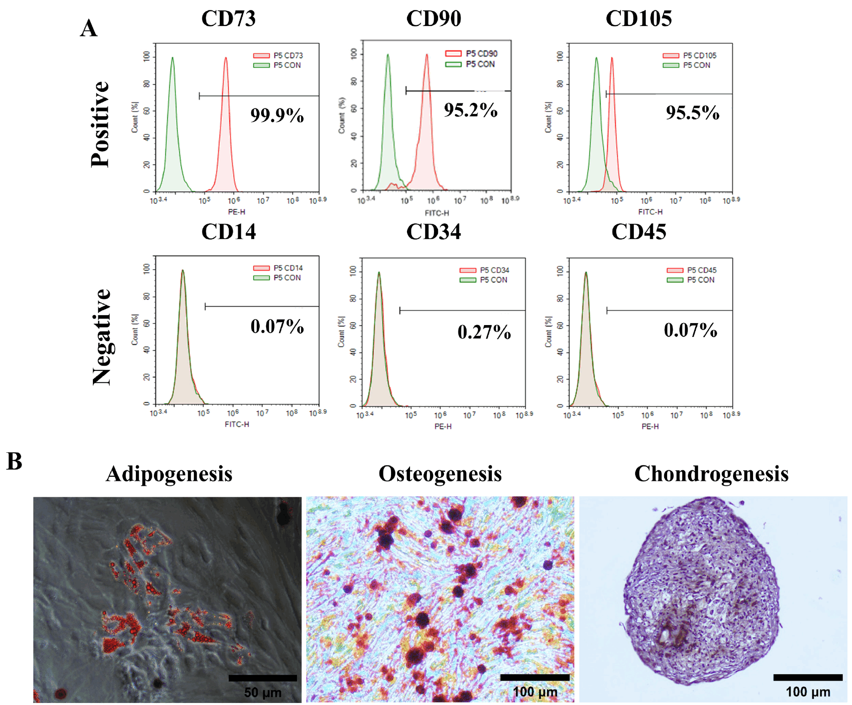
TMSCs exhibited plastic-adherent and spindle-shaped morphology, both of which are typical characteristics of MSCs (data not shown). Flow cytometric analysis confirmed the mesenchymal phenotype of TMSCs (Fig. 2A). TMSCs retained high expression of MSC-specific positive markers (top, from left), including CD73 (99.9%±0.02%), CD90 (95.23%±1.84%), and CD105 (95.57%±1.61%), but had low expression of negative markers CD14 (0.07%±0.03%), CD34 (0.27%±0.16%), and CD45 (0.07%±0.03%) (bottom, from left).
To evaluate their mesodermal differentiation potential, TMSCs were cultured in commercially available lineage specific induction media for 14∼21 days. The TMSCs were differentiated into adipogenic, osteogenic and chondrogenic lineages (from left), as evidenced by intracellular lipid droplet accumulation (Oil Red O), calcium deposits (Alizarin Red S), and sulfated proteoglycans (Alcian Blue), respectively (Fig. 2B). These results demonstrate that TMSCs fulfill the minimal criteria for MSCs, exhibiting adherence to plastic, a mesenchymal immunophenotype, as reflected by high expression of CD73, CD90, and CD105, and low expression hematopoietic markers CD14, CD34, and CD45, as well as trilineage mesodermal differentiation potentials.
Histological changes following nerve transection-induced vocal fold atrophy
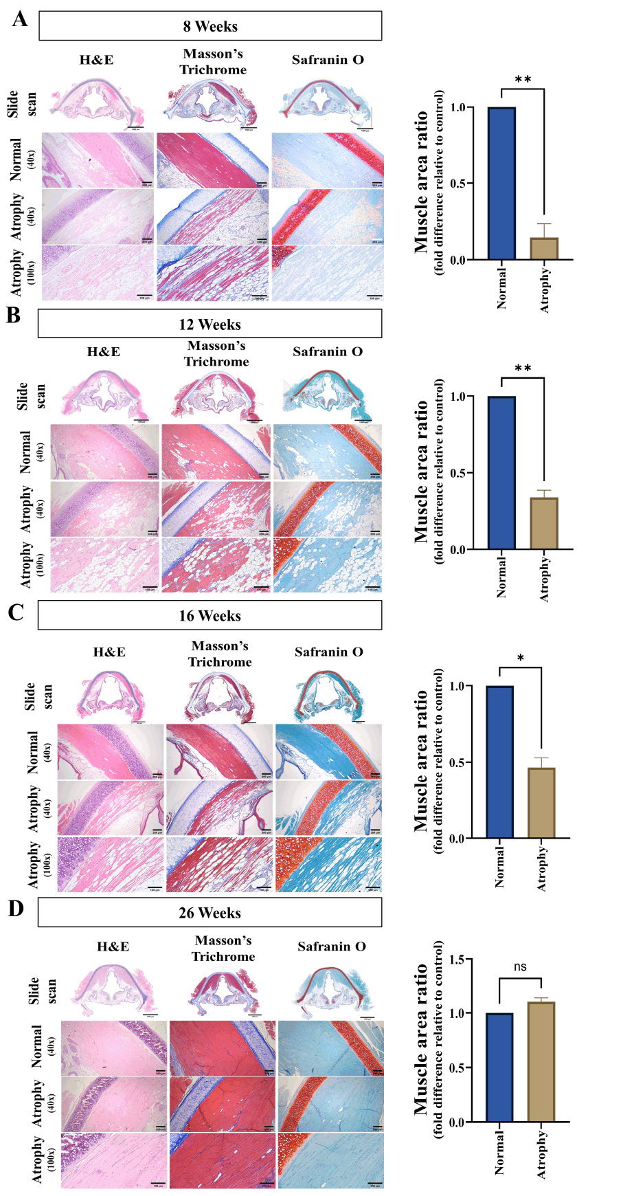
Animals were sacrificed at 8, 12, 16, and 26 weeks postoperatively, and further histological analyses were performed to observe the progression of vocal fold atrophy (Fig. 3A-3D). Histological evaluation using H&E, Masson’s trichrome stains and safranin O stain revealed (from left) significant changes in vocal fold tissue over the period at each time point after RLN injury using unilateral nerve transection.
At 8 weeks after RLN injury, H&E staining revealed significant in the vocal fold atrophy, characterized by decreased muscle fiber (pink) size and disorganization, indicating a substantial loss of muscle mass and structural integrity. The vocal fold appeared thinner and less organized compared to the intact vocal folds (Fig. 3A), consistent with features of severe vocal fold atrophy. Quantitative analysis of H&E staining confirmed that the muscle mass of the damaged (atrophic) site reduced to 20.9%±0.07% of the normal control site (Fig. 3A), indicating marked muscle loss 8 weeks after RLN injury. Masson’s trichrome staining demonstrated increased collagen (blue) deposition within the thin muscle fiber (red), indicating onset of fibrosis. Safranin O staining showed a slight decrease in cartilage (red), along with a marked reduction of the ECM (blue) compared with the normal control site.
At 12 weeks after the nerve transection, partial recovery of muscle structure was evident (Fig. 3B). H&E staining revealed slightly more organized muscle fibers compared with those at week 8. indicating the initiation of spontaneous recovery in the atrophied muscle fiber (pink). Quantitative analysis of H&E staining confirmed that the muscle mass of the damaged (atrophic) site reduced to 33.9%±0.04% of the normal control site (Fig. 3B), indicating 13.1%±0.1% higher muscle mass compared to that of 8 weeks. However, the muscle fibers and mass were still remained to be smaller and less densely distributed than those in the normal control site. Despite this improvement, Masson’s trichrome staining collagen (blue) deposition remained elevated, suggesting ongoing fibrosis, even though the severity was somewhat reduced compared to that of week 8. Safranin O staining showed no further reduction in cartilage (red), and ECM (blue) composition was slightly improved compared with those at week 8.
At 16 weeks after RLN injury, H&E staining revealed partial reorganization of muscle fibers, with continued mild atrophy relative to the normal control site (Fig. 3C), suggesting progressive recovery of the muscle fibers (pink). Quantitative analysis of H&E staining confirmed that the muscle mass of the damaged (atrophic) site reduced to 46.5%±0.05% of the normal control site (Fig. 3C), indicating 25.7%±0.02% and 12.6%±0.09% higher muscle mass compared to those of 8 and 12 weeks, respectively (Fig. 3C). Masson’s trichrome staining still showed the persistence of intermuscular (red) fibrosis, even though collagen (blue) deposition was significantly reduced compared with those at 8 and 12 weeks, suggesting partial resolution of fibrosis. Safranin O staining confirmed increased cartilage (red) content and ECM (blue) restoration compared with those at 12 weeks.
The damaged muscle tissue recovery was almost recovered by 26 weeks after the RLN injury (Fig. 3D). H&E staining showed that the size and structure of muscle fibers (pink) were restored to a state similar to those of normal control site, confirming near complete structural recovery. The decreased muscle fiber observed (Fig. 3A) at earlier time points 8 weeks was almost completely recovered. Quantitative analysis of H&E staining confirmed that the muscle mass of the damaged (atrophic) site reached to 111.0%±0.03% relative to the normal control site, corresponding 9.9%±0.04%, 23.0%±0.06%, and 35.6%±0.03% higher muscle mass compared to those of 8, 12, 16 weeks, respectively (Fig. 3D). Masson’s trichrome staining showed collagen (blue) deposition similar to that of normal control site, even though some residual fibrosis still persisted. Safranin O staining demonstrated the complete restoration intact of cartilage (red) content and ECM (blue) structure, with staining intensity similar comparable to that of the normal control site, indicating almost complete recovery of cartilage matrix at the injury site.
These results demonstrate that vocal fold atrophy induced by RLN injury in the rabbit model was the most prominent at 8 weeks after injury, accompanied by muscle fiber disorganization, ECM remodeling, and fibrosis. Spontaneous recovery began by 12 weeks, and the tissue was almost completely healed by 26 weeks from RLN injury. Therefore, 8 weeks from RLN injury was selected as the optimal time frame for evaluating the efficacy of CaHA and TMSC+HA treatments in regenerating the atrophied vocal fold, because muscle atrophy and fibrosis were most severe at this stage, allowing assessment of therapeutic potential before the onset of natural recovery.
Assessing regenerative effects of CaHA and TMSC+HA in vocal fold atrophy
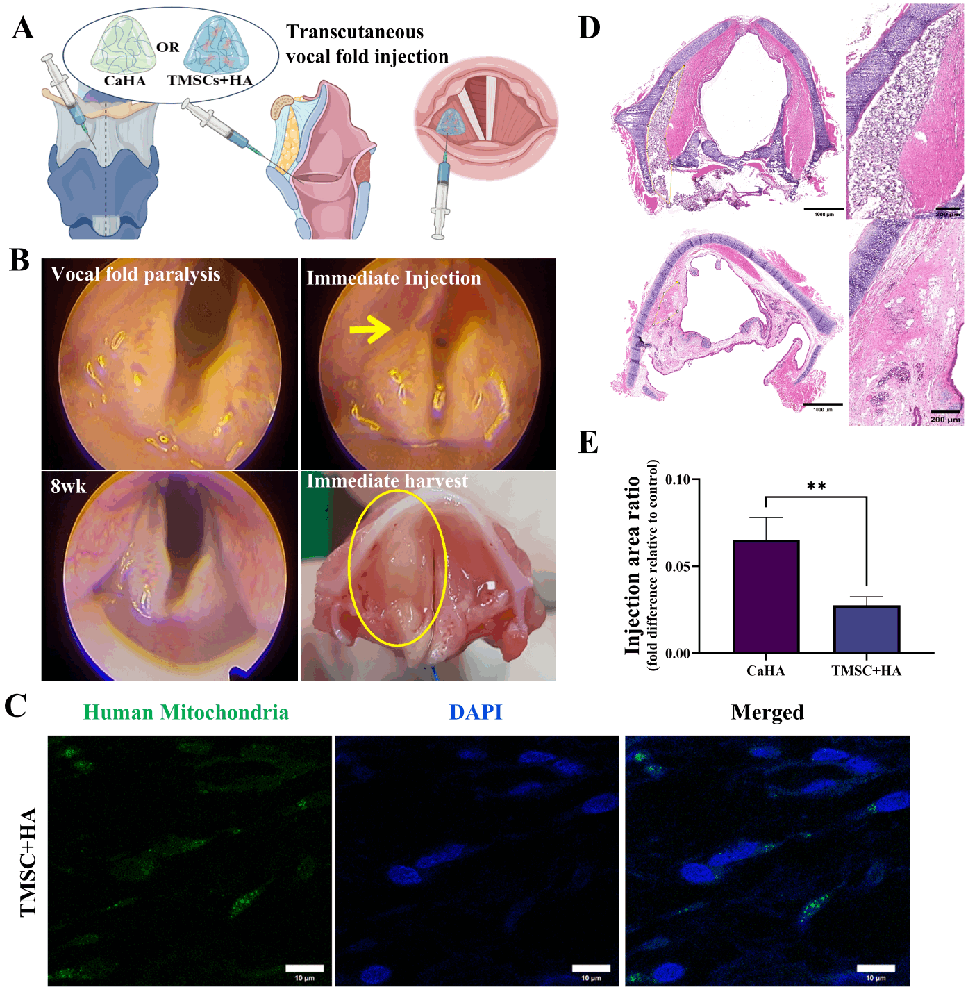
The injection needle was inserted percutaneously through the thyroid cartilage area. The tip of a 22-gauge needle was bent at approximately a 30-degree angle. It was gently advanced along the edge of the thyroid cartilage into the paraglottic space surrounding the vocal cords. Once in position, CaHA or TMSC+HA was injected while endoscopic monitoring ensured proper restoration of the vocal cords to their midline volume (from the left) (Fig. 4A). Endoscopic evaluation of vocal fold atrophy prior to injection (top left) and observation immediately after injection (top right) demonstrated sufficient medialization of the atrophied vocal fold (yellow arrow), which was maintained for up to 8 weeks post-injection (bottom left). Gross examination of the larynx harvested at 8 weeks after RLN injury confirmed (bottom right) the persistence of CaHA within the glottic area (yellow circle) (Fig. 4B). Immunofluorescence of human mitochondria demonstrates successful implantation of TMSCs at the Injected site (atrophied muscle) of the vocal fold (Fig. 4C). CaHA at 8 weeks after injection were still persisted within the Injected site (atrophied muscle) as visualized by porous whitish purple materials, correcting the medialization of the vocal fold (Fig. 4D, top). In contrast, significant recovery of muscle fiber regeneration was observed as evidenced with cell nuclei at the site of TMSC+HA in Injected site (atrophied muscle) (Fig. 4D, bottom). These results validate their localization and persistency at the injection site (Fig. 4D).
Quantitative analysis revealed clear differences in residual material remaining in the injected area between the CaHA and TMSC+HA groups. Histological examination of H&E staining confirmed that the amount of the residual material in the injected site (atrophied muscle) was 6.57%±0.85% and 2.69%±0.56% relative to the total tissue area for CaHA and TMSC+HA, respectively (Fig. 4E). Consequently, CaHA resulted in 3.89%±0.56% higher residual material remaining at the injection site compared to that of TMSC+HA (Fig. 4E).
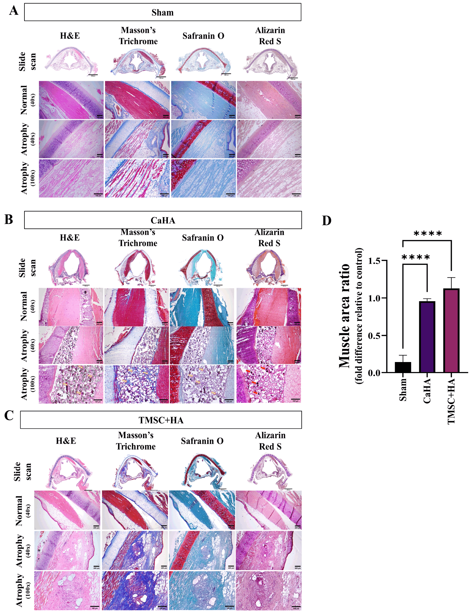
Histological examination with H&E staining revealed no evidence of CaHA absorption or migration; however, cellular infiltration was observed around the injected CaHA particles (yellow arrow) (Fig. 5B). Adjacent tissues, including muscle and cartilage, were unaffected, and the interstitial spaces between muscle fibers (pink) appeared similar to those of the contralateral normal control site (normal intact muscle), in contrast to the Sham group (Fig. 5A). Masson’s trichrome staining further confirmed that adjacent tissues, including muscle (red), were unaffected by CaHA, even though a localized foreign body giant cell reaction (yellow arrows) was observed, suggesting potential inflammatory reaction from the CaHA at the injection site. Safranin O staining demonstrated porous regions corresponding to the injected particles, but no neocartilage formation was detected. Cartilage (red) and ECM (blue) contents remained unaltered in the CaHA group (Fig. 5B). Alizarin Red S staining validated the presence of granular deposits (reddish-brown) within the injected site (atrophied muscle) of CaHA, as indicated by red arrows (Fig. 5B).
Histological analysis with H&E staining demonstrated that, TMSC+HA significantly improved restoration of muscle fibers, which were characterized by narrower interstitial spaces between muscle fibers (pink), compared to those from Sham and CaHA groups (Fig. 5C). Unlike the CaHA, the TMSC+HA penetrated the vocalis muscle without forming a fibrous capsule, indicating superior biocompatibility and tissue integration compared to CaHA. Masson’s trichrome staining showed pronounced formation of muscle fibers (red) and decreased collagen (blue) deposition. Safranin O staining showed that, the TMSC+HA did not induce neocartilage formation similar to that from CaHA. Alizarin Red S staining confirmed the absence of calcium deposition (reddish brown) within the TMSC+HA Injected site (atrophied muscle). In contrast with CaHA, TMSC+HA did not produce any granular deposits, indicating low immunoreactivity from the implantation.
Quantitative analysis of H&E staining confirmed that the injection of CaHA and TMSC+HA resulted in 95.6%±0.02% and 112.7%±12.61% of muscle mass at the atrophic site relative to the normal control site (normal intact muscle) (Fig. 5D). Compared to the atrophic site without any treatment (Sham), the muscle masses of atrophic site injected with CaHA or TMSC+HA was calculated to be 81.9%±0.06% and 98.2%±0.13%, respectively. The injection of TMSC+HA resulted in 16.3%±0.12% higher muscle mass compared to that of CaHA (Fig. 5D).
Discussion
In this study, we employed a glottic insufficiency model that mimics RLN injury-induced atrophy to reproduce the pathophysiological mechanism underlying human vocal fold atrophy using a rabbit model. Rabbit vocal folds share structural similarities with human folds, including a multilayered structure, organized tissue, and the arrangement of key fibers, making them an ideal model to simulate vocal fold pathologies and post-injury functional performance. In the contralateral normal TA muscle of rabbit, fibers were well-organized, densely packed with peripheral nuclei and minimal interstitial space. At the eight weeks after RLN injury, histological changes in the vocal fold tissues were most pronounced, with significant muscle atrophy evident on the H&E staining. Reduced muscle fiber size and disorganization of the fibers were observed, markedly compromising the structural integrity of the vocal folds and resulting in thinning and disruption of tissue architecture. Masson’s Trichrome staining also revealed increased collagen deposition in the atrophic vocal fold, confirming the progression of fibrosis. Endoscopic evaluation similarly demonstrated that the concave contour of the membranous vocal fold, resulting from the loss of TA muscle bulk, led to incomplete glottic closure and dysphonia, further validating successful induction of vocal fold atrophy in the RLN injury rabbit model.
Interestingly, the atrophied vocal fold began to regenerate spontaneously at 16 weeks after RLN injury, and by 26 weeks, muscular characteristics were almost completely restored. This regenerative capacity contrasts with that of human vocal folds, which lack the ability to recover once atrophy and fibrosis are established (22). Therefore, the 8-week time point appears to represent a critical window for evaluating therapeutic interventions in rabbits, as it precedes spontaneous regeneration while reflecting maximal tissue alteration. This observation aligns with previous reports: Svistushkin et al. (23) identified 8 weeks post-injury as a pivotal period for observing structural improvements and ECM remodeling in vocal folds treated with human mesenchymal stem cells. Similarly, Hertegård et al. (22) reported that 8 weeks post-scar excision in a rabbit model corresponded to significant changes in viscoelasticity and collagen content. Collectively these findings support the selection of 8 weeks post-RLN injury as optimal time for assessing the efficacy of interventions such as CaHA or TMSC+HA, as determined in the present study.
In this study, a rabbit model of RLN injury-induced vocal fold atrophy was used to evaluate and compare the efficacy of CaHA and TMSCs combined with an HA scaffold in restoring the structure and function of vocal fold under clinically relevant conditions. CaHA (RadiesseⓇ) is a long-lasting, biocompatible material that provides structural support and durability, making it effective for medialization in vocal fold atrophy (24). It is one of the most widely used materials for clinical applications in various medical procedures, such as facial and vocal fold augmentation. In cases of vocal fold atrophy, CaHA offers a durable solution owing to its slow resorption rate, allowing restoration of volume and maintenance of glottic closure for up to a year or longer. Our results demonstrated that CaHA injection provided improved repositioning of the vocal fold muscle structure compared with untreated atrophy. Moreover, CaHA maintained tissue bulk and mechanical support over the 8-week observation period, contributing to improved glottic competence. CaHA significantly improved the muscle mass at the injection area of the atrophic site, showing 82% higher muscle mass compared to untreated Sham group. CaHA is known to have limited viscoelasticity and lack of biological activity to promote tissue regeneration (ref), and therefore, this effect may result from the immediate injection of CaHA, which likely functioned as an effective filler to prevent further atrophic progress, thereby maintaining muscle content. CaHA also induced a local inflammatory response at the injection site, characterized by immune cell infiltration, as previously described by Tanna et al. (25), which may increase the risk of foreign body reaction and limit long-term integration of the material.
In contrast, HA (TissuefillⓇ) closely mimics the viscoelastic properties of the vocal folds and integrates well into the ECM, providing more physiologic vibration and improved voice quality upon CaHA injection. HA remains a preferred injectable material for vocal fold augmentation because of its viscoelastic compatibility, reversibility, clinical safety, and ease of injection (26). Despite these advantages, HA has a relatively short duration of effect, often degrading before sufficient regeneration occurs. Consequently, recent strategies have focused on enhancing HA-based therapies by combining them with regenerative agents such as stem cells.
Building on these considerations, our study evaluated the efficacy of combining TMSCs with HA in promoting regeneration of atrophied vocal folds. The results showed that the injection of TMSCs suspended in HA hydrogel achieved successful integration into the atrophic TA muscle, demonstrating superior regenerative potential compared with untreated and CaHA-treated groups. Histological analysis at 8 weeks post-treatment revealed that TMSC+HA injection increased muscle fiber density, improved fiber alignment, and reduced interstitial spacing relative to untreated atrophic tissue; suggesting partial restoration of muscle fibers from RLN-induced atrophy and attenuation of fibrosis. Furthermore, quantitative analysis also validated higher muscle mass formation by the TMSC+HA compared to CaHA at the injection area of the atrophic vocal fold. These finding demonstrates that the regenerative effects of TMSC+HA, likely driven by the MSCs and their paracrine signaling, contributing to significantly higher muscle regeneration than the volumetric augmentation provided by CaHA alone. In addition, TMSC+HA injection did not elicit an inflammatory response, indicating superior biocompatibility compared to CaHA. These findings suggest that TMSC+HA therapy promotes structural restoration of both cellular and extracellular components, aligning with the ideal characteristics of injectable material; biocompatibility, minimal immune response, and the capacity to recover both structure and function. Moreover, the increased fiber density and regular alignment observed after TMSC+HA injection reflect viscoelastic properties comparable to native vocal fold tissue, which are essential for proper vocal fold vibration and voice production (27). Histological analysis revealed regeneration of vocal fold muscle, including reduced fibrosis and reorganization of muscle fibers. These structural improvements provide important evidence of the vocal fold regeneration (28) but they do not necessarily indicate functional recovery, which will need to be addressed in future studies.
In addition to favorable histological outcomes, HA combined with the regenerative potential of TMSCs represents a clinically practical therapeutic strategy, fulfilling key criteria of ideal injectable materials, including ease of use, stability, and cost effectiveness. Our findings are consistent with previous studies demonstrating that stem cell implantation enhances reinnervation and improves intrinsic laryngeal muscle function in RLN injury models (29). For example, autologous muscle progenitor cell implantation improved adductor and abductor function in canine RLN transection models, whereas MSC injection laryngoplasty restored vocal fold bulk and vibratory properties in paralysis models (26). In line with these findings, we observed that TMSCs embedded HA hydrogel persisted in situ and interacted with host fibers, providing a supportive microenvironment for engraftment and tissue regeneration. These results underscore the potential of TMSCs based injectable therapy not only to augment tissue volume but also to promote substantial structural of the vocal fold, offering a promising strategy for improving the structural integrity and potential functional restoration capacity of the laryngeal musculature in vocal folds atrophy.
Nevertheless, several limitations should be acknowledged. The sample size (n=2∼3 per group) was determined based on the feasibility of using the rabbit model for assessing vocal fold muscle regeneration (30), and it showed consistent histological patterns across animals, supporting the reproducibility of the results. First, the observation period was limited to 8 weeks, corresponding to the time window before spontaneous regeneration typically occurs in the rabbit model. Therefore, our findings should be interpreted as evidence of early therapeutic efficacy, as longer-term assessments would likely be confounded by natural recovery processes. Second, despite histological improvement, functional voice outcomes, such as phonatory parameters and acoustic measures were not directly assessed, limiting translational relevance. Finally, the underlying mechanism of regeneration by TMSC+HA mediated regeneration require further elucidation through molecular and cellular analyses. While our study focused on structural regeneration of the vocal fold muscle, it did not include detailed analyses of immune responses and long-term engraftment. Future studies are warranted to evaluate host immune responses and long-term safety to further support the potential clinical translation of TMSC+HA. This study should be considered a proof-of-concept investigation focusing on histological outcomes, and further studies with larger sample sizes are required to validate the findings.
Despite these limitations, it is noteworthy that unlike CaHA, which exhibits limited regenerative potential, our results support TMSC+HA as a safer and more effective approach for vocal fold regeneration. Previous studies have highlighted that HA hydrogels, particularly when supplemented with growth factors, can guide endogenous stem cells toward sites of injury and facilitate tissue regeneration (24). The ability to deliver or culture stem cells within HA-based scaffolds has generated substantial interest, as it may enable in vivo cell differentiation following implantation. Consistent with these reports, our findings suggest that the combination of HA and MSCs, even in their undifferentiated state, is safe and promotes cell survival and tissue restoration, thereby reducing morbidity and enhancing therapeutic efficacy.
In summary, this study provides evidence that injection of TMSCs suspended in HA (TissuefillⓇ) can restore atrophic TA muscle structure and potentially improve glottic closure in vocal fold muscle atrophy. By combining the established safety and viscoelastic properties of HA with the regenerative potential of TMSCs, this approach offers a promising therapeutic avenue for the management of atrophied vocal folds from RLN injury.
Supplementary Materials
Supplementary data including one video can be found with this article online at https://doi.org/10.15283/ijsc25106

 XML Download