



 PDF
 Citation
 Print


Introduction
Stem cells are defined by their ability to self-renew while maintaining a stable gene expression profile and their potential to differentiate into various human cell types (1, 2). These properties make human pluripotent stem cells (hPSCs) invaluable tools for regenerative medicine, disease modeling, drug discovery platforms, and advancing our understanding of human development. Self-renewal and pluripotency collectively define “stemness” and provide the biological basis for cell fate determination and tissue homeostasis. Human stem cells are categorized into pluripotent, multipotent, or unipotent stem cells based on their differentiation potential. Among them, hPSCs, including human embryonic stem cells (hESCs) derived from the blastocyst and human induced pluripotent stem cells (hiPSCs) reprogrammed by OCT4, SOX2, KLF4, and MYC (OSKM), can self-renew over long periods and differentiate into the three germ layers: ectoderm, mesoderm, and endoderm (3-12). Owing to these unique properties, hPSCs hold great promise for applications in regenerative medicine, disease modeling, drug screening, tissue engineering, and developmental biology (13-18).
A notable feature of hPSCs is their unusually short cell cycle compared to somatic cells. hPSCs have an extended S phase and a shortened G1 phase, accelerating their proliferation (19-21). This accelerated cell cycle is not merely a consequence of pluripotency but appears to be mechanistically linked to stemness maintenance, as cell cycle perturbations can trigger unwanted differentiation programs (22-24). Multiple intrinsic signaling pathways, including Wnt, TGF-β, and fibroblast growth factor (FGF), regulate stemness and the cell cycle. Wnt signaling regulates cell proliferation, adhesion, and migration (25, 26), whereas TGF-β signaling modulates differentiation, proliferation, and apoptosis (27). The FGF pathway activates extracellular signal-regulated kinase/mitogen-activated protein kinase (ERK/MAPK) signaling, which supports hPSC proliferation, survival, migration, and differentiation (28, 29). Although the role of ERK/MAPK signaling in maintaining pluripotency has been extensively studied in murine pluripotent stem cells (PSCs), which are typically maintained in a naïve state in vitro, the precise molecular mechanisms by which this pathway contributes to the maintenance of hPSC stemness remain less well defined. Human PSCs are predominantly maintained in a primed, post-implantation-like state, and the upstream regulatory factors linking ERK/MAPK signaling to the core pluripotency network in this context are still largely unknown.
In pursuit of identifying novel regulators that control the pluripotent state through modulation of the cell cycle, we focused on two mitochondrial inner membrane proteins: prohibitin (PHB) and stomatin-like protein 2 (STOML2). PHB, a highly conserved protein, has been localized to multiple intracellular compartments, including the mitochondria, nucleus, and plasma membrane, and is known to participate in diverse cellular functions such as energy metabolism, cell proliferation, and apoptosis regulation (30-35). In particular, within the mitochondrial inner membrane, PHB1 and PHB2 form heterodimeric ring-shaped complexes that are critical for mitochondrial genome stability and structural maintenance. In mouse embryonic stem cells (ESCs), PHB2 is highly expressed in the undifferentiated state, and its depletion leads to increased apoptosis. Conversely, PHB2 overexpression not only enhances ESC proliferation in the presence of leukemia inhibitory factor (LIF) but also significantly suppresses differentiation toward neuronal (TuJ1+) and endodermal (Foxa2+) lineages (36).
STOML2, localized to the mitochondrial inner membrane, functions as an integral protein supporting mitochondrial biogenesis, energy metabolism, and the suppression of apoptosis (37-40). Notably, it binds cardiolipin and interacts with the PHB complex to maintain mitochondrial structural integrity and assemble respiratory chain complexes. This interaction is thought to optimize mitochondrial efficiency, meeting the high energy demands required for sustained cell proliferation. In cancer cell lines such as PANC1 and BxPC3, STOML2 overexpression increases cell proliferation by over 40%, whereas its knockdown (KD) reduces colony-forming capacity by more than 70% (39). These effects are attributed to enhanced oxygen consumption rate, increased ATP production, and activation of proliferative signaling pathways. Based on these findings, the PHB–STOML2 axis is proposed to support the energetic and biosynthetic demands of PSCs, conferring the functional resilience necessary for long-term self-renewal (41).
Despite these insights, the roles of PHB and STOML2 in regulating hPSC identity and somatic cell reprogramming remain largely unexplored. We hypothesize that STOML2, acting in concert with PHB, facilitates ERK/MAPK signaling to maintain stemness and support reprogramming in hPSCs (42, 43). In the present study, we investigated the functional contribution of STOML2 to sustaining the undifferentiated state and regulating the cell cycle in hPSCs. We aimed to identify whether STOML2 acts as a candidate upstream modulator of pluripotency-related transcription factors and ERK/MAPK signaling, as well as to evaluate its effects on stem cell marker expression, cell cycle progression, and spontaneous differentiation.
Materials and Methods
Maintenance of hPSCs
Human ESCs (H9; WiCell Research Institute) and hiPSCs (iPSC-CMC009; Korea Centers for Disease Control and Prevention) were maintained under feeder-free conditions using either mTeSR-1 or TeSR-E8 medium (STEMCELL Technologies). Cells were cultured on dishes pre-coated with either vitronectin XF (STEMCELL Technologies) or growth factor-reduced Matrigel (Corning) according to the manufacturer’s instructions and a previous study (44). The culture medium was supplemented with 0.1% penicillin-streptomycin (Gibco) and was replaced daily. For routine passaging, cells were enzymatically dissociated using TrypLE Express (Gibco) and replaced every 4∼5 days at a 1:6∼1:10 ratio based on colony density and confluency. Cultures were maintained at 37℃ in a humidified incubator with 5% CO2. Morphology and colony integrity were routinely monitored under a phase-contrast microscope (CKX53; Olympus) to ensure pluripotent characteristics and eliminate spontaneously differentiated cells (45).
All experimental procedures involving hESCs were approved by the Shared Institutional Review Board for hESC research [Approval No. P01-202309-03-001].
Induction of spontaneous differentiation of hPSCs via embryoid body (EB) formation
To induce spontaneous differentiation, EBs were generated from hPSCs. When hESCs or hiPSCs reached approximately 70%∼80% confluency, colonies were mechanically detached into small clumps using sterile glass pipettes under a dissecting microscope (CKX53; Olympus) to avoid single-cell dissociation. The clumps were subsequently transferred to non-adherent bacterial-grade Petri dishes to prevent attachment and promote three-dimensional aggregation (46). Cells were cultured in a differentiation medium composed of Dulbecco’s Modified Eagle Medium/Nutrient Mixture F-12 (DMEM/F12; Gibco) supplemented with 20% KnockOutTM Serum Replacement (Gibco), 1 mM L-glutamine (Gibco), 1% non-essential amino acids (NEAA; Gibco), 0.1 mM β-mercaptoethanol (Gibco), and 1% penicillin–streptomycin (Gibco). No exogenous growth factors, such as basic fibroblast growth factor (bFGF), were added. The medium was replaced daily for the duration of the differentiation period. EBs were typically harvested at various time points (e.g., days 2, 4, 6, and 8) for downstream analyses, including RNA extraction, immunostaining, or protein expression studies, to evaluate differentiation into the three germ layers.
Reprogramming of human inducible fibroblast-like cells expressing ectopic human telomerase (hiF-T cells) secondary system cells
hiF-T cells, a doxycycline-inducible secondary reprogramming system (originally provided by Dr. Davide Cacchiarelli, Broad Institute) (45), were maintained in high-glucose DMEM supplemented with 10% fetal bovine serum (Corning), 1% NEAA (Gibco), 1% GlutaMAXTM Supplement (Gibco), 0.1 mM β-mercaptoethanol (Gibco), 0.1% penicillin–streptomycin (Gibco), and 4 ng/mL bFGF (PeproTech). Cells were subcultured every 4∼5 days using TrypLETM Express Enzyme (1×, Gibco). For reprogramming, cells were seeded at 20%∼30% confluency and treated with 5 μg/mL puromycin (Gibco) for 24 hours to select for transgene-positive cells. Following selection, the culture medium was changed to TeSRTM-E7TM Reprogramming Medium (2-Component, STEMCELL Technologies) supplemented with 2 μg/mL doxycycline hyclate (Sigma-Aldrich) and cultured on Vitronectin XF–coated dishes (STEMCELL Technologies). In some conditions, 0.1 μM LSD1 inhibitor (EMD Millipore) was also added to enhance reprogramming efficiency. The medium was changed daily, and cultures were maintained under standard conditions (37℃, 5% CO2). Emerging iPSC colonies were observed from day 12 onwards and manually picked for expansion between days 20∼25 (45).
RNA extraction, complementary DNA (cDNA) synthesis, and reverse transcription quantitative polymerase chain reaction (RT-qPCR)
Total RNA was extracted from cultured cells using easy-BLUETM reagent (iNtRON Biotechnology) according to the manufacturer’s protocol. RNA concentration and purity were measured using a NanoDropTM 2000 spectrophotometer (Thermo Fisher Scientific), and only samples with A260/280 ratios between 1.8 and 2.1 were used for downstream applications. For cDNA synthesis, 1 μg of total RNA was reverse-transcribed using the CellScript All-in-One 5× First Strand cDNA Synthesis Master Mix (CellSafe) in a T100TM Thermal Cycler (Bio-Rad), following the manufacturer’s instructions. RT-qPCR was performed using TOPrealTM qPCR 2X PreMIX (SYBR Green with low ROX) (Enzynomics) on a CFX ConnectTM Real-Time PCR Detection System (Bio-Rad). Each reaction was run in triplicate using 20 μL reaction mixtures containing 10 μL 2× PreMix, 1 μL cDNA template, and 0.4 μM of each forward and reverse primer. PCR conditions were as follows: initial denaturation at 95℃ for 10 minutes, followed by 40 cycles at 95℃ for 15 seconds and 60℃ for 1 minute. Gene expression levels were normalized to β-actin as the internal control, and relative expression was calculated using the 2−ΔΔCt method. All primer sequences were designed using the IDT PrimerQuest Tool (https://www.idtdna.com) and validated for amplification efficiency and specificity. Melt curve analyses were performed to confirm PCR product specificity. The primer sequences used in this study were as follows: β-actin, forward 5’-CAT GTA CGT TGC TAT CCA GGC-3’ and reverse 5’-CTC CTT AAT GTC ACG CAC GAT-3’; OCT4, forward 5’-GAC AGG GGG AGG GGA GGA GCT AGG-3’ and reverse 5’-CTT CCC TCC AAC CAG TTG CCC CAA AC-3’; NANOG, forward 5’-CAG TCT GGA CAC TGG CTG AA-3’ and reverse 5’-CTC GCT GAT TAG GCT CCA AC-3’; STOML2, forward 5’-GCC AAG GAC TCC AAC ACT ATC-3’ and reverse 5’-CCT TGG TGA GGG CTC CAT ATA C-3’; PHB, forward 5’-CGT GGT GAA CTC TGC CTT AT-3’ and reverse 5’-TAC CAC AAT GTC CTG CAC TC-3’; BMP4, forward 5’-GGA GAT GGT AGT AGA GGG ATG T-3’ and reverse 5’-CGT GTG TGT GTG GTG TAT GT-3’; Hand1, forward 5’-CGC CTA GCC ACC AGC TAC ATC-3’ and reverse 5’-CGC CAT CCG CCT TCT TGA GTT-3’; for GATA6, forward 5’-TCC ACT CGT GTC TGC TTT TG-3’ and reverse 5’-CCC TTC CCT TCC ATC TTC TC-3’; for SOX17, forward 5’-CGC TTT CAT GGT GTG GGC TAA GGA CG-3’ and reverse 5’-TAG TTG GGG TGG TCC TGC ATG TGC TG-3’; for NR2F2, forward 5’-GAA GAT CTC TCC CTT CAC CTT TC-3’ and reverse 5’-GAA TCT CCT CCT CGG TGT TTA TC-3’; for PAX6, forward 5’-GCG GAA GCT GCA AAG AAA TAG-3’ and reverse 5’-GGG CAA ACA CAT CTG GAT AAT G-3’.
Western blot analysis
Total cellular proteins were extracted using RIPA lysis buffer (Thermo Fisher Scientific) supplemented with a 100× protease and phosphatase inhibitor cocktail (Cell Signaling Technology; #5872S). Cells were lysed on ice for 20 minutes, followed by centrifugation at 13,000×g for 20 minutes at 4℃ to remove cellular debris. The supernatant containing total protein was collected, and protein concentrations were quantified using the PierceTM BCA Protein Assay Kit (Thermo Fisher Scientific; #23227) according to the manufacturer’s protocol. Equal amounts of protein (20∼40 μg) were resolved on 8%∼12% sodium dodecyl sulfate–polyacrylamide gels and transferred to 0.2 μm polyvinylidene difluoride membranes (Amersham) using a wet transfer system (buffer tank method) at 100 V for 90 minutes at 4℃. Membranes were blocked with 5% skim milk (BD DifcoTM) in TBS-T buffer (20 mM Tris-HCl, 150 mM NaCl, 0.1% Tween-20) for 30 minutes at room temperature. After blocking, membranes were incubated overnight at 4℃ with primary antibodies (1:1,000) diluted in TBS-T containing 1% bovine serum albumin (BSA). Membranes were subsequently washed three times for 10 minutes with TBS-T and incubated with horseradish peroxidase (HRP)-conjugated secondary antibodies (1:2,500) diluted in 1% BSA/TBS-T for 2 hours at room temperature. Protein signals were detected using the PicoEPDTM Western Blot Detection Kit (Elpis Biotech; #EBP-1073) or the ECL Prime Western Blotting Detection Reagent (Amersham, #RPN2232), and visualized using a chemiluminescence imaging system. Band intensities were quantified using ImageJ software, and β-actin was used as the internal loading control for normalization. The following primary antibodies were used for Western blotting: anti-β-actin (Santa Cruz Biotechnology, sc-47778), anti-OCT3/4 (Santa Cruz, sc-5279; and Cell Signaling Technology, #2890S), anti-NANOG (Cell Signaling Technology, #4893S), anti-STOML2 (Santa Cruz, sc-376165), anti-PHB (Invitrogen, MA5-15899), anti-phospho-ERK1/2 (Santa Cruz, sc-7383), and anti-ERK1/2 (Santa Cruz, sc-514302). HRP-conjugated secondary antibodies used were anti-mouse IgG (Santa Cruz, sc-516102) and anti-rabbit IgG (Santa Cruz, sc-2357).
Immunocytochemistry (ICC)
Cells were cultured on confocal imaging dishes (SPL Life Sciences) and fixed with 4% paraformaldehyde (PFA; Biosesang) for 20 minutes at 4℃. After fixation, cells were washed twice with phosphate-buffered saline (PBS) and permeabilized using 0.1% Triton X-100 (Sigma-Aldrich) for 5 minutes at 4℃. The samples were subsequently washed twice with PBS and incubated in a blocking solution containing 5% BSA in PBS for 1 hour at room temperature to prevent nonspecific antibody binding. Following blocking, cells were incubated overnight at 4℃ with primary antibodies diluted in PBS containing 1% BSA. After three washes with 0.05% Triton X-100 in PBS, cells were incubated with species-specific Alexa Fluor–conjugated secondary antibodies (1:2,000 dilution; Thermo Fisher Scientific) in 1% BSA/PBS for 2 hours at room temperature in the dark. After incubation, cells were washed another three times with 0.05% Triton X-100 and subsequently counterstained with 4’,6-diamidino-2-phenylindole (DAPI) (1 μg/mL; Thermo Fisher Scientific) for nuclear visualization. Stained samples were stored at 4℃ in PBS and imaged using a confocal laser scanning microscope (LSM 710; Carl Zeiss). Fluorescence intensity and localization were analyzed using ImageJ (NIH).
Establishment of STOML2-KD hPSC lines using lentiviral short hairpin RNA (shRNA) transduction
To establish STOML2-KD hPSC lines, H9 hESCs and iPS-CMC009 were transduced with STOML2-targeting shRNA lentiviral particles (STOML2 shRNA (h) Lentiviral Particles, sc-76593-V; Santa Cruz Biotechnology) or a Scramble control shRNA lentivirus (sc-108080; Santa Cruz Biotechnology). Cells were seeded on vitronectin-coated 6-well plates at a density of 1×10⁵ cells/well and cultured in mTeSRTM-1 medium (STEMCELL Technologies) supplemented with 10 μM Y-27632 (Tocris) for 24 hours prior to transduction. For each well, 1 mL of serum-free medium containing 2×10⁶ TU of lentiviral particles and 8 μg/mL polybrene (Merck) was added, and the cells were incubated for 24∼48 hours. After transduction, the medium was replaced with a fresh mTeSRTM-1 medium. The KD efficiency of STOML2 was verified at the transcript and protein levels using RT-qPCR, western blotting, and ICC. Only clones exhibiting consistent and significant KD of STOML2 expression were used for subsequent experiments.
Cell cycle and pluripotency analysis via flow cytometry
For cell cycle analysis, approximately 1×10⁶ cells from each experimental group (Scramble control and STOML2-KD hESCs and hiPSCs) were collected, washed with PBS, and fixed in 4% PFA (Biosesang) at 4℃ for 30 minutes. Fixed cells were permeabilized using 0.1% Triton X-100 (Sigma-Aldrich) for 5 minutes at 4℃ and subsequently treated with RNase A (10 mg/mL; Enzynomics) at 37℃ for 1∼2 hours. After washing, cells were stained with DAPI (1 μg/mL; Thermo Fisher Scientific) for 10 minutes in the dark. Cell cycle distribution was measured by quantifying DNA content using DAPI fluorescence detected in the Pacific Blue channel (excitation 405 nm, emission 450 nm) on a BD fluorescence-activated cell sorting (FACS) CantoTM II flow cytometer (BD Biosciences). Data analysis was performed using FlowJo software (BD Biosciences). To assess pluripotency marker expression, cells were stained with PE-Cy7–conjugated anti-human stage-specific embryonic antigen-4 (SSEA-4) antibody (BD Pharmingen) for 30 minutes at 4℃ in the dark. For STOML2 detection, cells were incubated with a mouse anti-STOML2 primary antibody (sc-376187; Santa Cruz Biotechnology), followed by a FITC-conjugated secondary antibody (sc-2012; Santa Cruz Biotechnology) for 30 minutes at room temperature in the dark. Fluorescence was detected in the FITC channel (excitation, 488 nm; emission, 530 nm) for STOML2 and the PE-Cy7 channel (excitation, 488 nm; emission, approximately 785 nm) for SSEA-4. Compensation was adjusted using single-stained and unstained controls. The percentage of double-positive (SSEA-4+/STOML2+) cells was calculated using FlowJo software.
Statistical analyses
All experiments were independently performed at least three times, and each condition was assessed in triplicate. Quantitative data are presented as the mean±standard deviation. Statistical comparisons between the two groups were performed using an unpaired, two-tailed Student’s t-test. p-values <0.05 were considered statistically significant (p<0.05, p<0.01, p<0.001, p<0.0001). All statistical analyses and graphical visualizations were conducted using GraphPad Prism 8 (GraphPad Software).
Results
Transcriptomic profiling identifies STOML2 as a candidate regulator of cell cycle and stemness in hPSC
To explore key transcriptional programs involved in regulating the cell cycle and stemness in hPSCs, we analyzed publicly available microarray data (GSE54186) comparing undifferentiated hESCs (H1 and H9) and differentiated EBs derived from the same cell lines (46). Uniform manifold approximation and projection analysis revealed distinct clustering between undifferentiated and differentiated populations, indicating substantial transcriptional divergence (Supplementary Fig. S1A). Differential gene expression analysis identified 5,146 genes significantly upregulated in undifferentiated hPSCs, compared to those in EBs. Gene ontology (GO) enrichment analysis of these genes revealed strong associations with cell cycle progression, G2/M transition regulation, DNA replication, and telomerase activity—hallmarks of stem cell maintenance (Supplementary Fig. S1B, top). Kyoto Encyclopedia of Genes and Genomes (KEGG) pathway analysis further highlighted multiple signaling pathways, including ERK/MAPK, PI3K-Akt, and p53, with ERK/MAPK emerging as the most prominently enriched pathway linked to cell cycle regulation (Supplementary Fig. S1B, bottom). Visualization of differentially expressed genes mapped onto the ERK/MAPK pathway showed significant transcriptional key node changes to regulate cell cycle and pluripotency (Supplementary Fig. S1C). A protein–protein interaction (PPI) network analysis was conducted to explore potential regulators that may bridge ERK/MAPK signaling and core stemness factors. Although STOML2 and PHB were not centrally positioned in the network, both exhibited connectivity to MYC, a pluripotency-associated transcription factor that also interacts with ERK signaling components. This observation suggests a possible indirect link between STOML2/PHB and key pluripotency transcription factors, including OCT4, NANOG, KLF4, and MYC, through shared signaling intermediates such as MYC (Supplementary Fig. S1D). Although PHB has been previously implicated in regulating stemness-related functions through epigenetic and transcriptional mechanisms, STOML2’s role in hPSCs remains unexplored (33). These findings suggest that STOML2 may serve as a novel upstream modulator of ERK/MAPK signaling and stemness in hPSCs.
STOML2 and PHB are specifically expressed in the undifferentiated state of hPSCs alongside core stemness genes
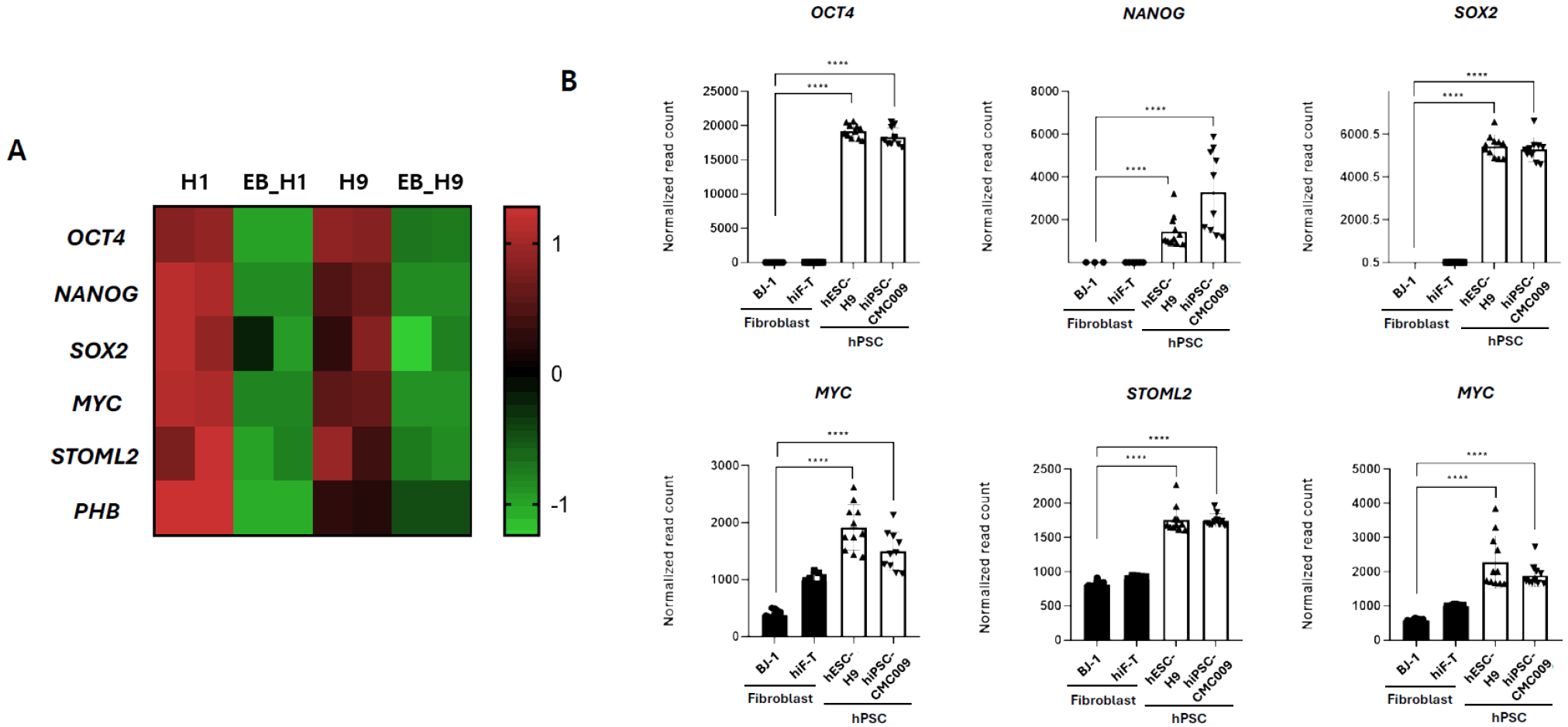
To evaluate the expression patterns of STOML2 and PHB relative to canonical stemness markers, we performed transcriptome analysis using RNA sequencing (RNA-seq) data generated in our laboratory. Heatmap visualization (Z-score normalized) revealed that pluripotency-associated transcription factors—including OCT4, NANOG, SOX2, and MYC—were highly expressed in undifferentiated hESC-H9 and hiPSC-CMC009 but were significantly downregulated in hESC-EBs and human inducible fibroblast-like TERT cells (hiF-T). Notably, STOML2 and PHB exhibited expression patterns similar to those of core pluripotency markers, showing preferential expression in undifferentiated hPSCs and marked downregulation in differentiated or somatic cell types (Fig. 1A). To validate these findings, RT-qPCR was performed to quantify gene expression in human fibroblast (BJ1 and hiF-T) and undifferentiated hPSC lines (H9 hESCs and iPSC-CMC009 hiPSCs). Consistent with the RNA-seq analysis, OCT4, NANOG, SOX2, MYC, STOML2, and PHB were all expressed at significantly higher levels in hESCs and hiPSCs compared to fibroblasts (Fig. 1B). Differences in expression across all genes were statistically significant (p<0.0001). These results confirmed that STOML2 and PHB, in a manner comparable to established pluripotency factors, are specifically enriched in undifferentiated hPSCs and may help to maintain stem cell identity.
STOML2 and PHB are progressively downregulated during differentiation and share an expression pattern with core pluripotency genes
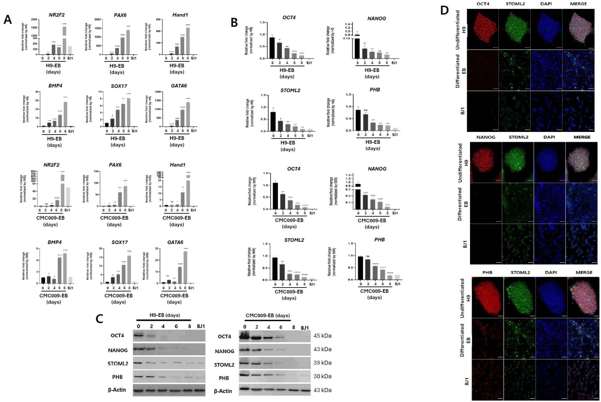
To validate the transcriptional patterns of STOML2 and PHB identified in bulk RNA-seq data, their expression was examined during spontaneous EB differentiation of hPSCs. EBs were generated from hESC-H9 and hiPSC-CMC009 and harvested at 2-day intervals for RT-qPCR analysis, western blotting, and ICC. RT-qPCR analysis demonstrated that as differentiation progressed, the mRNA levels of lineage-specific markers—including NR2F2 and PAX6 (ectoderm), Hand1 and BMP4 (mesoderm), and SOX17 and GATA6 (endoderm)—increased in a time-dependent manner, confirming successful induction of 3-germ layer differentiation (Fig. 2A). Conversely, the expression of the core pluripotency markers OCT4 and NANOG progressively declined during differentiation. Notably, STOML2 and PHB also showed a significant and gradual decrease in expression, mirroring the decline in canonical stemness genes (Fig. 2B). Western blot analysis further confirmed these trends at the protein level. In H9 and CMC009 cells, OCT4, NANOG, STOML2, and PHB proteins were abundantly expressed at day 0; however, they progressively diminished from day 2 onwards. By day 8, protein levels were nearly undetectable, similar to those observed in BJ1 fibroblasts (Fig. 2C). This consistency between mRNA and protein expression levels supports the stem cell–specific nature of STOML2 and PHB expression. To visualize the spatial localization and co-expression of STOML2 and PHB with known stemness markers, immunocytochemical staining was performed for OCT4–STOML2, NANOG–STOML2, and PHB–STOML2 in H9 hESCs (day 0), differentiated H9 EBs (day 8), and BJ1 fibroblasts. In undifferentiated hESCs, OCT4 and NANOG strongly co-localized with STOML2 within the same cells, and STOML2 and PHB were also robustly co-expressed, indicating that these proteins are selectively enriched in the pluripotent state. In contrast, differentiated EBs and fibroblasts exhibited negligible staining for all four proteins, with merged images confirming their loss of expression upon differentiation (Fig. 2D). Collectively, these results demonstrate that STOML2 and PHB are co-expressed with core pluripotency factors and are specifically enriched in undifferentiated hPSCs, supporting their potential roles as stemness-associated regulators.
STOML2 and PHB expression is progressively upregulated during reprogramming and parallels core pluripotency gene reactivation
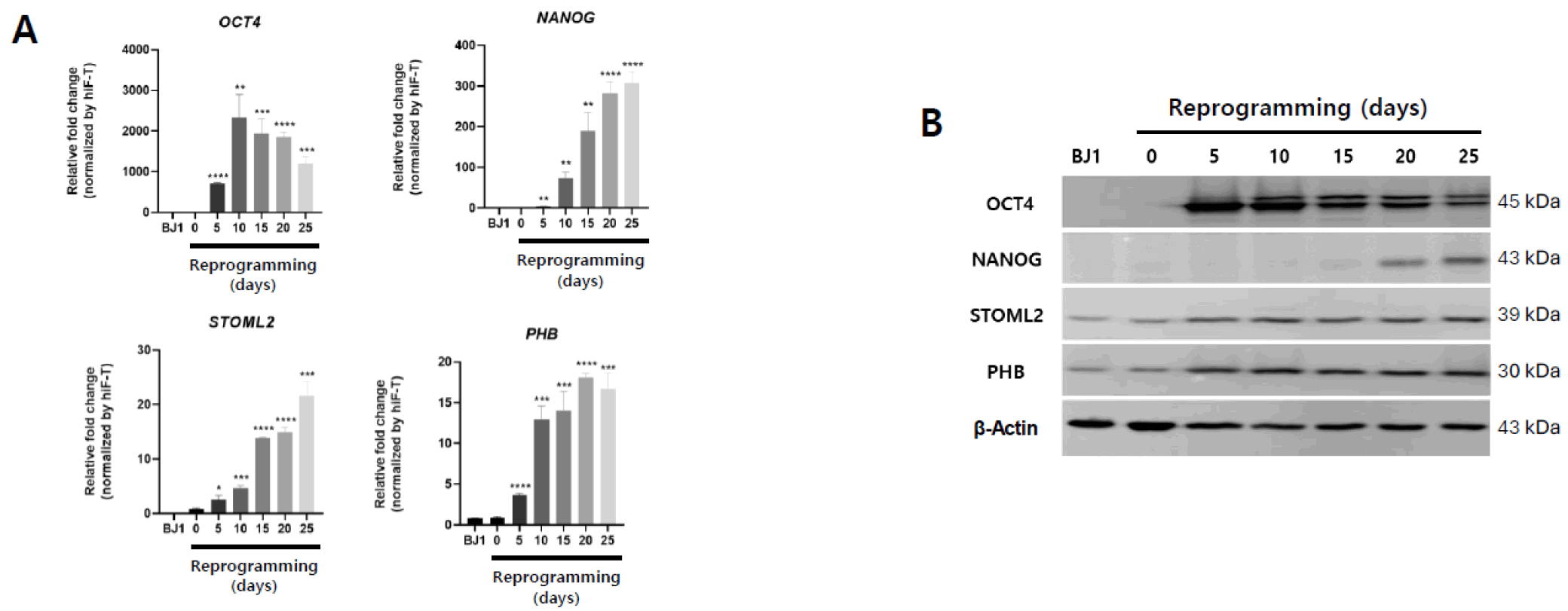
To assess whether STOML2 and PHB exhibit parallel expression patterns during the acquisition of pluripotency, we analyzed their transcriptional dynamics during reprogramming in a hiF-T secondary reprogramming system (45). Cells were harvested every 5 days from day 0 to day 25 following reprogramming induction, and expression of OCT4, NANOG, STOML2, and PHB was assessed via RT-qPCR and western blot analysis (Fig. 3A, 3B). RT-qPCR analysis revealed a gradual and coordinated increase in the mRNA levels of all four genes over time. Notably, STOML2 and PHB expression increased markedly from day 5, reaching high levels by day 25, with STOML2 showing approximately a 20-fold elevation and PHB exhibiting a similar induction of approximately 18-fold relative to baseline (Fig. 3A). These trends closely paralleled the reactivation of canonical pluripotency markers OCT4 and NANOG, suggesting that STOML2 and PHB are induced as part of the reprogramming transcriptional program. At the protein level, western blot analysis confirmed the progressive accumulation of STOML2 and PHB throughout reprogramming. Similar to OCT4 and NANOG, their protein levels markedly increased between day 5 and 25, reaching comparable expression levels by the late reprogramming stage (Fig. 3B). In contrast, BJ1 fibroblasts showed negligible expression of all four proteins, further supporting STOML2 and PHB expression specificity in the pluripotent state. These results indicate that STOML2 and PHB are highly expressed in established hPSCs and actively upregulated during reprogramming, suggesting their role in re-establishing stem cell identity and activating pluripotency-associated gene expression networks.
STOML2 KD impairs stemness maintenance in hPSCs and downregulates pluripotency markers and ERK/MAPK signaling
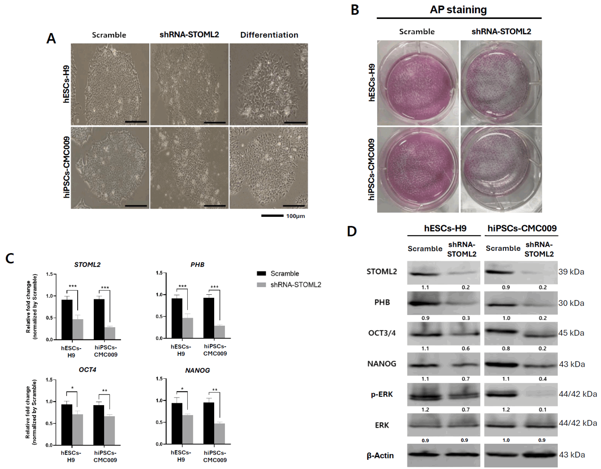
To investigate STOML2’s role in maintaining pluripotency, shRNA-mediated KD of STOML2 was performed in hESC-H9 and hiPSC-CMC009, followed by morphological, transcriptional, and protein analyses. Phase-contrast imaging showed that control (Scramble) cells maintained typical hPSC colony morphology, characterized by tightly packed cells and well-defined colony borders. In contrast, STOML2-KD cells displayed flattened, loosely organized colonies with irregular edges and an expanded cell footprint, exhibiting mesenchymal-like features consistent with early differentiation (Fig. 4A). Alkaline phosphatase (AP) staining further supported this observation: Scramble colonies displayed strong AP activity, whereas STOML2-KD colonies showed reduced staining intensity, alongside decreased colony number and size (Fig. 4B). RT-qPCR analysis confirmed efficient STOML2 KD in both cell lines, with transcript levels reduced by >50% relative to controls (p<0.001). Expression of PHB, a downstream target of STOML2, was also significantly decreased (p<0.001), suggesting a regulatory relationship. Furthermore, the expression of core stemness genes OCT4 and NANOG was markedly reduced in STOML2-KD cells (Fig. 4C). Western blot analysis corroborated these findings at the protein level. STOML2 protein expression decreased by approximately 80% in H9 and 75% in CMC009 cells relative to that of the controls. PHB protein levels also significantly declined following STOML2 KD. Consistently, OCT4 and NANOG protein levels were significantly reduced in STOML2-KD cells (Fig. 4D). ICC was performed to visualize the spatial localization and co-expression of these proteins. In Scramble cells, STOML2 was strongly co-expressed with OCT4, NANOG, and PHB in hESCs and hiPSCs, confirming their enrichment in the pluripotent state. However, STOML2-KD cells exhibited markedly reduced STOML2 staining, accompanied by heterogeneous and diminished OCT4, NANOG, and PHB expression. In some colonies, nuclear OCT4 and NANOG signals were nearly undetectable. Additionally, morphological disintegration of colonies with peripheral cell dispersion was observed (Supplementary Fig. S2).
Notably, STOML2 KD resulted in a strong reduction of phosphorylated ERK (p-ERK), indicating impaired ERK/MAPK pathway activity. Total ERK levels remained unchanged between groups, suggesting that STOML2 regulates pathway activation rather than ERK protein abundance. These data collectively indicate that STOML2 is essential for maintaining PHB and pluripotency gene expression and sustaining ERK signaling activity in hPSCs (Fig. 4D).
STOML2 is required for proper cell cycle progression and suppression of spontaneous hPSC differentiation
To elucidate the functional significance of STOML2 in hPSCs, GO and KEGG pathway enrichment analyses were performed based on transcriptomic data (Supplementary Fig. S1). These analyses revealed that STOML2 is functionally associated with cell cycle regulation genes and ERK/MAPK signaling. Therefore, we next evaluated whether STOML2 modulates cell proliferation and cell cycle progression in hPSCs.
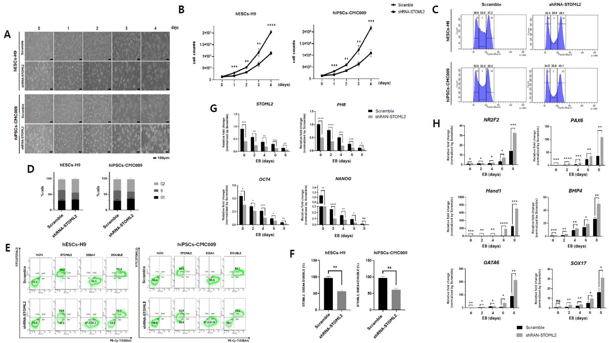
STOML2 KD was induced in hESC-H9 and hiPSC-CMC009 using shRNA, and cellular proliferation was assessed over a 4-day period. A phase-contrast microscope showed normal hPSC colony expansion, whereas STOML2-KD cells displayed visibly smaller colonies and lower cell density, especially apparent on days 3 and 4 (Fig. 5A). Quantitative cell counts confirmed significantly slower proliferation in STOML2-KD cells, reaching <1 million cells by day 4, compared to >2 million in controls (p<0.01∼0.0001, Fig. 5B). We further investigated whether this reduced proliferation was accompanied by alterations in cell cycle distribution. Flow cytometry analysis revealed that approximately 32% of Scramble control cells were in the S phase, consistent with the typical high hPSC replication activity. In contrast, STOML2-KD cells showed a marked reduction in the S phase population (approximately 20.9%) and a corresponding increase in G2/M phase cells (from approximately 37% to >45%), with no significant difference observed in the G1 phase (Fig. 5C, 5D). These changes were consistently observed in hESCs and hiPSCs, suggesting that STOML2 is essential for maintaining the rapid cell cycle dynamics characteristic of pluripotent cells.
Given that STOML2-KD impaired ERK/MAPK signaling and reduced expression of stemness-associated genes (OCT4, NANOG, and PHB), whether STOML2 expression correlates with stem cell identity at the single-cell level was evaluated. Dual immunostaining of STOML2 and the surface pluripotency marker SSEA-4, followed by FACS analysis, revealed that >90% of H9 and 009 hPSCs were STOML2+/SSEA-4+ under control conditions. However, in STOML2-KD cells, this double-positive population was significantly reduced to 54.5% (H9) and 59.2% (CMC009) (Fig. 5E). Quantification showed a >40% reduction in STOML2+/SSEA-4+ cells upon KD (p<0.01, Fig. 5F), strongly suggesting that STOML2 is selectively expressed in pluripotent cells and required for maintaining their identity.
We examined whether STOML2 is involved in maintaining stemness and suppressing spontaneous differentiation. Expression of pluripotency markers (STOML2, PHB, OCT4, and NANOG) and lineage-specific differentiation markers, NR2F2 and PAX6 (ectoderm), Hand1 and BMP4 (mesoderm), and GATA6 and SOX17 (endoderm), was assessed during spontaneous EB differentiation over 8 days. In Scramble controls, pluripotency genes gradually declined, whereas in STOML2-KD cells, they decreased more rapidly—particularly STOML2 and PHB, which significantly dropped from day 2, reaching less than half the expression level of controls by day 8 (Fig. 5G). Conversely, lineage markers increased significantly faster and reached higher levels in STOML2-KD cells. By day 8, several markers were expressed at two to five times the level observed in controls, indicating accelerated spontaneous differentiation upon STOML2 depletion (Fig. 5H). Collectively, these data demonstrate that STOML2 is a crucial regulator of hPSC-specific cell cycle progression and contributes to the maintenance of stemness, whereas its depletion is associated with enhanced differentiation.
Discussion
Human PSCs are defined by their capacity for self-renewal and their potential to differentiate into all three germ layers. These properties render hPSCs invaluable for regenerative medicine, disease modeling, and developmental biology. Pluripotency is maintained via precise regulation of transcriptional networks, signaling cascades, and cellular metabolism. Notably, hPSCs exhibit a uniquely short G1 phase and an extended S phase, allowing for their characteristic rapid proliferation. Disruption of this cycle is closely linked to the loss of stemness and differentiation onset. However, the molecular mechanisms that coordinate cell cycle progression with stem cell maintenance remain incompletely understood.
In this study, we identify STOML2 as a regulator that integrates mitochondrial organization with nuclear signaling to maintain pluripotency in hPSCs. Although previous studies associated STOML2 with metabolic adaptation and proliferative behavior in non-stem cell contexts, they did not address its relevance to stem cell identity. Our data suggest that STOML2 regulates hPSC self-renewal and inhibits spontaneous differentiation via the PHB–ERK/MAPK axis. Transcriptomic analysis and PPI network modeling revealed that STOML2 and PHB form a signaling hub connecting ERK and core stemness regulators such as OCT4 and NANOG. STOML2 and PHB were highly expressed in undifferentiated hPSCs, downregulated upon differentiation, and upregulated during reprogramming, highlighting their dynamic regulation across pluripotent state transitions. ICC and flow cytometry confirmed that STOML2 was co-expressed with stemness markers in hPSCs, but not in differentiated or somatic cells.
Functionally, STOML2 KD in hPSCs induced considerable loss of pluripotency, as evidenced by morphological changes, reduced AP activity, and decreased expression of pluripotency-associated genes. Notably, p-ERK levels significantly decreased upon STOML2 depletion, whereas total ERK remained unchanged, demonstrating that STOML2 sustains ERK signaling activation—likely via PHB stabilization. This identifies STOML2 as a key upstream modulator of a canonical pluripotency-related pathway.
A particularly novel aspect of our findings is the impact of STOML2 on cell cycle progression. STOML2 KD significantly reduced the proportion of S phase cells and increased G2/M phase accumulation, correlating with a slower growth rate. This hPSC cycle disruption suggests that STOML2 is required for maintaining the unique cell cycle configuration that supports self-renewal. STOML2 also prevents premature differentiation, acting as a molecular gatekeeper beyond maintaining stemness. Loss of STOML2 accelerated lineage-specific gene expression from all three germ layers and simultaneously suppressed stemness marker expression. Notably, this effect was observed as early as day 2 of spontaneous differentiation, implying that STOML2 actively suppresses differentiation cues even under basal conditions. The dual role of STOML2—promoting pluripotency while inhibiting lineage commitment—suggests it may serve as a binary switch in stem cell fate regulation. Notably, our results highlight a novel STOML2–PHB–ERK/MAPK axis linking mitochondrial signaling to nuclear gene expression to sustain hPSC identity. Although PHB regulates pluripotency genes via epigenetic mechanisms, this study is the first to identify STOML2 as its upstream modulator in hPSCs. As ERK plays context-dependent roles in promoting or suppressing differentiation, STOML2 likely fine-tunes this pathway to favor stemness maintenance.
STOML2 aids in maintaining pluripotency by contributing to stem cell–specific cell cycle progression and the self-renewal transcriptional network. By modulating PHB levels and sustaining ERK activity, STOML2 promotes pluripotency-associated gene expression and delays premature differentiation. These findings suggest that STOML2 may function as a mediator linking mitochondrial activity to nuclear gene regulation in hPSCs. STOML2 and PHB are robustly upregulated during pluripotency acquisition and maintenance, with expression dynamics paralleling those of core stemness regulators. Unlike canonical transcriptional markers such as OCT4, NANOG, and SOX2, however, their contribution likely lies in mitochondrial organization and ERK–PHB signaling, which support the metabolic state required for pluripotency. This also explains the residual expression observed in differentiated cells, where mitochondrial stability remains essential. Although the direct upstream regulators of STOML2 in the pluripotent state have not been fully elucidated, several pathways may provide mechanistic links. In other cellular contexts, STOML2 expression responds to IL-6–JAK–STAT3 signaling, hypoxic cues via HIF-1α, and inflammatory or metabolic stress, suggesting that environments favoring PSC reprogramming or naïve-like maintenance could activate STOML2 through similar routes. Moreover, the role of STOML2 in regulating mitochondrial metabolism and mitophagy in cancer stem cells, a context in which it participates in metabolic remodeling, raises the possibility that pluripotent cells may leverage analogous mitochondrial axes to fine-tune cell fate decisions. Further studies are needed to determine the upstream regulatory mechanisms of STOML2, its interactions with other mitochondrial pathways, and its potential in disease modeling or stem cell-based therapeutic strategies.
Supplementary Materials
Supplementary data including two figures can be found with this article online at https://doi.org/10.15283/ijsc25092
 XML Download