



 PDF
 Citation
 Print


Introduction
Huntington’s disease (HD) is an autosomal dominant neurodegenerative disorder caused by an unstable cytosine-adenine-guanine (CAG) repeat expansion in the huntingtin (HTT) gene. This genetic aberration results in the production and toxic intracellular aggregation of mutant huntingtin (mHTT), triggering a multi-faceted pathological cascade (1). A hallmark of HD is the selective degeneration of striatal medium spiny neurons (MSNs), which underlies the progressive motor, cognitive, and psychiatric impairment, disruption of proteostatic clearance mechanisms, and chronic neuroinflammation (2, 3).
Although mHTT-lowering strategies, such as antisense oligonucleotide and gene-editing approaches, are being actively developed for early intervention, these modalities are inherently limited in their capacity to restore degenerated neural circuitry in symptomatic stages of the disease (4, 5). In this context, cell-based therapies using human pluripotent stem cell-derived neural precursors have emerged as a promising regenerative strategy. Such approaches offer the potential to replace lost MSN populations, reconstitute damaged striatal circuitry, and provide sustained trophic support within the diseased microenvironment (6). In particular, transplanted neural precursors may serve as a source of neurotrophic factors, including brain-derived neurotrophic factor (BDNF), which is markedly reduced in the HD striatum and is essential for neuronal survival and synaptic maintenance (7). Consistent with this concept, preclinical studies have demonstrated that intrastriatally transplanted neural precursors can survive long-term, differentiate into neuronal lineages, and ameliorate motor deficits in experimental models of HD (6, 8).
In the present study, we assessed the therapeutic effects of clinical-grade human induced pluripotent stem cell (iPSC)-derived neural precursor cells (s513-NPCs) using two complementary rodent models of HD: the rapidly progressing R6/2 transgenic fragment model, which exhibits an aggressive disease phenotype, and the YAC128 mouse model, which harbors the full-length human HTT gene and recapitulates more protracted pathological features (9-12). In addition to evaluating functional outcomes following transplantation, we examined associated molecular and cellular changes within the host striatum, including alterations related to proteostasis and neuroinflammatory status. By comparing these effects across distinct disease contexts, this study aims to further inform the therapeutic potential of iPSC-derived neural precursor transplantation as a neuroprotective strategy for HD.
Materials and Methods
Human iPSC culture and neural differentiation
The clinical-grade, HLA (human leukocyte antigen)-homozygous 513-iPSC line (YZWJ s513; CiRA Foundation) was maintained under feeder-free conditions on iMatrix-511 (Nippi)-coated plates in StemFit Basic 03 medium supplemented with 80 ng/mL basic fibroblast growth factor (bFGF; Ajinomoto). As previously described (13, 14), iPSCs were dissociated into single cells using TrypLE Select (Gibco) and seeded at a density of 6,000 cells per microwell in an AggreWell 800 plate (STEMCELL Technologies) for neural induction. Differentiation was initiated via a dual-SMAD inhibition protocol for 8 days in neural induction medium containing 100 nM LDN-193189 and 10 μM SB431542. On day 9, resulting embryoid bodies were dissociated with Accutase (Gibco) to obtain s513-NPCs, which were then expanded in neural maintenance medium supplemented with 2% B-27 Supplement without vitamin A (Gibco) and 1% N2 supplements (Gibco) in a 1:1 mixture of DMEM/F12 (Gibco) and Neurobasal (Gibco).
Flow cytometric characterization
To confirm lineage purity, s513-NPCs were dissociated using 0.5 mM EDTA and incubated with phycoerythrin (PE)-conjugated antibodies against polysialylated neural cell adhesion molecule (PSA-NCAM) and human natural killer-1 (HNK-1), and an allophycocyanin (APC)-conjugated antibody against CD271 (Miltenyi Biotec). Specificity was verified using isotype-matched controls (IgG/IgM). Data were acquired on a BD AccuriTM C6 Plus and analyzed with BD Accuri software (version 1.0.34.1). Flow cytometric analysis confirmed high neural lineage purity. In addition, residual pluripotency markers (OCT4, SOX2, TRA-1-60, and SSEA4) were not detected during clinical-grade release testing and internal validation of the same s513 cell line.
Transgenic mouse models and stereotactic transplantation
All procedures were approved by the CHA University IACUC (No. 220047). All mice were housed and maintained within the centralized animal facility at the CHA Bio Complex. To support xenograft survival, cyclosporine A (5 mg/kg, Sigma) was administered intraperitoneally starting three days prior to transplantation and continued daily until sacrifice (4 weeks for R6/2; 3 months for YAC128). We utilized two models: the R6/2 fragment model (B6CBA background) and the YAC128 full-length genomic model (FVB/N background). Symptomatic 6-month-old YAC128 mice received bilateral striatal injections of a total of 4×10⁵ s513-NPCs in 4 μL (anterior-posterior [AP] +0.5 mm, medio-lateral [ML] ±1.8 mm, dorso-ventral [DV] −3.0 mm and −4.0 mm), while 5-week-old R6/2 mice received a total of 2×10⁵ s513-NPCs in 2 μL (AP +0.5 mm, ML ±2.0 mm, and DV −3.25 mm), reflecting differences in disease progression rate and striatal volume between models. Vehicle controls received an equivalent volume of saline.
Behavioral assessments
Motor performance was evaluated through a battery of assays including accelerating rotarod (0∼40 rpm), grip strength, open field activity (EthoVision XT 11.5; Noldus), hindlimb clasping, and the pole test as previously described (15, 16). Testing was conducted weekly for R6/2 mice and at 4-week intervals for YAC128 mice post-transplantation.
Immunostaining
Following the completion of all behavioral assessments, all animals were euthanized and transcardially perfused with 4% paraformaldehyde (PFA) in phosphate-buffered saline (PBS). The excised brains were post-fixed and cryoprotected before being coronally sectioned at 30 μm using a cryotome (CM3050, Leica). For both fixed cells and brain slices, samples were incubated for 1 hour at room temperature in a blocking buffer composed of 1% bovine serum albumin (BSA) and 0.1%∼0.3% Triton X-100 in PBS. Primary antibodies were applied and incubated overnight at 4℃. Primary antibodies used: KU80 (goat, AF5619, 1:500), Nestin (rabbit, ab105389, 1:500), MAP2 (rabbit, ab32454, 1:500), GFAP (rabbit, z0334, 1:500), Olig2 (mouse, mabn50, 1:500), and MW8 (DSHB, 1:100). Following washing steps, species-appropriate fluorescent dye-conjugated secondary antibodies were incubated for 1 hour at room temperature. Nuclei were visualized by counterstaining with 4,6‐diamidino‐2‐phenylindole (DAPI, ThermoFisher Scientific, 1:1,000 dilution). Fluorescent images were systematically captured and analyzed using a confocal microscope (LSM880, Zeiss).
Histological analysis and quantification
Quantitative assessments of graft survival, lineage differentiation, and host pathological responses were performed on serial coronal brain sections. For each animal, three representative sections (30 μm thickness) spanning the striatal graft core (AP range: +1.4 mm to −0.4 mm relative to bregma) were systematically selected for analysis. To evaluate in vivo differentiation, the profile was quantified by calculating the percentage of the human-specific nuclear KU80-positive donor cells that co-expressed specific neural lineage markers. The MW8 antibody was utilized to detect aggregated huntingtin inclusions. Regions of interest (ROIs) were strictly defined within the dorsal striatum and cortex. The mHTT burden was quantified as the percentage of the total ROI area occupied by MW8-immunoreactive aggregates (% Area) (17). All images were processed using a constant thresholding algorithm to segment aggregates from background noise, and measurements were executed using Fiji/ImageJ software.
Western blot analysis
Cortical and striatal tissues were independently dissected and processed for western blot analysis, and region-specific data are presented accordingly in the corresponding figures. Cortical and striatal tissues were lysed in radioimmunoprecipitation assay (RIPA) lysis buffer supplemented with a protease inhibitor cocktail (Roche). Protein concentrations were determined by the bicinchoninic acid (BCA) protein assay. Equivalent protein amounts were separated by 8, 10 or 12% SDS-PAGE, transferred to polyvinylidene fluoride (PVDF) membranes, and probed for targets including iNOS (rabbit, PA1-036), IL-1β (rabbit, SC-7884), Arginase-1 (ARG; mouse, SC-166920), LC3 (rabbit, 2775s), and ubiquitin (mouse, SC-8017). Immunoreactive bands were detected using enhanced chemiluminescence (ECL, Millipore) and imaged using a Bio-Rad imaging system. Band intensities were normalized to β-actin as the loading control for all immunoblot analyses. Densitometric analysis was performed using Image Lab software (Bio-Rad) to quantify the relative intensity of each band.
Statistical analysis
All quantitative data are reported as the mean±standard error of the mean. Statistical significance was determined using unpaired t-test or one-way ANOVA (analysis of variance) with Bonferroni post-hoc correction (α=0.05), as appropriate for the experimental design, using GraphPad Prism software (v5.01).
Results
High lineage fidelity and striatal potential of s513-NPCs
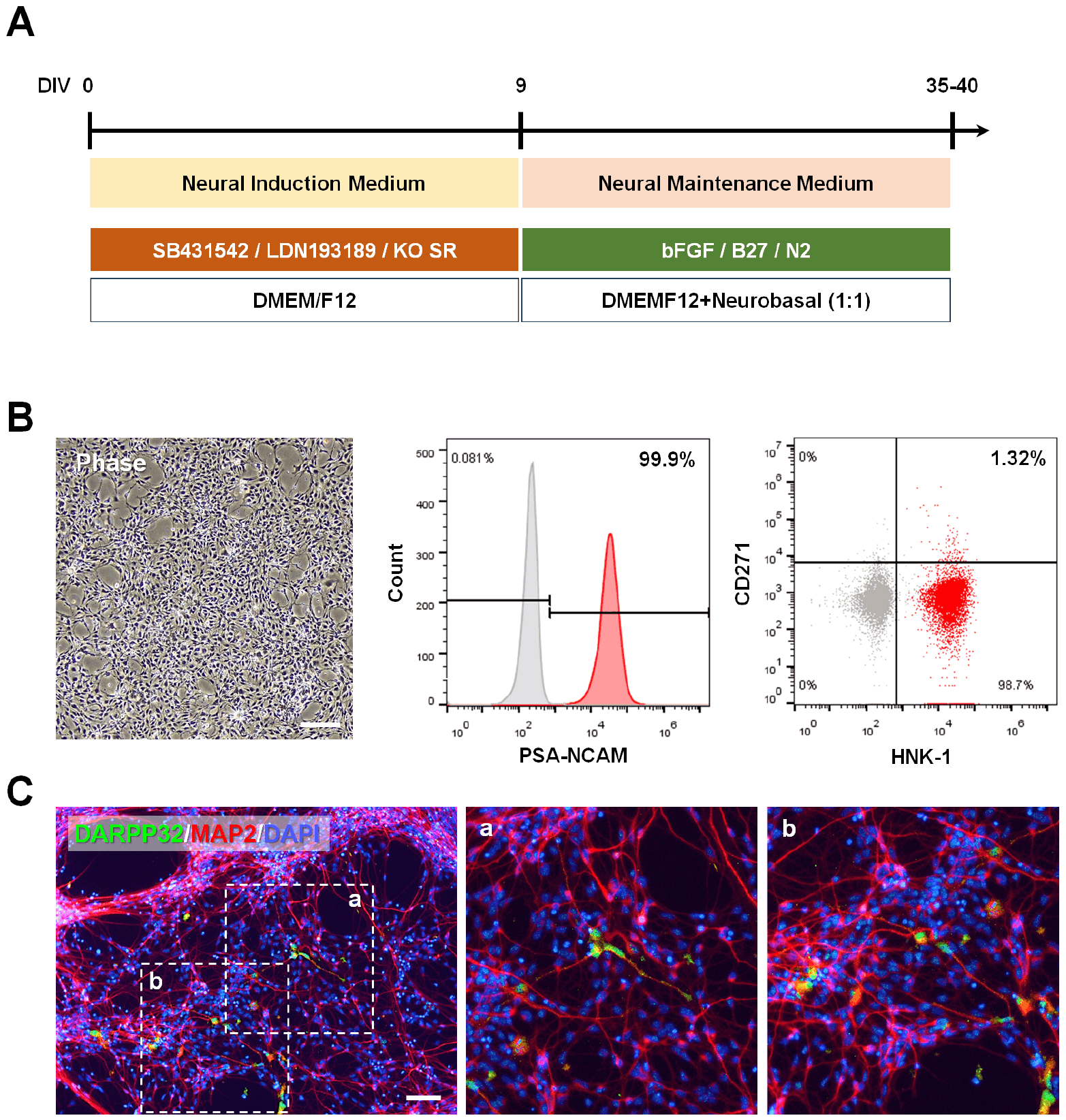
To establish a scalable and standardized cell source for transplantation, clinical-grade s513-iPSCs were differentiated into NPCs using a microwell-based dual-SMAD inhibition protocol (Fig. 1A). Flow cytometric analysis confirmed the high purity and lineage fidelity of the resulting NPC population, characterized by >99% PSA-NCAM expression and a minor population (<3%) of HNK-1+/CD271+ neural crest derivatives (Fig. 1B). Upon in vitro terminal differentiation for 21 days, s513-NPCs demonstrated robust neurogenic capacity, yielding a mature MAP2+ neuronal population. Notably, a significant subpopulation co-expressed the striatal-specific marker DARPP-32 (Fig. 1C), confirming the intrinsic competency of s513-NPCs to generate MSNs, the primary cell type lost in HD.
s513-NPC transplantation ameliorates motor deficits across diverse Huntington’s disease phenotypes
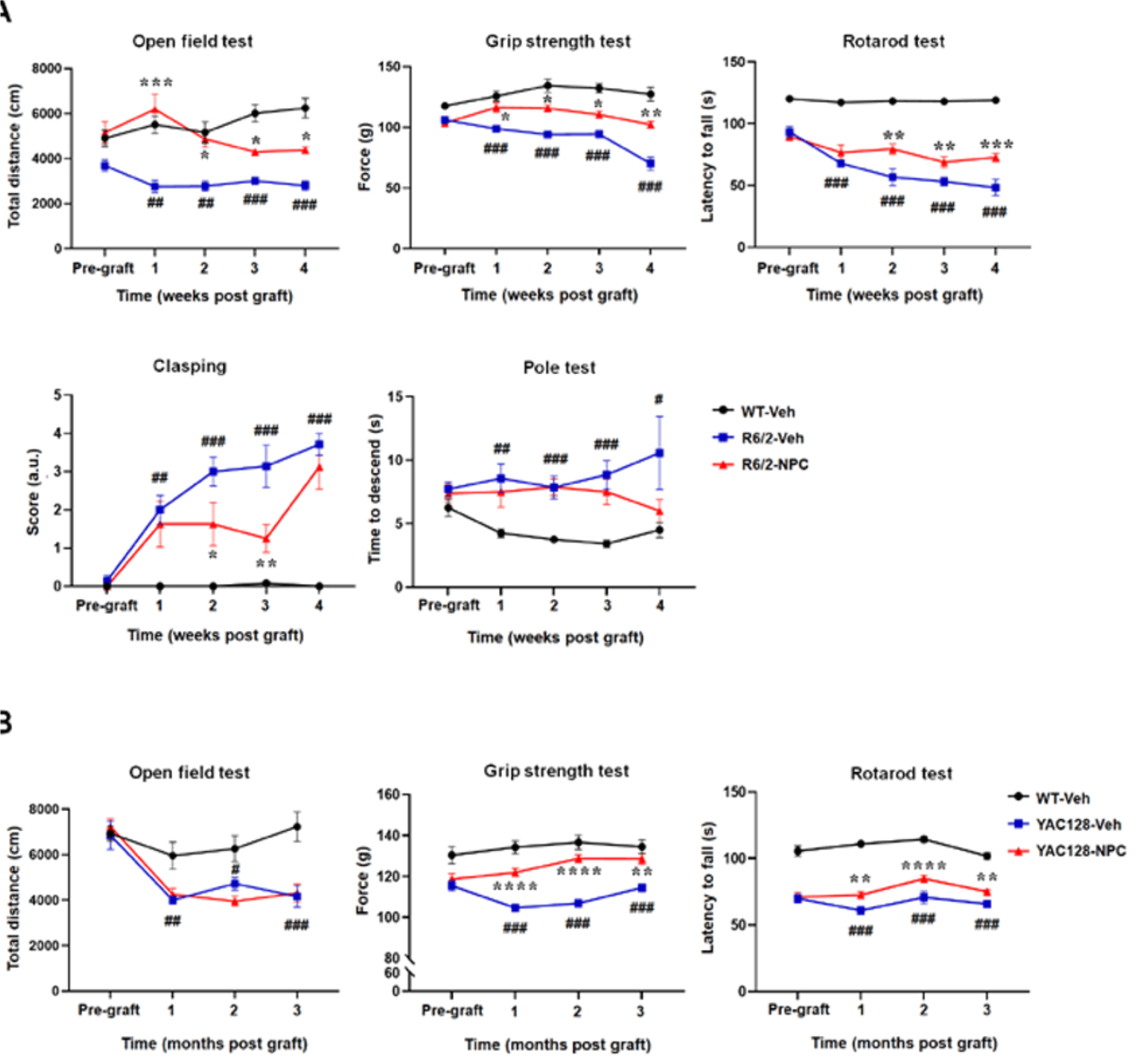
To evaluate functional efficacy, we performed longitudinal behavioral phenotyping in two complementary HD models: the rapidly progressing R6/2 fragment model and the protracted YAC128 full-length genomic models. In the R6/2 model, NPC-transplanted mice (R6/2-NPC) exhibited rapid and sustained functional recovery. Significant improvements in forelimb grip strength and spontaneous locomotor activity were observed as early as 1 week post-transplantation and persisted throughout the 4-week study period (Fig. 2A; p<0.05). Furthermore, R6/2-NPC mice displayed superior motor coordination on the rotarod (weeks 2∼4; p<0.01) and a marked reduction in the severity of dystonic hindlimb clasping (weeks 2∼3; p<0.05) (Fig. 2A). In the YAC128 model, s513-NPC transplantation (YAC128-NPC) provided durable symptomatic relief over a chronic period. Enhanced grip strength and rotarod latency emerged at 1 month post-grafting and remained significantly improved throughout the 3-month evaluation (Fig. 2B; p<0.01). Spontaneous exploratory activity in the open field remained comparable between the YAC128-NPC and vehicle groups, suggesting that the therapeutic effects were dominantly motor-specific in this model (Fig. 2B). Collectively, these findings indicate that s513-NPCs provide robust functional amelioration across distinct pathological contexts of HD.
In vivo engraftment and advanced neuronal maturation of transplanted s513-NPCs
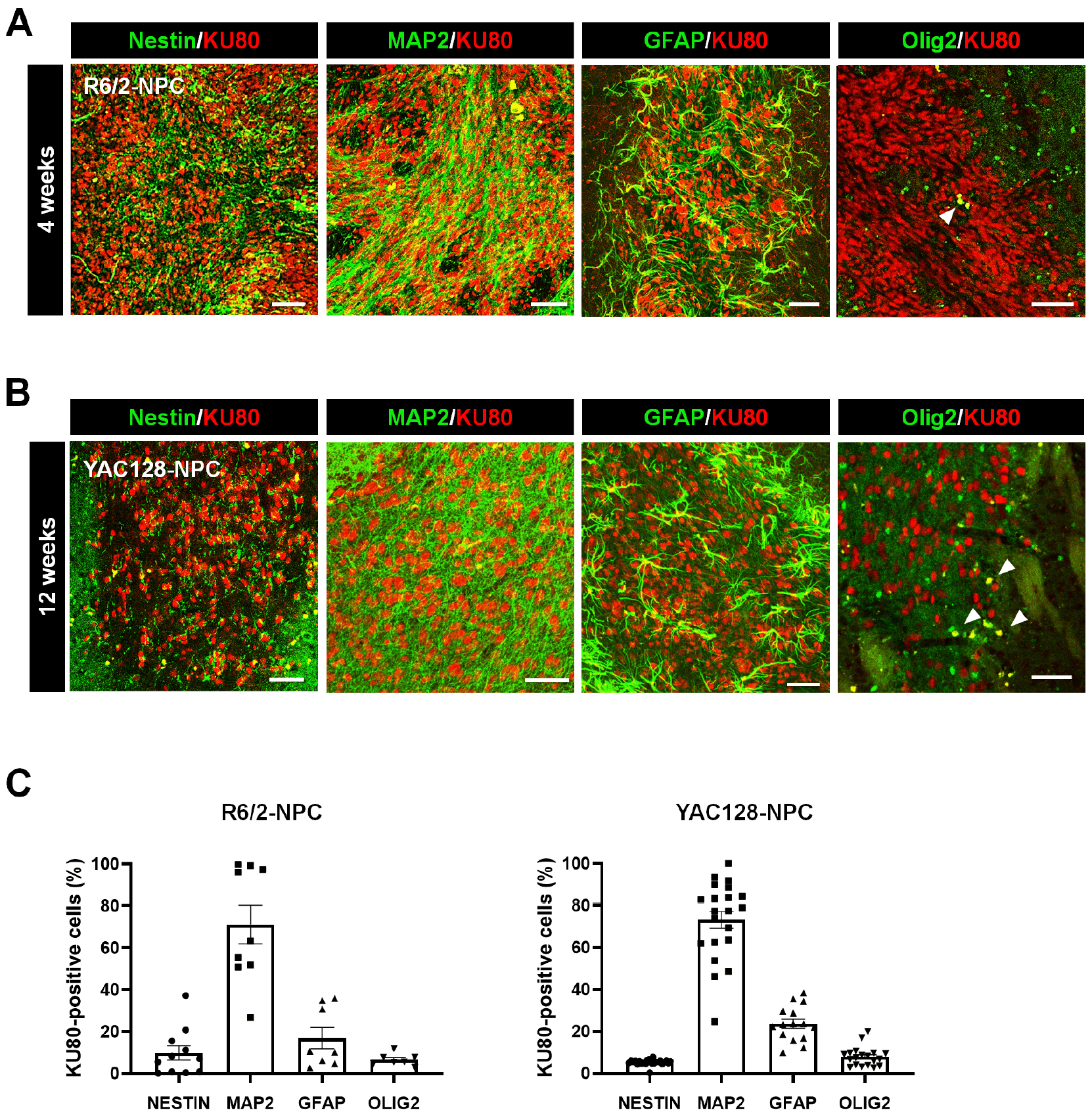
We used the human-specific nuclear marker, KU80 to delineate the in vivo trajectory of the grafted cells. In the R6/2 model at the 4-week endpoint, 71.11%±8.61% of KU80-positive donor cells had progressed to mature neuronal fate expressing MAP2 (Fig. 3A, 3C). Only a minor fraction (9.80%±3.25%) retained the progenitor marker, Nestin (Fig. 3A, 3C). Multilineage potential was evidenced by differentiation into astrocytic (GFAP+, 16.91%±4.72%) and oligodendrocytic (OLIG2+, 6.63%±1.00%) lineages (Fig. 3A, 3C). Long-term evaluation in the YAC128 model at 3 months confirmed stable engraftment and sustained maturation. Similar to R6/2 data, the graft composition was predominantly neuronal (MAP2+, 73.10%±3.96%), with a minimal population for undifferentiated precursors (Nestin+, 5.37%±0.25%) (Fig. 3B, 3C). The glial contribution in this chronic environment included astrocytic GFAP+ cells of 23.56%±2.10% and oligodendrocytes Olig2+ cells of 7.84%±1.02% (Fig. 3B, 3C). These data indicate that s513-NPCs predominantly undergo terminal neuronal differentiation within the host striatum.
s513-NPC transplantation is associated with reduced mHTT burden and altered proteostasis-related markers
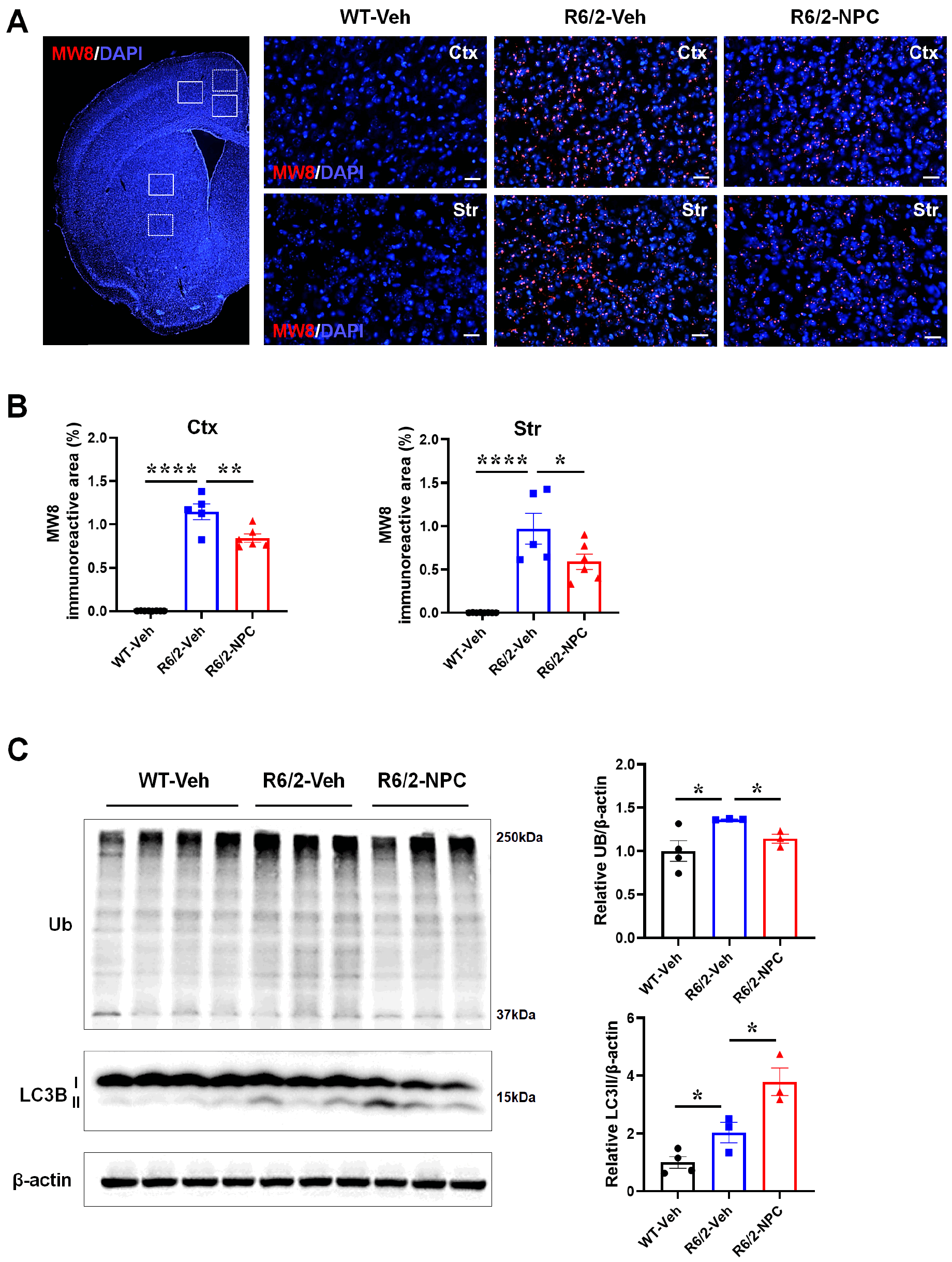
We next examined whether the observed functional improvement was accompanied by changes in mHTT-associated pathology. Quantitative analysis using the MW8 antibody revealed a significant reduction in mHTT aggregate area within both the cortex and striatum of NPC-transplanted R6/2 mice (Fig. 4A, 4B; p<0.05). Vehicle-injected R6/2 mice exhibited a marked accumulation of polyubiquitinated protein conjugates, consistent with impaired proteostatic processing (Fig. 4C; p<0.05). In contrast, s513-NPC transplantation was associated with a significant reduction in ubiquitinated protein levels (Fig. 4C; p<0.05). In parallel, increased LC3-II levels were observed in the striatum of NPC-transplanted mice (Fig. 4C; p<0.05). Together, these findings indicate that s513-NPC transplantation is accompanied by coordinated changes in proteostasis-related markers concurrent with reduced mHTT burden.
Context-dependent modulation of the BDNF-TrkB signaling axis
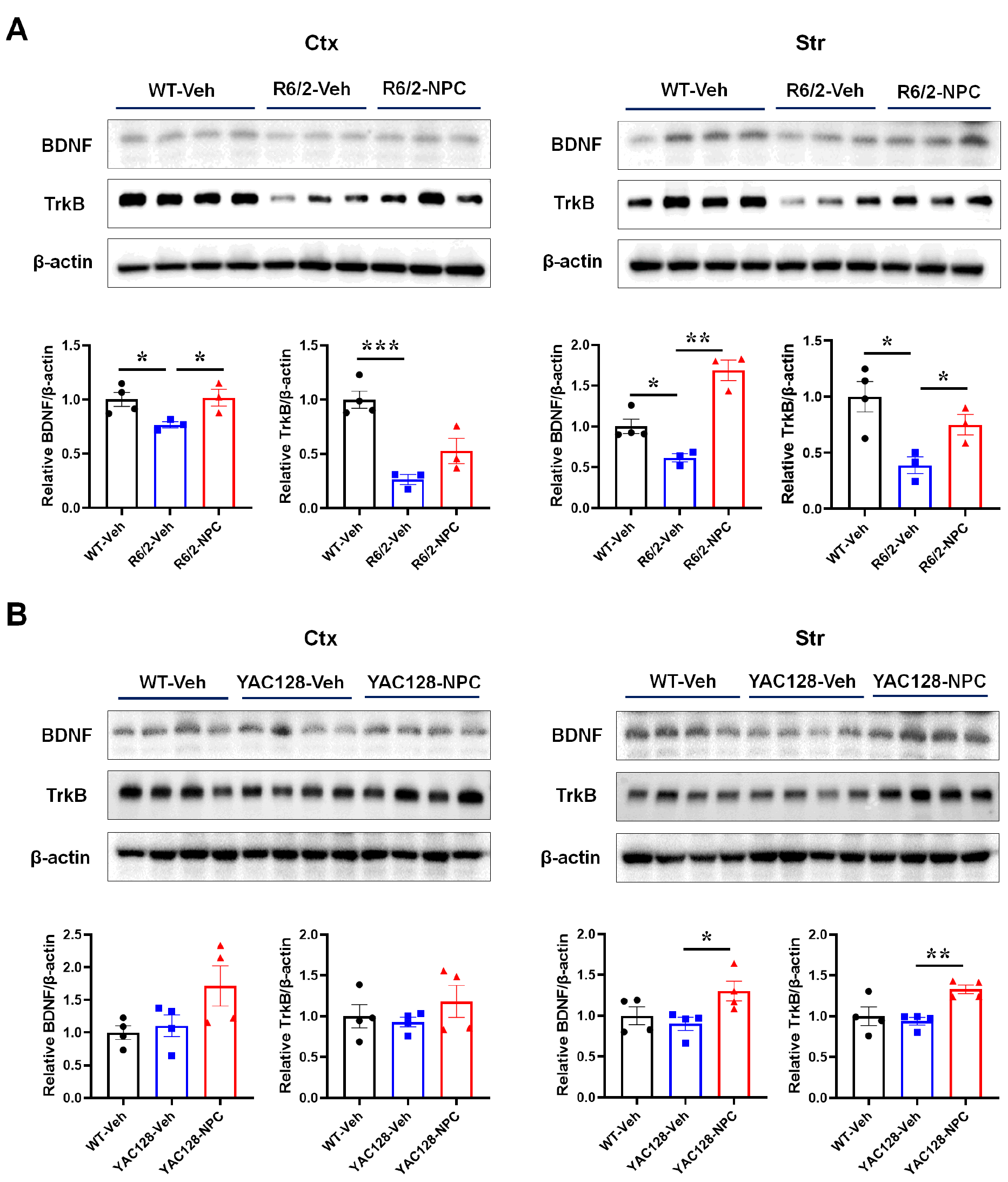
Given that BDNF depletion is closely associated with striatal vulnerability in HD, we examined changes in neurotrophic signaling following NPC transplantation. In the R6/2 model, characterized by aggressive progression and severe neurotrophic deficits, NPC grafting significantly rescued both BDNF and TrkB protein expression within the striatum (Fig. 5A; p<0.05). Cortical BDNF levels were similarly restored (Fig. 5A; p<0.05), whereas cortical TrkB expression showed only a non-significant upward trend, suggesting that receptor-level modulation is most robust at the primary site of engraftment. In contrast, within the YAC128 model, striatal BDNF and TrkB levels in vehicle-injected mice remained comparable to wild-type levels at the examined time point (Fig. 5B). Despite this preserved baseline, s513-NPC transplantation induced a significant localized augmentation of both BDNF and TrkB specifically within the striatum (Fig. 5B; p<0.05), with no significant changes observed in the cortex. These findings suggest that s513-NPC transplantation is associated with pathology-dependent modulation of local neurotrophic signaling.
Spatially distinct modulation of neuroinflammatory responses
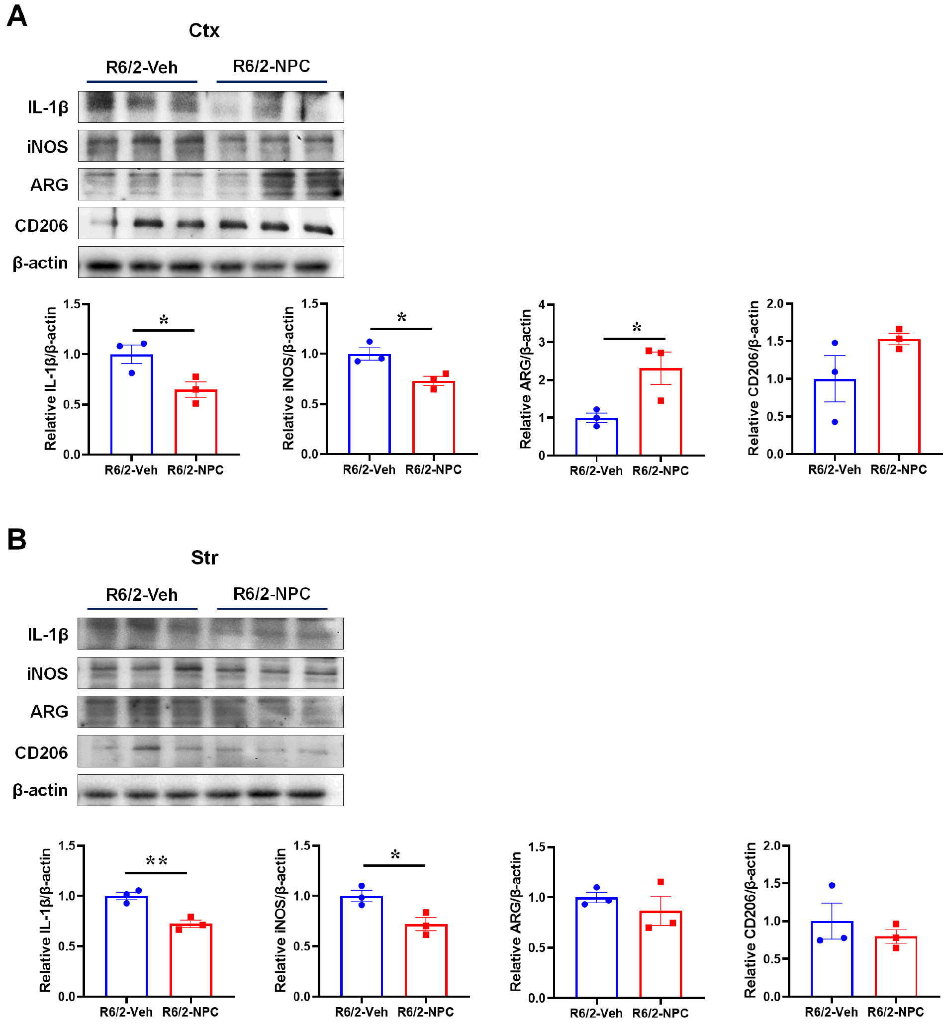
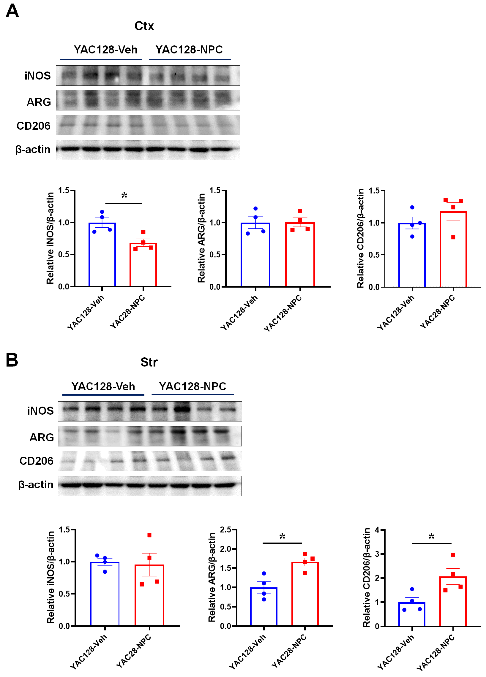
To determine the immunomodulatory influence of s513-NPCs, we quantified representative pro- and anti-inflammatory mediators in both HD models. IL-1β and iNOS were selected as representative pro-inflammatory markers, whereas ARG and CD206 were assessed as indicators of reparative immune states, consistent with established microglial activation frameworks. In the R6/2 model, NPC transplantation markedly suppressed the pro-inflammatory milieu, as evidenced by a significant attenuation of IL-1β and iNOS expression within both the cortex and striatum (Fig. 6A, 6B; p<0.05). The induction of reparative anti-inflammatory signaling exhibited distinct regionality. ARG was significantly upregulated in the cortex (Fig. 6A; p<0.05), while striatal anti-inflammatory factors remained largely unaffected at this stage (Fig. 6B). A divergent pattern of immunomodulation was observed in the YAC128 model. While pro-inflammatory suppression was more restricted, with iNOS expression significantly reduced only in the cortex (Fig. 7A; p<0.05), the anti-inflammatory response was specifically enhanced within the striatum, where levels of reparative factors were significantly elevated compared to vehicle controls (Fig. 7B; p<0.05). Collectively, these results indicate that s513-NPC transplantation is associated with region- and context-dependent modulation of neuroinflammatory responses.
Discussion
In this study, we show that intrastriatal transplantation of clinical-grade, HLA-homozygous s513-NPCs is associated with sustained improvements in motor performance and attenuation of key pathological features in both the R6/2 and YAC128 models of HD. The observation of therapeutic effects across fragment and full-length genomic models suggests that the benefits of s513-NPC transplantation are preserved across distinct disease contexts. The efficient differentiation of donor cells into DARPP-32–expressing MSN–like populations supports the notion that s513-NPCs can provide an appropriate cellular substrate within the degenerated striatum, consistent with previous reports demonstrating functional integration of human neural stem cells in HD models (6, 18, 19).
A prominent finding in the R6/2 model was the reduction in neurotoxic mHTT aggregates following NPC transplantation, which was accompanied by changes in markers related to proteostatic processing. Given prior evidence linking BDNF depletion to impaired protein quality control in the HD striatum (20-22), the observed modulation of BDNF–TrkB signaling and autophagy-related markers may contribute to the reduction of aggregate burden. While BDNF is well recognized for its role in synaptic maintenance under physiological conditions (23), accumulating evidence suggests that neurotrophic signaling may assume additional roles in stressed or degenerating neural environments, including the regulation of protein turnover pathways (24-29). Further studies will be required to determine the extent to which these processes are causally linked.
The distinct immunological responses observed across disease models further highlight the context-dependent nature of NPC–host interactions. In the rapidly progressing R6/2 model, transplantation was associated with suppression of pro-inflammatory markers, whereas in the more protracted YAC128 model, features consistent with a reparative immune milieu were observed within the striatum. These findings suggest that transplanted NPCs may interact dynamically with the host immune environment, with outcomes shaped by regional pathology and disease stage, in line with emerging concepts of microglial plasticity in neurodegenerative disorders (30-34).
From a translational perspective, the use of HLA-homozygous, clinical-grade iPSC lines may help address important challenges related to manufacturing scalability and immunological compatibility, with the potential to reduce the need for long-term immunosuppression (35-38). Nevertheless, immunosuppressive treatment with cyclosporine A was employed in the present study as a standard measure to support xenograft survival in murine models, and further studies will be required to determine the extent to which HLA matching alone can mitigate immune responses in clinical settings. The stable lineage commitment of s513-NPCs and the absence of ectopic overgrowth in both models further support the feasibility of this approach (39, 40). Taken together, our findings indicate that transplantation of iPSC-derived neural precursor cells confers reproducible neuroprotective effects in HD models and supports further investigation of this strategy as a cell-based therapeutic approach for preserving striatal function and neural integrity in HD.
 XML Download