



 PDF
 Citation
 Print


Introduction
Arts syndrome (OMIM: 301835) is a rare X-linked recessive neurodevelopmental disorder characterized by profound sensorineural hearing loss, intellectual disability, early-onset ataxia, hypotonia and recurrent infections (1). The condition is caused by loss-of-function mutations in the PRPS1 gene (phosphoribosyl pyrophosphate synthetase 1) (2), which is essential for the de novo and salvage pathways of purine and pyrimidine nucleotide synthesis. The PRPS1 gene encodes phosphoribosyl pyrophosphate synthetase 1, a critical enzyme catalyzing the synthesis of phosphoribosyl pyrophosphate (PRPP), which is a central substrate for nucleotide and nicotinamide adenine dinucleotide (NAD+) biosynthesis (3, 4). The necessity of PRPS1 for rapid proliferation and high energy demand makes its dysfunction particularly detrimental to the central nervous system (CNS), where efficient nucleotide metabolism is crucial for development, repair, and neurotransmission (5).
While the genetic basis is established, the specific cellular and molecular mechanisms by which PRPS1 mutations affect human neural development remain poorly characterized. Current therapeutic strategies are limited to supportive care. A more complete understanding of the disease pathogenesis in relevant human neural cells is necessary to identify targeted, disease-modifying therapies. The use of patient-derived induced pluripotent stem cells (iPSCs) and their subsequent differentiation into neural lineages offers a critical platform to model human neurodevelopmental disorders in vitro.
In experimental models, nicotinamide mononucleotide (NMN), another NAD+ precursor, has been shown to restore mitochondrial function and reduce senescence in mesenchymal stem cells (6), as well as to improve neurite complexity and survival in motor neurons from amyotrophic lateral sclerosis (ALS) models (7, 8). These findings suggest that NAD+ precursor therapy may have broader therapeutic potential in disorders linked to nucleotide metabolism.
In this study, we established iPSCs from an Arts syndrome patient carrying a PRPS1 missense mutation (p.V42L) to investigate disease-specific cellular phenotypes in neural stem cells (NSCs) and mature neurons. We assessed their pluripotency, neural differentiation, and cellular phenotypes compared with controls. Patient-derived NSCs exhibited reduced proliferation, abnormal nuclear morphology, and increased senescence, despite relatively preserved mitochondrial structure. Interestingly, intracellular adenosine triphosphate (ATP) levels were significantly elevated in mutant NSCs, suggesting altered energy utilization or disrupted biosynthetic flux. Furthermore, neurons derived from patient NSCs displayed shortened neurites and reduced branching complexity, reflecting abnormal neural maturation.
Finally, we explored NMN supplementation as a potential therapeutic approach. NMN, a precursor of NAD+, has been shown to restore cellular energy and improve mitochondrial and nuclear function in several metabolic and neurodegenerative models (8, 9). Our data demonstrate that NMN treatment partially rescued proliferation and morphological abnormalities, suggesting that NAD+ metabolism restoration may mitigate PRPS1-related neurodevelopmental deficits.
Materials and Methods
Ethics statement
Peripheral blood mononuclear cells (PBMCs) were obtained from a patient carrying the PRPS1 c.124G>C (p.V42L) mutation with informed consent under protocols approved by the research ethics committee of Pusan National University Yangsan Hospital (approval number: 11-2024-010). All procedures involving human-derived materials were conducted in accordance with institutional guidelines and regulations, and adhered to the principles of the Declaration of Helsinki.
Patient information
A 10-year-old male patient presented with global neurodevelopmental delay. The patient exhibited facial dysmorphisms and congenital nystagmus noted since infancy. He experienced progressive sensorineural hearing loss, ultimately requiring cochlear implantation. Visual impairment was also present, along with significant cognitive delay, with an intellectual disability level corresponding to an IQ score of 56. Neurologically, the patient demonstrated gait disturbances characterized by ataxia.
Trio-based whole-exome sequencing identified a hemizygous PRPS1 variant, c.124G>C (p.V42L), which is associated with PRPS1-related disorders, including Arts syndrome. This genetic finding is consistent with the patient’s clinical manifestations, particularly his neurodevelopmental deficits, sensorineural hearing loss, and motor coordination difficulties.
Generation and maintenance of induced pluripotent stem cells (iPSCs)
Patient PBMCs and control human PBMCs (Lonza Bioscience) were reprogrammed into iPSCs using the CytoTuneTM-iPS 2.0 Sendai Reprogramming Kit (Thermo Fisher Scientific) according to the manufacturer’s instructions. Emerging iPSC colonies with typical morphology were manually selected, expanded, and maintained under feeder-free conditions on plates coated with Vitronectin (Thermo Fisher Scientific) using Essential 8TM Medium (Thermo Fisher Scientific). Medium was changed daily, and cells were passaged every 5∼6 days using 0.5 mM EDTA solution.
Characterization of iPSCs
iPSCs were assessed for pluripotency by Pluripotent Stem Cell 4-Marker Immunocytochemistry Kit for SSEA4, OCT4, SOX2 and TRA-1-60. Karyotyping was performed using G-banding analysis by Dx&Vx, Inc. Pluripotency was further confirmed by trilineage differentiation assays. Short tandem repeat (STR) analysis of iPSCs and patient’s PBMCs was assessed in all 18 alleles by Cosmo gene technology.
Trilineage differentiation assay
The pluripotency of iPSC lines was confirmed using the STEMdiffTM Trilineage Differentiation Kit (STEMCELL Technologies) according to the manufacturer’s instructions. Briefly, iPSCs were seeded onto Vitronectin-coated 24-well plates at a density of 2.5×10⁴ cells/cm2 in Essential 8TM medium supplemented with 10 μM Y-27632 (STEMCELL Technologies) for the first 24 hours. The following day (day 0), the culture medium was replaced with lineage-specific induction media provided in the kit to direct differentiation into ectoderm, mesoderm, or endoderm. Cells were maintained for 5 days under standard culture conditions (37℃, 5% CO2), with daily medium changes. On day 5, cells were fixed in 4% paraformaldehyde for 15 minutes at room temperature, permeabilized with 0.2% Triton X-100, and blocked with 3% bovine serum albumin. Immunocytochemistry was performed using antibodies specific for each germ layer: SOX17 (Cell Signaling Technology) and FOXA2 (Abcam) for endoderm, Brachyury (Cell Signaling Technology) and α-SMA (Abcam) for mesoderm, and PAX6 (Thermo Fisher Scientific) and Beta-Tubulin III (STEMCELL Technologies) for ectoderm. Secondary antibodies conjugated to Alexa Fluor dyes (Invitrogen) were applied for 1 hour at room temperature, and nuclei were counterstained with 4’,6-diamidino-2-phenylindole (DAPI). Fluorescence images were acquired with a Nikon ECLIPSE Ti microscope.
Differentiation of iPSCs into neural stem cells (NSCs) and neurons
NSCs and Neurons were generated from iPSCs using GibcoTM PSC Neural Induction Medium, Neuronal Differentiation Medium and B-27TM PLUS Neuronal Maturation Medium (Thermo Fisher Scientific) according to the manufacturer’s protocol. For neural induction, iPSCs at 60%∼80% confluence were switched to GibcoTM PSC Neural Induction Medium (Thermo Fisher Scientific) and cultured for 7 days with every other day medium changes. Cells were dissociated using Accutase to obtain NSCs and replated on Geltrex matrix-coated plate in Neural Expansion Medium. The induced NSCs expressed typical neural progenitor markers, including PAX6, SOX1, SOX2 and Nestin, as confirmed by immunocytochemistry. For neuronal differentiation, NSCs were dissociated by Accutase and the single cells were replated onto poly-D-Lysine (50 μg/mL) and laminin (3 μg/mL)-coated plates at a density of 0.5∼1×10⁵ cells/cm2 in NeurobasalTM medium supplemented with B-27TM Supplement, GlutaMAXTM, CultureOne Supplement and Ascorbic acid (200 μM). After 3 days, the medium was gradually replaced with NeurobasalTM medium containing B-27TM PLUS Neuronal Maturation Supplement, GlutaMAXTM, CultureOne Supplement and 200 μM Ascorbic acid to promote neuronal maturation. Cultures were maintained for an additional 2∼4 weeks with medium change every 2∼3 days. Differentiated neurons were characterized by the expression of TUJ1 and MAP2 exhibited extensive neurite outgrowth.
Immunocytochemistry
Cells were fixed in 4% paraformaldehyde for 15 minutes, washed with phosphate-buffered saline (PBS), permeabilized in 0.2% Triton X-100, and blocked in 5% donkey serum. Primary antibodies were incubated overnight at 4℃, followed by Alexa Fluor–conjugated secondary antibodies (Invitrogen). Nuclei were counterstained with DAPI (Invitrogen). Confocal images were obtained with a Zeiss LSM 710 microscope.
Quantitative real-time PCR analysis
Total RNA was isolated from iPSCs, NSCs using TRIzolTM Reagent (Invitrogen) according to the manufacturer’s instructions. RNA purity and concentration were measured using a NanoDrop 2000 spectrophotometer (Thermo Fisher Scientific). A total of 1 μg of RNA was reverse-transcribed into cDNA using SuperScriptTM VILOTM cDNA Synthesis Kit (Invitrogen) in a final volume of 20 μL. Quantitative real-time PCR (RT-qPCR) was performed using QuantiNova SYBR Green PCR Kit (Qiagen) on a Rotor-Gene Q real-time cycler (Qiagen). Each reaction contained SYBR Green Master Mix, 2 μL cDNA template, and 250 nM of each primer (Supplementary Table S1). The cycling conditions were as follows: initial denaturation at 95℃ for 10 minutes, followed by 40 cycles of denaturation at 95℃ for 10 seconds, annealing at 60℃ for 15 seconds, and extension at 72℃ for 30 seconds. Melt-curve analysis was performed to confirm amplification specificity. Relative gene expression was calculated using the 2–ΔΔCt method, with GAPDH serving as the internal control. All reactions were performed in triplicate.
NSCs proliferation assay
Proliferation of NSCs was evaluated using the Premix WST-1 Cell Proliferation Assay System (Takara Bio Inc.) according to the manufacturer’s instructions. Briefly, cells were seeded into 96-well plates at a density of 2×10³ cells per well and cultured under standard conditions. At the indicated time points, 10 μL of Premix WST-1 reagent was added to each well containing 100 μL of culture medium, followed by incubation at 37℃ for 3 hours. The absorbance was measured at 450 nm using a microplate reader with 690 nm as a reference wavelength.
Senescence-associated β-galactosidase (SA-β-gal) staining
Cellular senescence was evaluated using the Senescence β-Galactosidase Staining Kit (Cell Signaling Technology) according to the manufacturer’s protocol. Briefly, NSCs were seeded at subconfluent density and stained after 48 hours. Cells were washed twice with PBS and fixed with the provided fixative solution for 15 minutes at room temperature. After fixation, cells were incubated with β-galactosidase staining solution (pH 6.0) at 37℃ overnight in a dry incubator (without CO2). The following day, cells were examined under a light microscope, and SA-β-gal-positive cells displaying blue cytoplasmic staining were counted in randomly selected fields. The percentage of SA-β-gal-positive cells was calculated relative to the total number of cells.
Mitochondrial function and adenosine triphosphate measurement
Mitochondrial activity was assessed using MitoTrackerTM and MitoSOXTM Red (Invitrogen, Thermo Fisher Scientific) according to the manufacturer’s protocols. Cells were incubated with MitoTrackerTM (25 nM) for 30 minutes at 37℃ to visualize mitochondrial mass, followed by washing with pre-warmed PBS. For mitochondrial reactive oxygen species (ROS) detection, cells were loaded with MitoSOXTM Red (500 nM) for 30 minutes at 37℃ in the dark, washed twice, and imaged immediately under a fluorescence microscope using identical acquisition settings for all groups. Intracellular ATP levels were quantified using the ATP Determination Kit (Molecular Probes, Thermo Fisher Scientific). Cells were lysed, and luminescence was measured using a microplate luminometer following the manufacturer’s instructions.
Nicotinamide mononucleotide treatment
NMN (TargetMol) was dissolved in sterile H2O and added to culture medium at a final concentration of 250 or 500 μM, unless otherwise stated. NSCs and differentiating neurons were treated continuously with NMN during culture. Control groups received vehicle only.
Statistical analysis
All experiments were performed with at least three independent biological replicates. Data are presented as mean±standard error of the mean. Statistical analyses were conducted using GraphPad Prism 10. Comparisons between two groups were made using unpaired two-tailed Student’s t-test. Multiple group comparisons were performed using one-way or two-way ANOVA with Tukey’s post hoc test. Differences were considered significant at p<0.05.
Results
Generation and characterization of patient-derived iPSCs
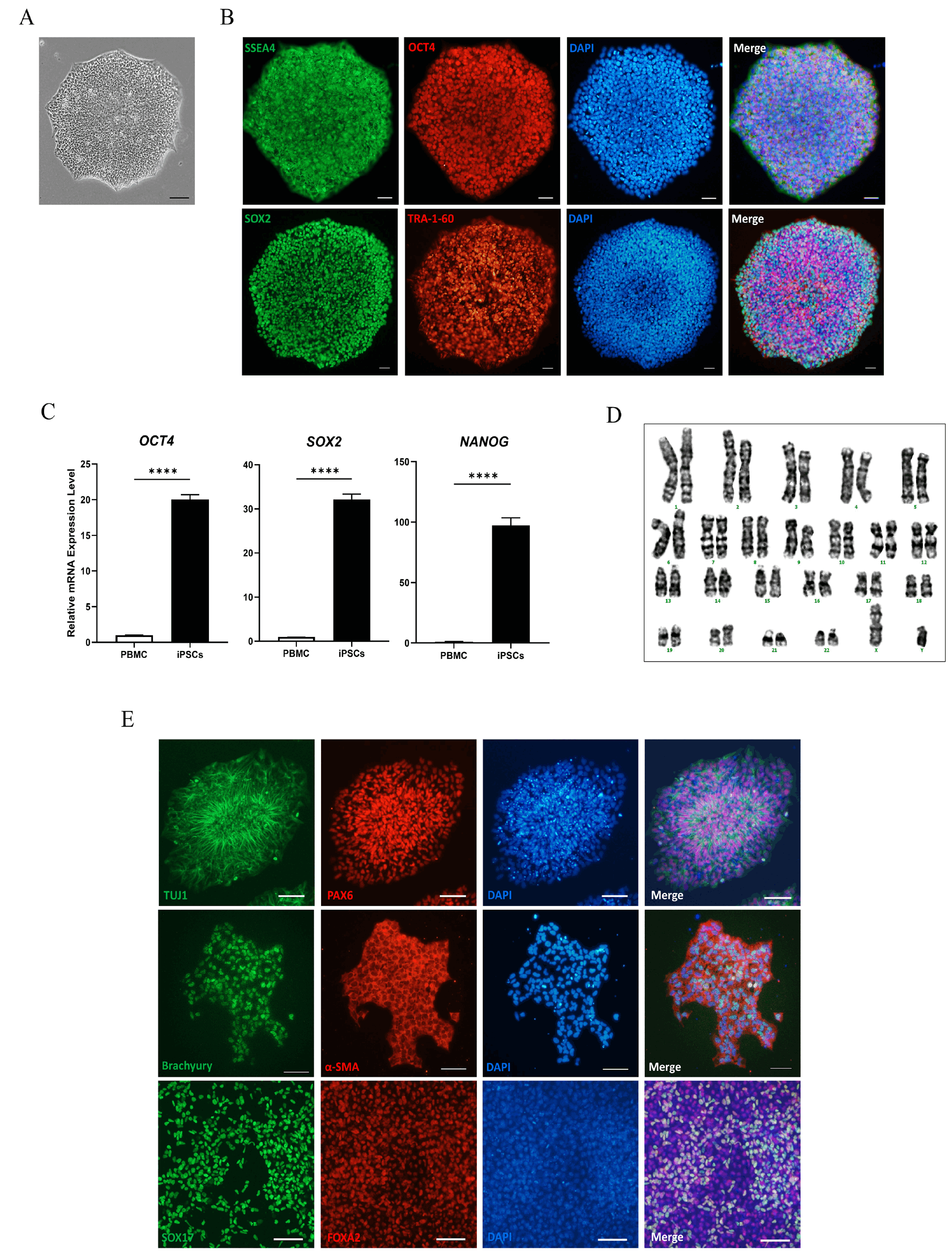
PBMCs from an Arts syndrome patient carrying the PRPS1 p.V42L variant were successfully reprogrammed into iPSCs using Sendai virus-based delivery of OCT4, SOX2, KLF4, and c-MYC. The established iPSC colonies exhibited typical human embryonic stem cell–like morphology with high nuclear-to-cytoplasmic ratios and well-defined borders (Fig. 1A). Immunocytochemical analysis confirmed robust expression of pluripotency markers OCT4, SOX2, SSEA4, and TRA-1-60 (Fig. 1B). RT-qPCR analysis further demonstrated high expression levels of OCT4, SOX2, and NANOG compared to donor PBMCs (Fig. 1C). Karyotype analysis confirmed genomic stability of iPSCs (Fig. 1D). Also, STR profiling verified a 100% genetic identity between patient PBMCs and derived iPSCs, confirming their origin (Supplementary Fig. S1A). Trilineage differentiation assays demonstrated the capacity of iPSCs to differentiate into ectoderm (TUJ1, PAX6), mesoderm (Brachyury, α-SMA), and endoderm (SOX17, FOXA2) lineages (Fig. 1E). Sendai-specific PCR confirmed clearance of Sendai virus reprogramming vectors in late-passage cultures (Supplementary Fig. S1B).
Differentiation into NSCs, defective proliferation, and senescence-like phenotype in patient-derived NSCs
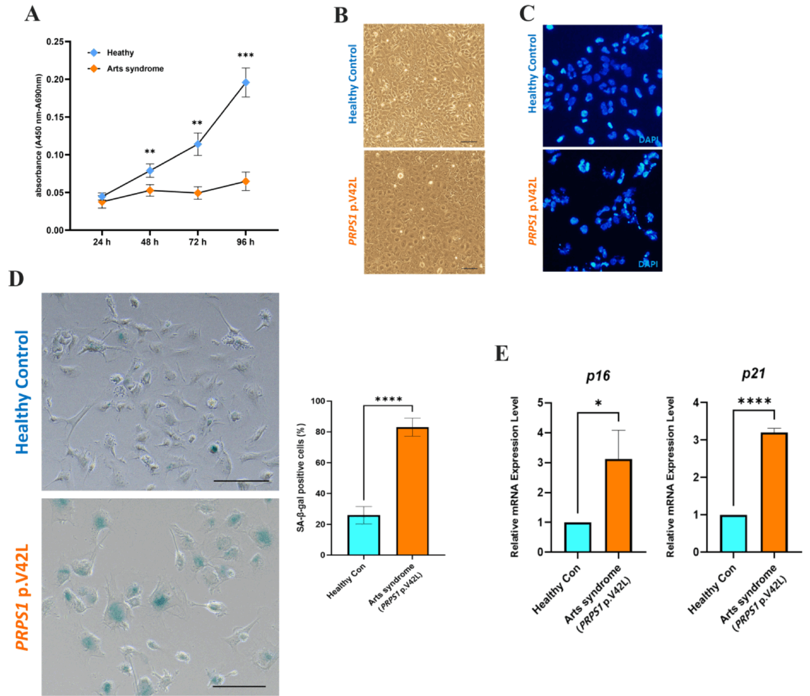
Both healthy control and PRPS1 p.V42L iPSCs efficiently differentiated into NSCs expressing neural progenitor markers SOX2, Nestin, SOX1, and PAX6 (Supplementary Fig. S2A). RT-qPCR analysis confirmed significantly increased expression of these markers in NSCs compared with their parental iPSCs, verifying successful neural induction (Supplementary Fig. S2B). Although the induction process appeared normal, PRPS1-mutant NSCs displayed significant proliferative and morphological defects relative to controls. WST-1 cell proliferation assays revealed a clear reduction in cell growth rate in patient-derived NSCs over multiple time points (Fig. 2A). Despite the lower cell number, live/dead staining showed no significant differences in viability between control and mutant NSCs (Supplementary Fig. S2C). Notably, ATP quantification revealed a significant increase in intracellular ATP levels in PRPS1-mutant NSCs compared with controls (Supplementary Fig. S2D), suggesting a potential metabolic imbalance. Mitochondrial morphology and ROS levels were also comparable (Supplementary Fig. S2E), indicating that the reduced proliferation is not due to overt cell death or mitochondrial failure. Phase-contrast microscopy demonstrated that patient-derived NSCs displayed flattened and enlarged cells compared to the compact, uniform morphology of healthy controls (Fig. 2B). DAPI staining revealed irregular nuclear shapes and chromatin condensation in mutant NSCs (Fig. 2C), consistent with cellular stress. Such nuclear distortions are indicative of disrupted nuclear integrity and cellular senescence processes, suggesting that the PRPS1 mutation may predispose NSCs to premature senescence, as supported by SA-β-gal staining and senescence-related gene expression in subsequent analyses. Furthermore, SA-β-gal staining indicated a pronounced increase in blue-stained cells in PRPS1-mutant NSCs, signifying enhanced cellular senescence (Fig. 2D). Consistently, RT-qPCR analysis demonstrated substantial upregulation of the cellular senescence genes p16 and p21 in mutant NSCs compared with controls (p<0.01) (Fig. 2E). Collectively, these results suggest that although the PRPS1 p.V42L mutation does not affect early NSC generation or mitochondrial metabolism, it leads to impaired proliferation, abnormal nuclear morphology, and premature senescence during subsequent NSC maintenance and expansion.
Impaired neuronal differentiation in patient-derived NSCs
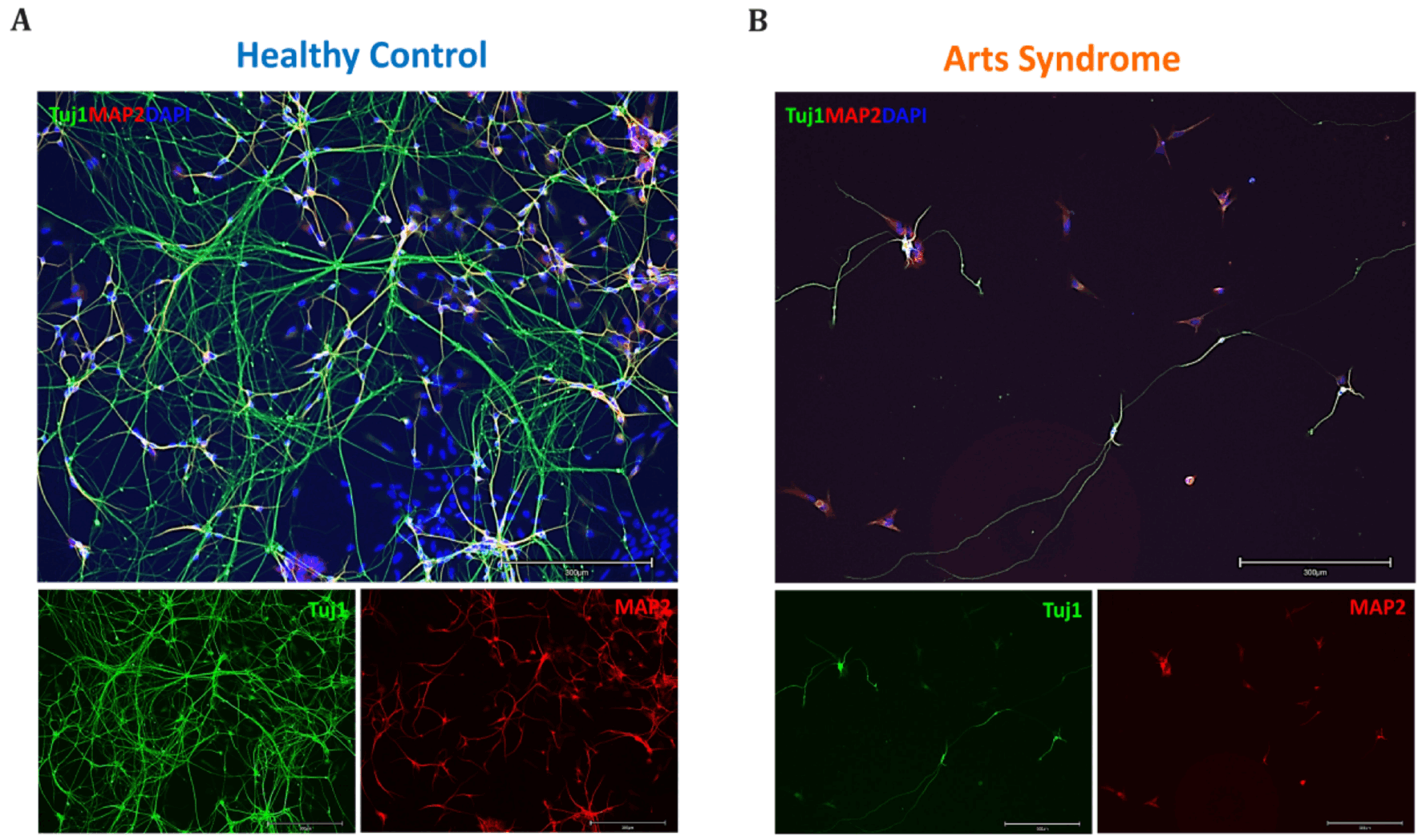
Following neural induction, both healthy control and PRPS1 p.V42L (Arts syndrome) NSCs were differentiated into neurons and immunostained for TUJ1 (βIII-tubulin) and MAP2 to evaluate neuronal morphology and maturation. In the healthy control group, extensive and highly interconnected neuronal networks were observed, with long, branching neurites forming dense mesh-like structures (Fig. 3A). In contrast, patient-derived neurons displayed markedly impaired neurite development (Fig. 3B). TUJ1+/MAP2+ cells were sparsely distributed, exhibiting fewer and shorter neuritic extensions with minimal branching. The overall neuronal network appeared disorganized, with reduced density and weak MAP2 expression relative to controls. These morphological defects indicate that the PRPS1 p.V42L mutation significantly compromises neuronal differentiation and maturation, leading to reduced neurite outgrowth and impaired network formation.
Nicotinamide mononucleotide supplementation restores neuronal differentiation and neurite outgrowth in PRPS1-mutant neurons
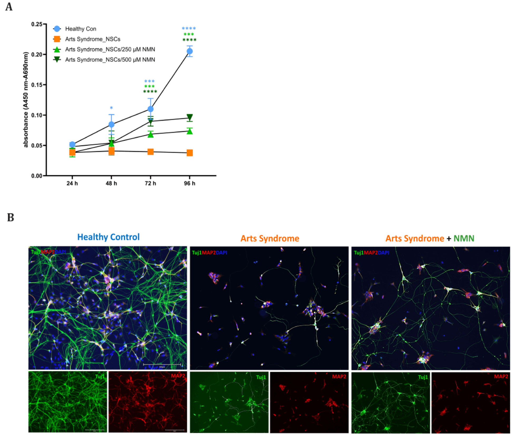
To evaluate the effect of NMN supplementation on cell proliferation and neuronal differentiation, WST-1 assays and immunocytochemical analyses were performed using healthy control and PRPS1 p.V42L (Arts syndrome) cell lines. Cell proliferation analysis using the WST-1 assay (Fig. 4A) revealed a significant reduction in metabolic activity in PRPS1-mutant NSCs compared with healthy controls, consistent with impaired proliferative capacity. Upon NMN treatment, patient-derived NSCs exhibited a clear increase in WST-1 absorbance across multiple time points, indicating a partial restoration of cell proliferation. Although proliferation levels in the NMN-treated group did not completely reach those of the control, the recovery was statistically significant, suggesting that NMN supplementation alleviates the proliferative defect associated with the PRPS1 mutation. Neuronal differentiation analysis (Fig. 4B) further demonstrated that these proliferative improvements translated into enhanced neuronal maturation. Remarkably, NMN supplementation markedly improved neuronal morphology in patient-derived neuron, promoting elongation and branching of neurites and increasing the density of TUJ1+/MAP2+ cells.
Together, these findings indicate that NMN enhances both proliferation and neuronal differentiation in PRPS1-mutant cells, suggesting that restoration of NAD+ metabolism may mitigate cellular dysfunctions caused by PRPS1 deficiency.
Discussion
This study provides a comprehensive analysis of PRPS1 p.V42L-associated neural defects using patient-derived iPSCs and their differentiation into NSCs and neurons. Our findings reveal that PRPS1 deficiency impairs NSC proliferation, induces abnormal nuclear morphology, and accelerates cellular senescence (Fig. 2). Furthermore, neurons derived from PRPS1-mutant NSCs exhibit markedly reduced neurite outgrowth and branching complexity, indicating compromised neuronal maturation (Fig. 3).
PRPS1 encodes phosphoribosyl pyrophosphate synthetase 1, which catalyzes the synthesis of PRPP - a central metabolite required for purine, pyrimidine, and NAD+ biosynthesis. Given the high metabolic demands of the CNS, even partial loss of PRPS1 function can disrupt neural development. Previous studies have linked PRPS1 mutations to a spectrum of X-linked disorders, including Arts syndrome, Charcot-Marie-Tooth disease-5, and nonsyndromic hearing loss (2, 3, 10). Our iPSC-based model extends these insights by demonstrating intrinsic defects in human neural lineages caused by PRPS1 dysfunction.
A key observation in our study is the reduced proliferation of PRPS1-mutant NSCs, accompanied by upregulation of senescence markers p16 and p21 (Fig. 2E). These findings suggest that impaired nucleotide synthesis leads to replication stress and premature cellular senescence. Interestingly, mitochondrial morphology and ROS levels were comparable between mutant and control NSCs (Supplementary Fig. S2E), indicating that the proliferative defect is not attributable to mitochondrial failure.
Notably, we observed a significant increase in intracellular ATP levels in PRPS1-mutant NSCs despite their reduced proliferation (Supplementary Fig. S2D). This paradox may be explained by impaired PRPP synthesis due to PRPS1 dysfunction. PRPP is a key substrate for nucleotide and NAD+ biosynthesis, both of which are ATP-consuming processes (11). A deficiency in PRPP could reduce ATP utilization in biosynthetic pathways, leading to intracellular ATP accumulation. This suggests that while mitochondrial energy production remains intact, the downstream metabolic demand for ATP is diminished due to disrupted nucleotide flux. These findings highlight a decoupling between energy production and utilization in PRPS1-deficient cells and underscore the central role of PRPP in coordinating metabolic and proliferative programs in NSCs.
Neurons derived from PRPS1-mutant NSCs displayed markedly reduced neurite outgrowth and branching complexity (Fig. 3B). Neurite extension requires coordinated energy metabolism and NAD+-dependent signaling, including SIRT1-mediated deacetylation (12, 13). Reduced NAD+ levels resulting from PRPP deficiency may alter these pathways, thereby hindering neuronal polarity and extension. This aligns with previous reports that NAD+ depletion limits axonal growth and synaptic plasticity in models of neurodegeneration and metabolic deficiency (14). The observed morphological abnormalities in patient-derived neurons thus likely reflect the downstream impact of impaired NAD+ synthesis caused by PRPS1 dysfunction.
Importantly, supplementation with NMN, a NAD+ precursor, partially restored proliferative capacity and neuronal morphology. One of the most notable findings in this study is the partial rescue of both proliferation and neuronal morphology following NMN supplementation (Fig. 4). NMN is a key NAD+ precursor that bypasses PRPP-dependent steps, thereby restoring NAD+ availability even under conditions of impaired PRPS1 activity (15, 16). Previous studies have demonstrated that NMN and related NAD+ precursors improve mitochondrial function, promote axonal regeneration, and alleviate neurodegenerative phenotypes in mouse models of ALS and peripheral neuropathy (17, 18). In our system, NMN treatment significantly increased WST-1-based proliferation in PRPS1-mutant NSCs and improved TUJ1+/MAP2+ neurite structure (Fig. 4B), indicating that NAD+ repletion supports both metabolic and structural recovery.
While NMN supplementation showed therapeutic promise, it did not fully restore normal phenotypes, indicating that PRPS1 loss affects additional PRPP-dependent pathways beyond NAD+ metabolism. These may include purine salvage, RNA synthesis, and other biosynthetic processes essential for neural development. Therefore, combination strategies targeting both nucleotide and NAD+ biosynthesis may be necessary for optimal correction of PRPS1-related defects.
In summary, our iPSC-derived neural model of Arts syndrome reveals that PRPS1 p.V42L mutation disrupts NSC homeostasis and neuronal development. NMN supplementation mitigated, but did not completely reverse, these abnormalities, highlighting both the therapeutic potential and mechanistic complexity of NAD+ restoration strategies. These findings provide new insights into PRPS1-related neurodevelopmental pathophysiology and support further exploration of metabolic interventions for Arts syndrome and related disorders.
Supplementary Materials
Supplementary data including one table and two figures can be found with this article online at https://doi.org/10.15283/ijsc25127
 XML Download