



 PDF
 Citation
 Print


Introduction
Drug-induced cardiotoxicity is a major cause of clinical trial failure and drug withdrawal (1, 2). Current preclinical assessments rely on ICH S7B/E14 guidelines using hERG assays and electrocardiogram (ECG) telemetry, which focus on QT prolongation. However, these have limited predictive value for complex arrhythmias due to interspecies differences and a single-ion channel approach (1, 2). As a promising alternative, human-induced pluripotent stem cell-derived cardiomyocytes (iPSC-CMs), which mimic human cardiac electrophysiology by reflecting multiple ion channel activities, are emerging as a valuable in vitro platform. The Comprehensive in vitro Proarrhythmia Assay (CiPA) initiative incorporates iPSC-CMs and ECG-like signal (field potential, FP) analysis to better predict pro-arrhythmic risk (1, 3). Currently, microelectrode array (MEA) is one of the primary tools used in CiPA for evaluating electrophysiological parameters. In line with these advances, the FDA Modernization Act 2.0 officially supports human cell-based nonclinical testing to overcome ethical and translational limitations of animal models, accelerating the shift toward human-relevant safety assessment platforms (1, 3, 4).
MEA systems are widely used to quantify electrophysiological signals from iPSC-CMs for evaluating cardiac parameters such as the QT interval (5, 6). These electrodes are typically composed of metals such as gold (Au), platinum (Pt), or titanium nitride (TiN), either individually or in combination, due to their excellent electrical conductivity, corrosion resistance, and mechanical stability (5-7). However, these metal electrodes are opaque, making it difficult to monitor morphological changes in cells or perform real-time fluorescence imaging during drug testing (8, 9). Moreover, the use of high-cost metals increases the overall manufacturing cost of MEA, limiting their feasibility for repeated toxicity testing (9, 10). To address these limitations, transparent electrodes made from indium tin oxide (ITO), which offer both optical transparency and high conductivity, have recently been introduced into MEA platforms. ITO-based MEAs support imaging, long-term recording, and low-cost fabrication, making them suitable for next-generation platforms (9, 11, 12). Nevertheless, studies utilizing ITO-based MEAs for cardiotoxicity evaluation remain limited, and further validation is needed to establish their applicability and reliability as a standardized assessment platform.
This study assessed an ITO-based MEA platform that enables both electrophysiological analysis and real-time optical observation, addressing limitations of traditional metal electrodes. iPSC-CMs were cultured on transparent ITO electrodes, and both morphological and electrophysiological signals were monitored. Drug responsiveness was evaluated under short- and long-term exposure to ion channel blockers and cardiotoxic agents. Drug effects were assessed by combining FP, cell viability, and morphological analysis. These findings suggest ITO-based MEAs as promising tools for integrated cardiotoxicity screening.
Materials and Methods
Generation of human iPSC-CMs
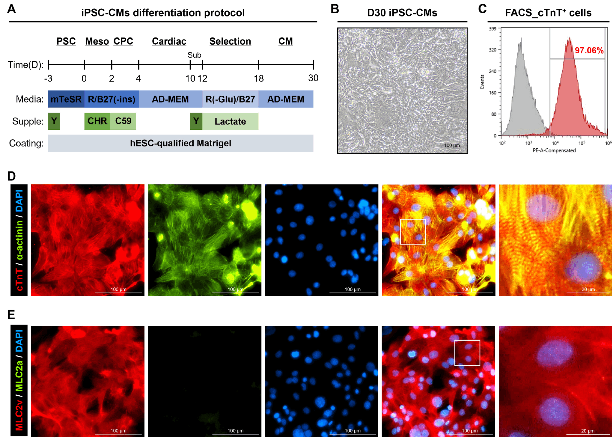
iPSCs were generated by reprogramming BJ fibroblasts (ATCC) using episomal vectors, as previously described (13). iPSCs were seeded on Matrigel (Corning Inc.)-coated dishes in mTeSRTM1 medium (STEMCELL Technologies Inc.) supplemented with 10 μM Y-27632 (Tocris Bioscience). Medium was changed daily until ∼90% confluency. Mesodermal differentiation was induced by treating cells with RPMI 1640 containing B27 minus insulin (B27(−); Gibco) and 6 μM and 3 μM CHIR99021 (Tocris Bioscience) for two days. Cardiac progenitors were induced with RPMI 1640+B27(−) containing 2 μM and 1 μM C59 (Selleckchem). From day 4 to 10, cells were cultured in Advanced Minimum Essential medium (AD-MEM; Gibco) with antibiotics and GlutaMAX (Gibco), with medium changes every two days until beating cells appeared. Beating cells were dissociated with TrypLE (Gibco) and replated on Matrigel. On day 12, metabolic selection using glucose-free RPMI with B27 and 4 mM sodium DL-lactate (Gibco) was performed for 6 days to enrich CMs. After purification, iPSC-CMs were maintained in fresh AD-MEM until day 30 for maturation (1, 14-16).
Flow cytometry analysis (fluorescence-activated cell sorting)
iPSC-CMs were fixed with 4% paraformaldehyde (PFA) for 20 minutes at room temperature (RT), washed, and resuspended in fluorescence-activated cell sorting (FACS) buffer. Cells were stained with PE-conjugated anti-cTnT antibody (1:50; Thermo Fisher Scientific) for 30 minutes at 4℃, then analyzed using a FACS Calibur flow cytometer with Cell Quest software (BD Biosciences).
Immunocytochemistry
iPSC-CMs were fixed with 4% PFA for 20 minutes at 4℃. Cells were then permeabilized and blocked in phosphate buffered saline (PBS) containing 0.1% Triton X-100 (Sigma-Aldrich) and 3% normal goat serum (Thermo Fisher Scientific) for 30 minutes at RT. Cells were incubated overnight at 4℃ with primary antibodies against cTnT (1:200; Abcam, ab45932), α-actinin (1:200; Sigma-Aldrich, A7811), MLC2v (1:200; Synaptic System, #310 003), and MLC2a (1:200; Synaptic System, #311 011). After washing, cells were incubated for 2 hours at RT on a shaker with secondary antibodies (Alexa Fluor 488 goat anti-mouse IgG and Alexa Fluor 594 goat anti-rabbit IgG [1:700; Invitrogen]). Nuclei were counterstained with DAPI (1:1,000; Thermo Fisher Scientific). Fluorescence images were acquired using a Nikon TE2000-U microscope (Nikon).
Live/dead assay
Cell viability was evaluated using the LIVE/DEADⓇ Viability/Cytotoxicity Kit (Invitrogen). iPSC-CMs were washed with PBS and incubated for 30 minutes in culture medium containing 2 μM calcein AM (C-AM) and 4 μM ethidium homodimer-1 (EthD-1). Fluorescence was detected using FITC (C-AM) and TRITC (EthD-1) channels under a fluorescence microscope. ImageJ was used to quantify fluorescence, and the ratio of C-AM to EthD-1 was calculated.
Preparation of chemicals
E-4031 (Sigma-Aldrich), nifedipine (Sigma-Aldrich), and doxorubicin (DOX, Sigma-Aldrich) were dissolved in dimethyl sulfoxide (DMSO; Sigma-Aldrich). Each drug was prepared at 10× the working dose in DMSO and diluted in culture medium before use. The final DMSO dose was kept below 0.1%, which is considered non-cytotoxic. For comparison with the experimental groups, 0.1% DMSO was designated as the control (CTL).
Electrophysiological signal analysis in ITO microelectrode array (ITO-MEA)
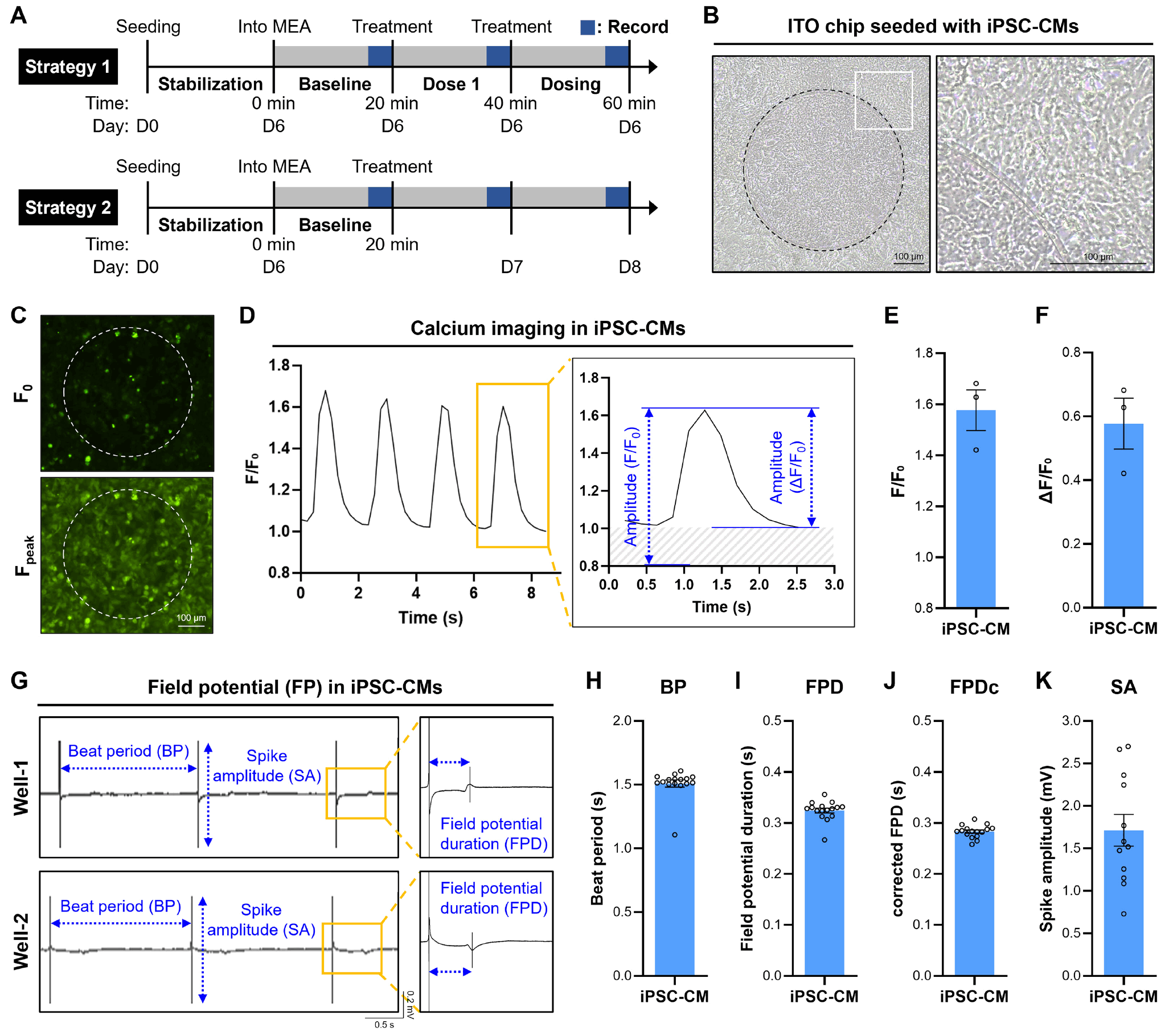
iPSC-CMs were seeded onto CFPS-ITO-16W2E MEA plates (Cellames). Prior to cell seeding, 5 μL of 50 μg/mL fibronectin (Sigma-Aldrich) was applied to each ITO electrode well and incubated for 1 hour in a CO2 incubator. After removing fibronectin, 5 μL droplets containing 50,000 cells were added to each well. Following a 2-hour incubation, 200 μL of fresh culture medium was added. Cells were cultured for 5∼6 days in a CO2 incubator to allow stabilization before MEA recordings. For FP analysis using the ITO MEA plates, the medium was replaced with AD-MEM 1 hour before recording. Plates were inserted into the CFPS-32 system (Cellames) within a CO2 incubator. After 17 minute-stabilization, baseline FP signals were recorded for 3 minutes. For Strategy 1 (acute drug response), 20 μL of medium was removed from each well and replaced with 20 μL of a 10× drug solution corresponding to the first dose (Dose 1), determined based on the reference value and Cmax. Subsequent dosing was performed stepwise with progressively increasing doses (E-4031: 3× and 10×; nifedipine: 10× and 100×). After each dosing step, cells were incubated for 17 minutes, followed by a 3-minute FP recording. For Strategy 2 (chronic drug response), 20 μL of medium was removed only on day 0 after stabilization in both the CTL and DOX groups and replaced with 20 μL of a 10× drug solution. FP was recorded on days 0, 1, and 2. All data were analyzed using the CFPS-32 software (Cellames).
Calcium transient imaging
To assess calcium transients in iPSC-CMs cultured on ITO chips, cells were incubated with 5 μM Fluo-4 AM (Invitrogen) and 0.02% Pluronic F-127 for 30 minutes. The cells were then washed once with fresh culture medium. Fluorescence signals were acquired using a Nikon TE2000-U microscope in the FITC channel, and videos were recorded around the ITO electrode region in each well. The acquired images were analyzed in ImageJ by selecting regions of interest (ROI) and extracting frame-by-frame fluorescence intensity. Calcium amplitude over time was quantified as F/F0 and ΔF/F0, where F represents the fluorescence intensity at each frame and F0 is defined as the baseline fluorescence intensity within the ROI across all frames (17).
Statistical analysis
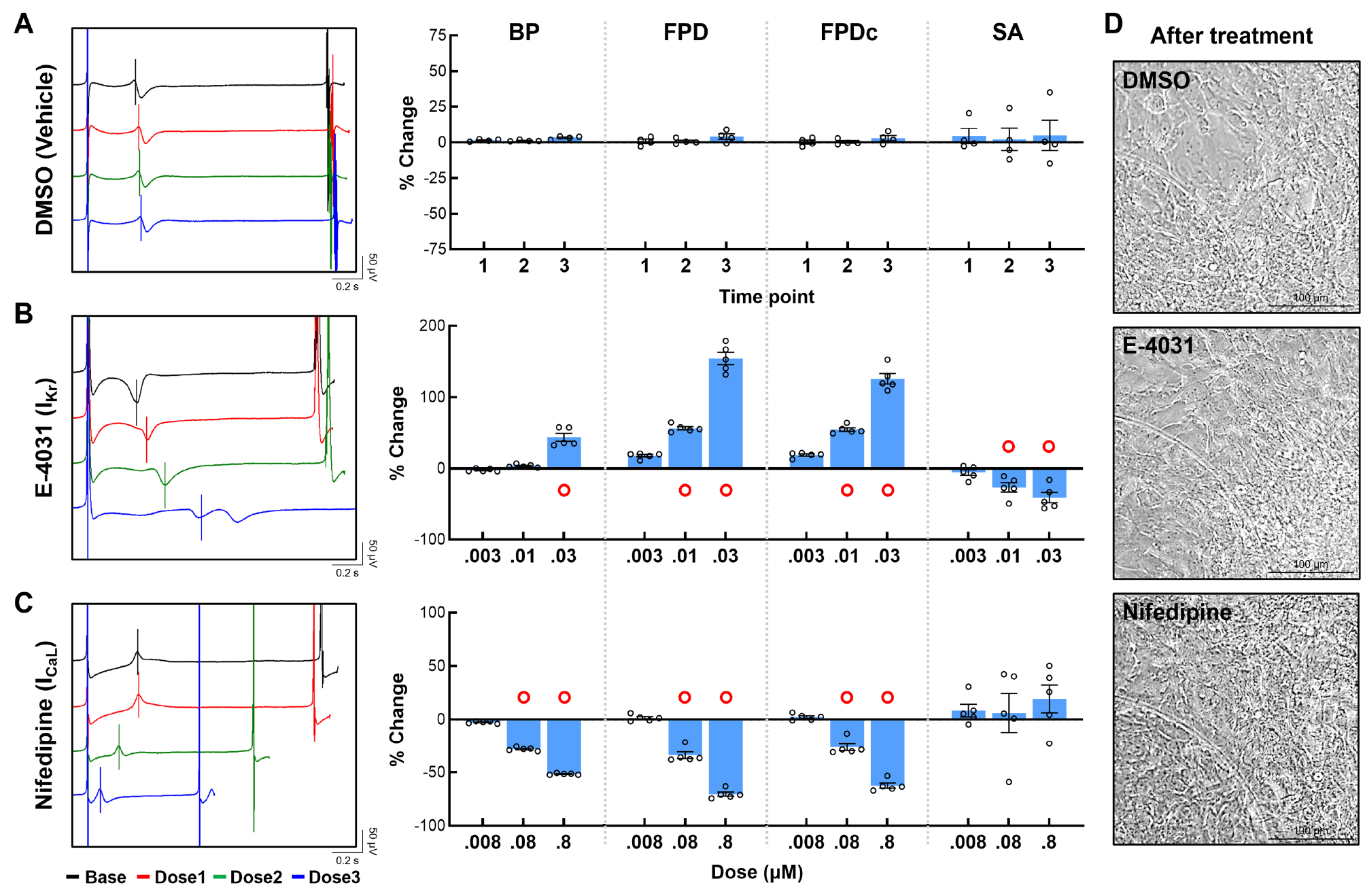
All the experiments were performed at least three times. Statistical analyses were performed with ANOVA one-way for comparisons between groups. Quantitative data are presented as the mean±standard error of the mean (SEM). A p-value (p<0.05, p<0.01, and p<0.001) was considered statistically significant. Based on previously published reports, a prolongation of corrected FPD (FPDc) exceeding 20% has been identified as falling within the pre-arrhythmic risk range (16). Accordingly, reflecting these findings, we defined a ≥20% change as the threshold for a meaningful response and indicated it with red circle (O). Data histograms were fit with GraphPad Prism, version 10.
Results
Characterization of iPSC-CMs
iPSC-CMs were generated using a previously established small molecules–based differentiation protocol (Fig. 1A) (1, 14-16). The D30 iPSC-CMs exhibited regular spontaneous beating (Fig. 1B and Supplementary Video S1), and FACS analysis revealed that 97.06% of the cells expressed the CM-specific marker cTnT (Fig. 1C). Furthermore, co-staining for cTnT and α-actinin (z-disc marker) demonstrated that iPSC-CMs co-expressed both markers and exhibited sarcomeric structures (Fig. 1D). To further assess whether the differentiated cells were of ventricular subtype, co-staining for MLC2v and MLC2a was performed. The results showed that most cells predominantly expressed MLC2v, with negligible expression of MLC2a (Fig. 1E). Collectively, these findings demonstrate that the in-house–generated iPSC-CMs not only express CM-specific markers but also differentiate into a ventricular subtype, thereby representing highly purified CMs suitable for cardiotoxicity assessment.
Optical and electrophysiological analysis of iPSC-CMs in ITO MEA
For electrophysiological signal-based drug screening, iPSC-CMs were first seeded onto ITO chips and stabilized for 5 days (Fig. 2A). The seeded iPSC-CMs showed clear morphological features even when positioned on the circular electrodes of the ITO chip (Fig. 2B and Supplementary Video S1). Subsequently, Fluo-4 AM-based calcium transient imaging was performed to assess the functional activity of iPSC-CMs (Fig. 2C). Clear intracellular calcium flux corresponding to the contraction–relaxation cycle of iPSC-CMs was observed (Fig. 2D). Quantification of calcium amplitude using F/F0 and ΔF/F0 metrics yielded values of 1.577±0.080 and 0.577±0.080, respectively (Fig. 2E, 2F). Prior to drug testing, the detection of FP signal was evaluated, and most wells exhibited stable depolarization/repolarization coupling in the FP raw waveforms, reflecting regular QRST-like patterns (Fig. 2G) (18). The primary parameters of FP analysis include the spike amplitude (SA), beat period (BP), field potential duration (FPD), and FPDc. SA represents the potential difference of ions in depolarization. BP represents the interval between two consecutive depolarizations, while FPD corresponds to the interval between depolarization and repolarization, which is analogous to the QT interval on an ECG. FPDc denotes the FPD corrected by Fridericia’s formula, the most widely used correction method in cardiotoxicity testing. In stabilized iPSC-CMs, the FP parameters were measured as follows: BP, 1.498±0.037 seconds; FPD, 0.320±0.006 seconds; FPDc, 0.280±0.003 seconds; and SA, 1.712±0.186 mV (Fig. 2H-2K). These results demonstrate that ITO electrodes enable not only the morphological monitoring of iPSC-CMs but also the acquisition of well-quantified electrophysiological signals.
Assessment of torsades de pointes (TdP) risk drugs induced cardiotoxicity using ITO MEA
Cardiac safety pharmacological evaluation using ion channel blockers was conducted via dosing method (Strategy 1 in Fig. 2A) (1, 19). To confirm that the observed effects were attributable to drug treatment rather than environmental factors, vehicle control was first applied using 0.1% DMSO, a non-cytotoxic dose. No significant changes were observed in the raw waveforms compared to baseline, and main FP parameters varied by less than 5% (Fig. 3A). Drug evaluation was performed using representative reference compounds, E-4031 and nifedipine (1, 19). E-4031, a class III antiarrhythmic agent known as a hERG channel blocker, was applied at doses of 0.003, 0.01, and 0.03 μM. BP was significantly increased by 43.715%±5.639% only at 0.03 μM, while FPD (0.003 μM, 17.920%±1.746%; 0.01 μM, 56.301%±2.356%; and 0.03 μM, 154.292%±8.537%) and FPDc (0.003 μM, 18.837%±1.604%; 0.01 μM, 54.560%±2.543%; and 0.03 μM, 125.798%±7.420%) were prolonged in a dose-dependent manner, as confirmed in the raw waveforms. In contrast, SA decreased in a dose-dependent manner (0.003 μM, −5.635%±4.080%; 0.01 μM, −26.535%±6.524%; and 0.03 μM, −40.936%±7.338%) (Fig. 3B). Nifedipine, an L-type Ca2+ channel inhibitor used for the treatment of hypertension and angina, was applied at Cmax (0.008 μM) and at higher doses of 0.08 μM and 0.8 μM. The results demonstrated a dose-dependent decrease in BP (0.08 μM, −28.174%±0.748%; and 0.8 μM, −51.567%±0.333%), FPD (0.08 μM, −33.879%±3.143%; and 0.8 μM, −70.417%±1.936%), and FPDc (0.08 μM, −26.280%±3.282%; and 0.8 μM, −62.439%±2.373%), consistent with observations in the raw waveforms. No significant difference was observed in SA (Fig. 3C). Morphological comparison after the highest dose treatment revealed changes in the beating pattern between groups, but no significant alterations in cell morphology were observed (Fig. 3D). In summary, ion channel blockers exerted pronounced effects on the electrophysiological function of iPSC-CMs even after short-term exposure, while having minimal impact on cellular morphology.
Monitoring cardiotoxic responses to DOX using ITO MEA
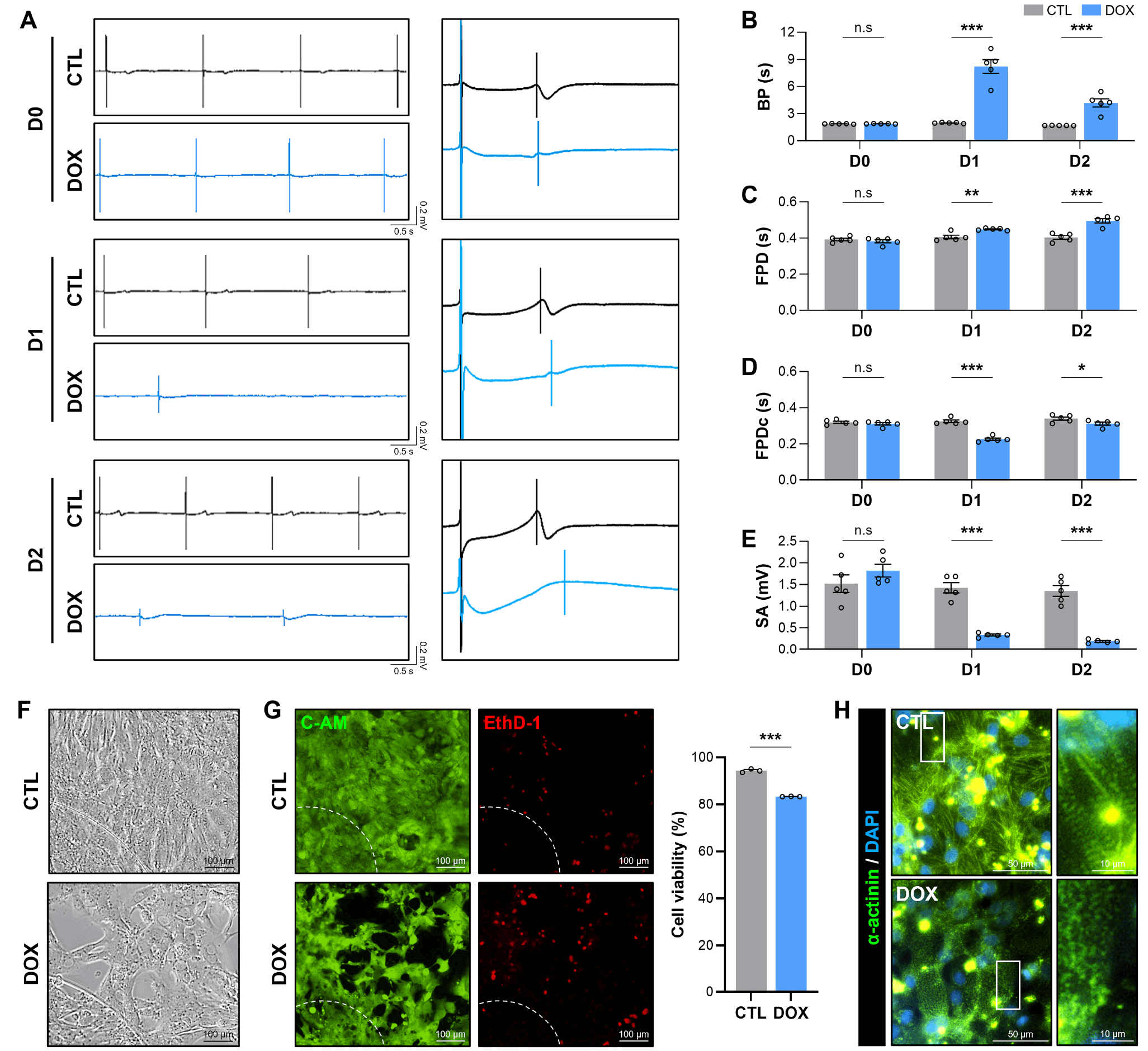
DOX, an anticancer agent, is known to induce apoptosis in CMs and is a representative drug that causes cardiotoxicity (20, 21). In this study, DOX was treated at a dose of 3 μM for 2 days, considering its half-life (Strategy 2 in Fig. 2A) (21). The CTL group was treated with 0.1% DMSO, and FP signals were monitored in both CTL and DOX groups at D0, D1, and D2 (Fig. 4A). In the CTL, no significant changes were observed in FP raw waveforms, and the T wave remained stable. In contrast, the DOX-treated group showed rapid waveform alterations over time, with a marked prolongation of the T wave (Fig. 4A). Quantitative analysis revealed that BP, FPD, FPDc, and SA remained stable across all time points in the CTL group (Fig. 4B-4E). However, in the DOX group, BP exhibited a sharp and significant increase at D1 (CTL: 1.961±0.023 seconds vs. DOX: 8.213±0.761 seconds), and although the extent of increase was slightly reduced at D2 (CTL: 1.678±0.005 seconds vs. DOX: 4.177±0.454 seconds), it remained significantly prolonged compared with CTL (Fig. 4B). FPD was significantly prolonged in a time-dependent manner in the DOX group (D0, 0.382±0.008 seconds; D1, 0.448±0.003 seconds; and D2, 0.495±0.011 seconds) relative to CTL (D0, 0.392±0.007 seconds; D1, 0.406±0.010 seconds; and D2, 0.403±0.010 seconds) (Fig. 4C), whereas FPDc showed a pronounced decrease at D1 due to the sharp BP prolongation and subsequently exhibited partial recovery at D2 (Fig. 4D). In addition, compared with CTL, DOX caused a sharp decrease in SA from D1, which persisted on D2 (Fig. 4E). Morphological analysis using the ITO chip revealed that, unlike ion channel blockers, DOX treatment led to a reduction in iPSC-CMs density (Fig. 4F). Furthermore, live/dead assays demonstrated increased EthD-1 expression and decreased cell viability in the DOX group compared with CTL (Fig. 4G). Immunostaining of α-actinin (a z-disc marker in CMs) showed that sarcomeric structures were well observed in the CTL, whereas sarcomeres were disintegrated in the DOX (Fig. 4H). Collectively, these results demonstrate that ITO chip–based monitoring can reveal that DOX-induced prolongation of BP and FPD is not limited to functional alterations but is accompanied by cytotoxicity and structural damage in CMs (Supplementary Video S2).
Discussion
Ensuring cellular quality is a fundamental prerequisite for reliable preclinical safety evaluation (1, 22). Since iPSC-derived cells require differentiation prior to use, lot-to-lot variation can occur depending on institution-specific protocols (23). In this study, in-house iPSC-CMs achieved >90% cTnT-positivity and ventricular-like morphology, closely resembling commercially available cells used in CiPA studies (Fig. 1) (23). Because iPSC-CMs intrinsically recapitulate electrophysiological activity, MEA platforms have become central to functional readouts (19, 22, 24). However, differences in MEA chip composition and recording hardware may alter absolute signal values (19, 22, 25). Establishing robust quality control (QC) thresholds is therefore indispensable. According to CiPA-reported QC criteria, MEA-based assays should satisfy: (1) spontaneous beat rate (BR) within 20∼90 bpm, (2) baseline BR values across wells within ±6 standard deviation of the mean, and (3) SA ≥0.3 mV (3, 22). Consistent with these standards, our iPSC-CMs exhibited SA ≥0.3 mV and a BP of 1.498±0.037 seconds (approximately 40 bpm), confirming that ITO-based MEA chips provide sufficient functional quality for reliable analysis (Fig. 2). These findings show that QC criteria are applicable across platforms, ensuring reproducible drug assessments.
Current cardiac safety pharmacology frameworks, including ICH S7B/E14 guidelines and the CiPA initiative, emphasize electrophysiological signal analysis (3, 26). The CiPA paradigm integrates multi-ion channel analysis, in silico modeling, ventricular iPSC-CMs, and in vivo ECG to assess torsadogenic risk associated with QT prolongation (3). Most MEA-based reports have validated short-term drug responses (25∼40 minutes exposure) across varying doses by monitoring FP alterations (1, 19, 22). Yet, the clinical reality is that many agents are administered chronically, and long-term cardiotoxicity remains underexplored (2, 27). For instance, pentamidine induces negligible FPD changes acutely, but manifests >100% prolongation after 72 hours (27). Chronic drug exposure may induce cytotoxicity, triggering apoptosis, mitochondrial dysfunction, and secondary electrophysiological changes (27-29). Clinically relevant tyrosine kinase inhibitors such as sunitinib and vandetanib exemplify this phenomenon, showing modest acute FPD changes (<40%) but pronounced FPD prolongation (>100%) and cytotoxicity after prolonged exposure (27). In line with this, our study demonstrated that 3 μM DOX treatment for 2 days induced apoptosis and disruption of CMs structure, accompanied by FP instability (Fig. 4). While FPD was continuously prolonged, BP exhibited a sharp increase on D1 followed by partial attenuation on D2 (Fig. 4). The terminal elimination half-life of DOX has been reported to be approximately 20∼48 hours, and previous studies have shown that toxic responses are alleviated in accordance with these pharmacokinetic characteristics (30, 31). In vivo studies have demonstrated that intracellular DOX uptake and apoptosis in CMs peak on the first day and decline thereafter. Considering this pharmacodynamic pattern, certain cardiotoxicity-related parameters may reflect a recovery phase following the initial acute toxicity peak, during which intracellular DOX accumulation and associated toxic signaling are partially alleviated. Notably, these responses on the ITO-MEA chip resemble clinical observations in DOX-treated patients, including CM apoptosis, bradycardia, and QT prolongation (20, 28). Collectively, these findings suggest that cardiotoxicity assessment should encompass not only electrophysiological alterations but also side effects such as cytotoxicity (28, 32, 33). Importantly, ITO chips present an attractive alternative to conventional systems, offering cost-effectiveness and enabling simultaneous electrophysiological and optical monitoring (9).
Despite these advantages, significant challenges remain. Conventional iPSC-CMs retain an immature phenotype, and 2D monolayers insufficiently capture tissue-level electromechanical interactions (34-36). Advances in maturation strategies and the adoption of 3D engineered cardiac tissues are critical to bridging this gap (15, 34-36). Furthermore, expanding beyond FP and optical monitoring to encompass contractility and Ca2+ handling would markedly enhance translational fidelity (22, 37). In summary, rigorous QC of iPSC-CMs combined with dual monitoring on ITO-based MEA chips offers a promising strategy for advancing cardiotoxicity assessment. Looking forward, integrating electrophysiological, optical, mechanical, and biochemical readouts into unified platforms could transform preclinical cardiac safety testing.
Supplementary Materials
Supplementary data including two videos can be found with this article online at https://doi.org/10.15283/ijsc25077
 XML Download