

Journal List > Int J Stem Cells > v.19(2) > 1516095781

Seok, Lee, and Seo: Investigating the Causal Links between the Aging Process and Alzheimer’s Disease Pathogenesis

Abstract

As global societies age, the prevalence of neurodegenerative disorders, such as Alzheimer’s disease, is rapidly increasing, intensifying the need to understand the mechanisms of aging and their contribution to these conditions. Consequently, the focus of aging research has shifted from the traditional concept of chronological age to a more nuanced understanding of biological age. This has spurred active investigation into robust biomarkers, including cellular senescence. However, the application of classical senescence markers to the brain presents a substantial challenge, as their validity in post-mitotic cells, such as neurons, remains unclear. In this review, we highlight the limitations of the current metrics for cellular senescence as indicators of biological aging, and propose a path forward focused on identifying and modeling cell-type-specific aging markers within the brain.

Introduction

Aging is a universal and inexorable biological process characterized by a progressive, time-dependent decline in physiological function, which heightens an organism’s susceptibility to internal and external stressors, thereby increasing the risk of disease and mortality. Aging is a multifactorial phenomenon driven by the accumulation of damage across multiple molecular and cellular levels. These “hallmarks of aging” include genomic instability, telomere attrition, epigenetic alterations, loss of proteostasis, mitochondrial dysfunction, and cellular senescence (1). However, for decades, in both clinical practice and biomedical research, the complexity of this process has been simplified into a single, convenient metric: chronological age (2). Defined simply as the passage of calendar years since birth, age has long served as a primary variable in an array of clinical and population-level analyses, for instance, in demographic studies to forecast societal healthcare needs and in epidemiological surveillance to track the incidence of age-related diseases, such as cancer and dementia (3, 4). The pervasive reliance on chronological age is based on the assumption that the aging process occurs uniformly across all individuals, overlooking significant variations. Nevertheless, its widespread use has historically stemmed from the absence of reliable indicators, measurement methods, and standards to quantify the progression of an individual’s unique aging process. Consequently, chronological age often serves as an imperfect proxy for functional capacity, resilience, and vulnerability to adverse health outcomes, providing an incomplete picture of the health status (4, 5). The heterogeneity observed in older adults highlights the limitations of chronological age. Individuals sharing the same chronological age often exhibit divergent physiological states: one may manifest high physical and cognitive resilience, whereas another suffers from multimorbidity and severe functional decline.
In contrast with its chronological counterpart, biological aging reflects the underlying rate of physiological deterioration and cumulative burden of molecular and cellular damage. It represents an individual’s functional age and provides a more accurate measure of their ability to maintain a stable internal environment and the risk of morbidity and mortality (1, 2). Consequently, gerontology and geriatric medicine need to adopt sophisticated and clinically relevant measures to capture the essence of biological aging. The development and validation of robust biomarkers for aging are paramount for advancing our understanding of this process and developing targeted interventions.
One of the most compelling and extensively validated measures of frailty is the Frailty Index (FI) (6-8). Based on the cumulative deficit model, the FI quantifies an individual’s state of vulnerability by systematically summing a diverse range of health deficits. These include diseases, symptoms, signs, laboratory abnormalities, and disabilities, with a typical index comprising 30∼70 variables. An individual’s score, calculated as the proportion of deficits present out of the total considered, provides a continuous, sensitive, and dynamic measure of biological age. Its strength lies in recognizing that aging is not the failure of a single organ system but a widespread loss of physiological complexity. Although FI is a powerful integrative tool, its application is challenging. Several of its components rely on variables that can be influenced by subjective patient reporting (e.g., feelings of fatigue) or clinical interpretation, rather than purely objective measurements. Furthermore, its accuracy depends on a thorough and precise clinical diagnosis of each deficit. This highlights a critical imperative for the field: whereas the FI provides an excellent high-level view of an individual’s frailty, its goal is to identify and validate precise molecular-level biomarkers. Such objective indicators would not only complement and refine FI but also enable us to probe the fundamental biological mechanisms of aging, transitioning from a description of vulnerability to a mechanistic understanding of its origins.
Ultimately, the transition from a chronological to a biological framework of aging is not merely an academic exercise but a necessity. This is an essential step toward achieving more precise, effective, and equitable healthcare for the rapidly growing global population of older adults, enabling us to move beyond extending lifespan to enhance healthspan and gain deeper insights into mechanisms of human aging.

Key Methodological Approaches to Measuring Biological Age

Numerous studies have utilized markers of cellular senescence to assess aging at both the cellular and organismal levels (9). Recently, the focus has expanded to epigenetic clocks as robust indicators of biological age (10). Furthermore, large-scale plasma proteomics studies have helped identify novel aging biomarkers and develop sophisticated models for disease prediction (11).

Cellular senescence

Cellular senescence is a biological program that actively contributes to the aging process. This is a state of irreversible cell cycle arrest, a characteristic first observed by Hayflick (12) in 1961, who noted that serially cultivated human cells gradually lost their proliferative capacity. Key indicators used to identify this senescent state include increased senescence-associated β-galactosidase (SA-β-gal) activity resulting from an elevation of lysosomal pH (from a normal pH of ∼4.0 to ∼6.0) (13, 14) and the robust expression of cell cycle inhibitors such as p16INK4a and p21Cip/Waf (15, 16). Although cell cycle arrest serves as a potent tumor-suppressive mechanism, it is largely regarded as a detrimental contributor to aging. The primary reason for this is the development of a senescence-associated secretory phenotype (SASP). Through the SASP, senescent cells secrete a potent cocktail of proinflammatory cytokines, chemokines, and proteases that can exert powerful and harmful effects on the surrounding tissues (17, 18). This can disrupt the normal tissue structure, promote chronic low-grade inflammation (inflammaging), and induce senescence in neighboring cells via paracrine signaling. However, traditional markers have several limitations. Because many are linked to replicative processes such as telomere attrition, their applicability to post-mitotic cells remains questionable. Moreover, the in vivo expression of these markers is inconsistent and highly context-dependent. For instance, high levels of SA-β-gal activity have been observed in the hippocampus of young mice, which challenges its specificity to aged tissues (19). Similarly, p16INK4a expression was not a consistent indicator of senescence. A comprehensive single-cell analysis by Omori et al. (20) revealed that p16-high cells exhibited remarkably heterogeneous phenotypes across different tissues, sometimes displaying a starkly different molecular profile from that of the classical senescent state. These findings underscore the critical need to develop biomarkers tailored to specific tissues or cell types and to validate cellular aging using multiple methodologies rather than relying on a single marker (21).
Cellular senescence is traditionally defined as the irreversible arrest of the cell cycle in proliferating cells. However, applying this definition to the brain presents a conceptual challenge because it primarily comprises post-mitotic cells, such as neurons. The origin of senescence markers, such as p16INK4a and p21Cip/Waf, is associated with their function as cell cycle inhibitors. Therefore, a critical discussion is warranted regarding whether these markers can be uniformly applied to non-dividing cells or whether they signify a distinct and uncharacterized state in the post-mitotic context. Although glial cells, such as astrocytes and microglia, retain some proliferative capacity and can undergo stress-induced senescence resembling the classical phenotype, neurons cannot (22, 23). Applying the same senescence criteria across these functionally and mitotically distinct cell types oversimplifies the intricate process of brain aging. In neurons, aging may be better characterized by progressive synaptic dysfunction, dendritic arbor simplification, impaired axonal transport, and metabolic decline rather than mitotic arrest. For astrocytes, an age-related shift toward a reactive, proinflammatory state may be a more relevant hallmark. Similarly, aging microglia often exhibit a dystrophic morphology and a primed, exaggerated inflammatory response (24-26). Therefore, it is imperative to move beyond the universal definition of senescence in the context of brain aging research. Future studies should aim to differentiate the age-related transformations in brain cells from the classical senescence observed in peripheral and mitotic tissues, identifying robust cell-type-specific biomarkers that accurately reflect functional decline.

Epigenetic clock

The epigenetic clock is a powerful model for quantitatively predicting biological aging based on the DNA methylation patterns. With aging, a general trend of hypomethylation is observed across the genome, whereas specific CpG islands in gene promoters tend to become hypermethylated (27). Because of its directional and predictable nature, DNA methylation has become a widely used tool for measuring biological age (10). This field was pioneered by Steve Horvath (28) in 2013, who developed a highly accurate age predictor based on the methylation status of 353 CpG sites. Since then, epigenetic clocks have evolved beyond estimating chronological age to reflect healthspan and disease risk. For example, GrimAge, a second-generation clock, incorporates the CpG sites associated with smoking status, body mass index, and plasma protein levels to predict mortality, cardiovascular disease, and cancer risk (29). Recently, clocks have been developed to quantify the pace of aging. For instance, the DunedinPACE clock reflects the individual rate of aging within a cohort of the same chronological age (30). The concept of epigenetic age acceleration (EAA), which is the difference between an individual’s epigenetic and chronological age, is used to predict health status and disease risk. For example, high EAA levels have been correlated with an increased incidence of neurodegenerative disorders, such as Alzheimer’s disease (AD) and Parkinson’s disease (PD) (31). However, current epigenetic clocks have limitations. They are based on cross-sectional data, which makes it difficult to determine whether changes in DNA methylation are a cause or a consequence of aging. Furthermore, because aging rates differ between tissues, it is challenging to generalize findings from accessible tissues, such as blood, to the entire organism (10).

Plasma proteomics

Recent studies have focused on using the plasma proteome to predict organ-specific biological age and understand the relationship between the age of individual organs and various diseases. The Global Neurodegeneration Proteomics Consortium conducted a large-scale proteomic analysis of approximately 35,000 biofluid samples from 23 cohorts. This study revealed that the ratio of cerebrospinal fluid to plasma proteins changes with age in healthy controls, with an increase in 848 proteins associated with coagulation, chemokines, and neurodegeneration. The increase in peripherally derived proteins also suggests age-associated blood-brain barrier disruption (32). Furthermore, the consortium identified common alterations in inflammatory responses, glycolysis, and extracellular matrix pathways across diverse neurodegenerative diseases, including AD, PD, and frontotemporal dementia (33). In a related study, a common proteomic signature was identified in carriers of APOE4 (coding Apolipoprotein E4), which is the most significant genetic risk factor for late-onset AD. This signature, characterized by changes in proinflammatory and infection-related proteins, has also been observed in the postmortem brain tissues. These findings suggest that APOE4 may not be a risk factor for a specific disease but a potent modifier of systemic immune-metabolic vulnerability that influences both aging and disease risk (34). Further research by the Tony Wyss-Coray group, which analyzed plasma proteomics from 44,498 individuals in the UK Biobank, showed that organ age prediction was sensitive to lifestyle and medication intake. The study revealed strong correlations between the predicted organ age and the incidence of diseases such as type 2 diabetes and AD. Notably, individuals with extreme brain aging exhibit a risk of AD comparable to that of heterozygous APOE4 carriers. This study also highlighted a significant association between brain and immune system aging and overall longevity, suggesting that accurately measuring the age of individual organs is critical for predicting disease and healthspan (35). Although these proteomic studies have been successful in identifying biomarkers associated with aging, they primarily observed phenomena within an already aged state. Therefore, it remains largely unclear whether the identified factors are causal drivers or consequences of the aging process.

A Distinct Trajectory between Healthy Aging and Alzheimer’s Disease

AD is the most common cause of dementia, pathologically characterized by the extracellular accumulation of amyloid-beta (Aβ) plaques and the intracellular formation of neurofibrillary tangles (NFTs), which are composed of hyperphosphorylated tau protein. Clinically, this neurodegenerative disorder is characterized by a progressive decline in memory and other cognitive functions, ultimately leading to a complete loss of independence (36). The prevalence of AD rises dramatically with increasing age. Furthermore, the observation of limited Aβ deposition and NFTs in the brains of cognitively normal older adults suggests a potential continuum between normative brain aging and AD pathology (37). Consequently, AD has historically been conceptualized as a form of “accelerated aging.” However, a substantial body of subsequent research has demonstrated that AD is not a mere extension of normal aging but a distinct and altered pathological process. Recent advances in single-cell analysis of human postmortem brain tissue have provided strong evidence supporting this modern perspective. In one landmark study, the reconstruction of a cellular manifold from 1.65 million single-nucleus transcriptomes, obtained from the postmortem brain tissue of 437 older adults, revealed that aging and the accumulation of AD pathology proceed along two independent trajectories (38). Notably, the trajectory representing progression to AD was characterized by distinct clusters of microglia and astrocytes exhibiting transcriptional changes in inflammatory and metabolic pathways that were not observed in normal aging. These findings suggest that AD is a pathologically transformed process at the cellular level. This divergence is also evident in epigenomic studies. Using multiregional single-cell ATAC-seq analysis, Liu et al. (39) demonstrated that AD progression was associated with extensive changes in chromatin accessibility and widespread epigenomic erosion. Conversely, some cognitively resilient older individuals exhibit suppression or delay of these epigenetic alterations, suggesting that AD is driven by the selective vulnerability of specific cell populations and dysregulation of key regulatory mechanisms (40).
More sophisticated aging models are required to delineate the differential pathways between aging and AD. To date, established aging clocks rely on DNA methylation, bulk transcriptomic profiles from RNA-seq, and proteomic and metabolomic markers in the blood. Although these approaches are valuable for estimating biological age at the organismal level, they inherently lack the resolution to capture cell-type-specific changes and heterogeneity at the tissue level. To overcome this limitation, Muralidharan et al. (41) recently constructed cell-type-specific aging clocks by analyzing single-nucleus transcriptome data from the postmortem prefrontal cortex of individuals aged 18∼94 years. This analysis revealed that major brain cell types, including neurons, astrocytes, microglia, and oligodendrocytes, exhibit distinct aging rates and divergent transcriptomic patterns. Specifically, the clocks of oligodendrocyte progenitor cells, microglia, and excitatory neurons demonstrated the highest concordance with the chronological age. These clocks demonstrated consistent predictive power when validated against independent, single-nucleus transcriptome datasets. This study confirmed the existence of cell-type-specific aging dynamics and showed that single-cell analysis can capture cellular-level differences that are obscured by bulk data. For example, the microglial clock identified age-related upregulation of genes involved in inflammatory responses and cytokine production, whereas neuronal and glial clocks captured changes in synaptic transmission pathways. When applied to a cohort of patients with AD, clocks predicted that microglia and oligodendrocytes would be significantly “older” than their chronological ages, providing direct evidence that specific glial cell populations are disproportionately vulnerable to pathology-associated aging.
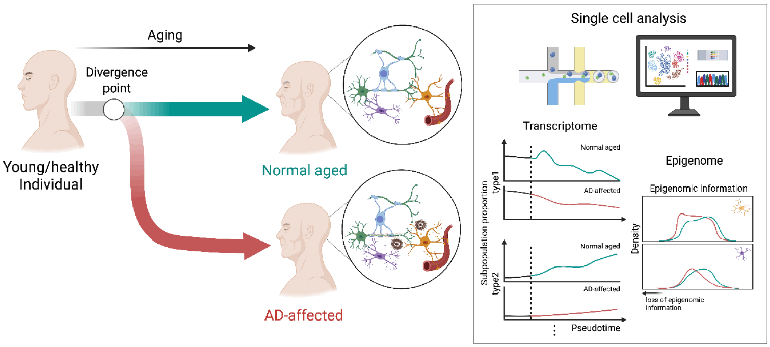
These findings indicate that AD is not a result of accelerated normal aging. Instead, it is a transformed pathological process that occurs when specific cell types become disproportionately vulnerable to age-related changes (Fig.1). Therefore, future research on AD must expand beyond models that merely recapitulate the accumulation of Aβ and tau. It is imperative to develop next-generation models that can precisely simulate the aging process and incorporate cell-type-specific vulnerabilities that underlie this devastating disease.

Limitations of Conventional Animal Models in Human Aging Research

Animal models have been foundational to aging studies. Invertebrate models, such as Caenorhabditis elegans and Drosophila melanogaster, offer the advantage of short lifespans, allowing for the rapid observation of aging phenotypes. They are also highly amenable to large-scale screening studies involving genetic manipulation or environmental changes. For instance, landmark studies in C. elegans have demonstrated that deletion of genes such as daf-2 or daf-16 can significantly extend lifespan (42). Similarly, experiments in C. elegans have provided early evidence that caloric restriction can prolong life, revealing the crucial roles of metabolic regulation and autophagy in longevity (43, 44). In D. melanogaster, the inhibition of transposable element activation has been shown to extend lifespan, suggesting that chromatin remodeling and epigenetic regulation are central mechanisms of aging and longevity (45). Despite their utility, invertebrate models have significant limitations in studying brain aging. Their nervous systems are less complex and lack the intricate cortical architecture of the human brain. This evolutionary divergence extends far beyond neuroanatomy. Fundamental differences in metabolism, distinct organization of organ systems, and the absence of an adaptive immune system, which is increasingly recognized as a critical modulator of neuroinflammation and cognitive function during aging, make it difficult to conclude that aging processes are directly analogous to those in humans. Consequently, mouse models have been widely used to study age-related diseases, including neurodegeneration. The advantages of the mouse model include the ability to easily observe age-related declines in performance in behavioral paradigms designed to assess cognitive domains (e.g., memory impairment), physical capacity (e.g., reduced muscle strength), and external appearance (e.g., changes in fur color and texture) (46). However, several significant challenges remain. Acquiring wild-type aged mice without genetic modifications requires at least 24 months, which is both time-consuming and resource-intensive. To bypass these logistical hurdles, some researchers have used strains developed for accelerated aging, such as senescence-accelerated mouse-prone 8 or progeroid models expressing mutated lamin A (progerin) (2, 47). However, a significant caveat is that these models are often closer to disease models than to true aging models. The cellular and tissue microenvironments in these animals can be dominated by aggressive pathological cascades that may differ substantially from the gradual multifactorial accumulation of deficits observed in natural physiological aging processes. Furthermore, fundamental interspecific differences cast doubt on whether these models accurately recapitulate the natural human aging process. Wild-type mice rarely develop AD and lack the genetic complexity of key human risk factors (48, 49). For example, humans carry three common alleles for APOE (APOE2, APOE3, and APOE4), with the APOE4 variant dramatically increasing the risk of disease. In contrast, mice possess only a single functionally distinct Apoe allele. This disparity necessitates the introduction of humanized genes, such as the various APOE variants, through genetic modifications to model this critical aspect of the disease (50). Models such as the 5xFAD mouse exhibit AD pathology at a very early age, Aβ accumulation by 2 months, and memory deficits by 4 months (51). Consequently, although AD is an age-related disease, these models often fail to reflect the underlying aging context.

Recent Advances in In Vitro Modeling of Human Brain Aging

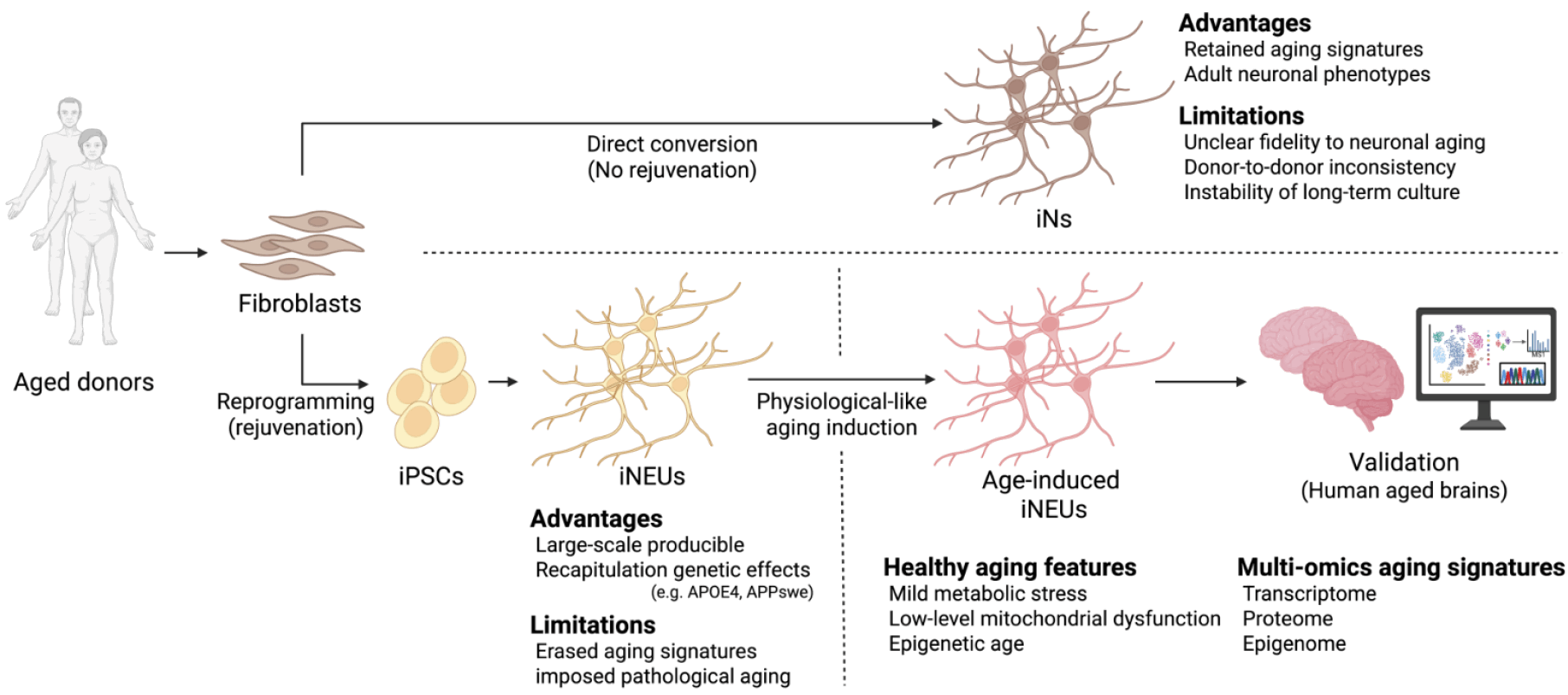
As recapitulating physiological aging in experimental models remains challenging, a more tractable strategy is to model one or more key factors or hallmarks of the aging process to investigate their pathological roles in AD (Table 1; 52-80). The development of such reductionist models often begins in vitro, where cellular and molecular variables can be precisely controlled and manipulated. This approach can start with establishing techniques to simulate age-associated phenotypes in isolated single-cell types and subsequently progress to more complex co-culture systems to study the impact of aging on cell-cell interactions. By creating robust in vitro platforms that mimic the defined aspects of aging, researchers can begin to elucidate the causal roles of these changes before undertaking more complex in vivo manipulations. Among the various techniques developed for this purpose, direct conversion has emerged as a powerful tool. Recent methodologies have enabled the direct generation of neurons from the skin cells of aged or diseased individuals, bypassing the induced pluripotent stem cell (iPSCs) stage. This process, known as direct conversion, involves the forced expression of key neuronal transcription factors, such as Ngn2 and Ascl1, or the use of specific miRNAs to drive neuronal identity (81-83). It is critical to distinguish between the concepts of ‘reprogramming’—the technical process of altering cell identity—and ‘rejuvenation’—the biological resetting of cellular age. iPSCs generation involves both cellular reprogramming (from the somatic to pluripotent state) and inherent rejuvenation, which erases age-associated signatures. In contrast, direct conversion achieves reprogramming of cell identity (e.g., from fibroblasts to neurons) without inducing cellular rejuvenation. Therefore, directly converted induced neurons (iNs) are uniquely advantageous for aging research because they decouple cell fate changes from age resetting, retaining the donor’s aging signatures (Table 2; 52, 53, 83-88). For example, iNs derived from the fibroblasts of older donors exhibit an increased epigenetic age compared to iPSC-derived neurons (iNEUs), which possess youthful characteristics owing to the rejuvenation that occurs during the iPSC reprogramming (84). Furthermore, iNs from patients with AD exhibit increased senescence markers (including SA-β-gal) and elevated levels of pathological markers, such as Aβ, phosphorylated tau, and mislocalized TAR DNA-binding protein 43, compared to age-matched controls (52, 53). Crucially, they also overcome a major hurdle in disease modeling related to the tau protein. Alternative splicing of the MAPT gene in the adult human brain produces a balanced 1:1 ratio of two major tau isoforms: those with three microtubule-binding repeats (3R) and those with four (4R). This balanced expression is considered a hallmark of mature adult neurons, and its dysregulation is believed to be central to a class of neurodegenerative disorders known as tauopathies (89). However, iNEUs, owing to their rejuvenated embryonic-like state, typically express only the fetal 3R isoform and fail to undergo a developmental switch to produce the 4R tau isoform. By retaining the epigenetic and maturational states of the donor cells, iNs express these adult 4R tau isoforms, making them an invaluable and physiologically relevant model for studying the mechanisms of adult-onset tauopathies (85).
Despite these advantages, the utility of iNs remains a matter of debate. Because they originate from fibroblasts, a cell type that may undergo a different aging process than neurons, it remains unclear whether the aged phenotypes observed in iNs truly reflect neuronal aging (90). Besides these conceptual challenges, significant practical hurdles have limited their widespread use. Primary fibroblasts enter replicative senescence after a limited number of divisions, severely restricting the total number of iNs generated from a single donor biopsy. This was compounded by issues related to quality control and reproducibility (90). For example, patient-derived fibroblast lines exhibit high variability in growth rate and neuronal conversion efficiency. This inherent inconsistency between donors makes it difficult to obtain standardized results and to compare findings across studies. Furthermore, iNs, as post-mitotic and highly specialized cells, are fragile and difficult to maintain in a healthy state for extended periods, which may be required to model the slow progression of age-related cellular decline. These limitations directly conflict with the increasing need for large-scale generation of neurons for modern biomedical research. A substantial and consistent supply of cells is essential for robust multiple experiments that are fundamental to rigorous scientific inquiry. More specifically, large cell numbers are a prerequisite for powerful research applications, such as high-throughput drug screening, which involves testing thousands of potential therapeutic compounds, and for comprehensive multiomics analyses (e.g., transcriptomics, proteomics, and metabolomics), which require significant starting material to generate reliable, high-quality data (91, 92). These large-scale approaches are critical for elucidating complex disease mechanisms and identifying novel therapeutic targets. Therefore, whereas iNs offer a unique window into aged cellular states, these limitations constrain their application in the studies necessary to advance the field.
iPSCs are generated from somatic cells, such as fibroblasts or blood cells, by expressing Yamanaka factors (93, 94). Given their theoretically unlimited proliferative potential, iPSCs are an excellent source for the large-scale production of neurons and iNEUs, which are also relatively stable in long-term cultures. However, as previously mentioned, the fundamental limitation of this model is the inherent rejuvenation that occurs during the reprogramming process. This effectively erases the aging signatures of the donor, including epigenetic alterations, and reverts the cells to an embryonic-like state (95). Consequently, whereas iNEUs derived from patients can recapitulate certain genetic aspects of a disease (96-98), the absence of a crucial aging context means that these phenotypes are often restricted to the earliest manifestations of the pathology, reflecting an asymptomatic or prodromal stage rather than a full-blown age-dependent clinical state. They fail to capture the complex interplay between genetic predisposition and decades of cellular stress that drive late-onset neurodegeneration, making their direct application in modeling age-related diseases problematic.
Various attempts have been made to intentionally induce an aging state in rejuvenated iNEUs. However, these strategies often introduce the very same conceptual limitations as those previously discussed for in vivo accelerated aging models. Rather than recapitulating physiological aging, these models tend to impose an aggressive pathology. For instance, introducing a progeria mutation into iPSC-derived dopaminergic neurons induces certain age-associated phenotypes, such as increased mitochondrial reactive oxygen species, but simultaneously triggers early features of PD, thereby confounding physiological aging with overt disease pathology (99). Similar outcomes were observed when telomere shortening was used to mimic premature aging in the same cell type (100). Although these models display specific phenotypes, they suffer from a fundamental confounding factor: mirroring the caveats of their in vivo counterparts, they represent a distinct pathological state rather than the gradual, multifactorial process of physiological aging. This makes it difficult to distinguish phenotypes driven by aging insults from those representing independent pathologies. Consequently, these models have limited utility in clarifying the causal relationship between physiological aging and the development of age-related diseases (Fig. 2).

Conclusions

The preceding sections underscore the profound limitations inherent in the current models of physiological aging, from the temporal and financial costs of aging animal cohorts to the correlational nature of human studies. These challenges have created a critical bottleneck, hindering our ability to move beyond identifying associations to establishing the causal mechanisms that drive age-related neurological decline. To overcome this impasse, the future of aging research may not lie in perfecting any single model but in developing an integrative framework that synergistically combines advances in bioinformatics, sophisticated in vitro systems, and large-scale human data to create and validate in vivo models with unprecedented physiological relevance. A foundational pillar of this framework is the continued advancement of high-resolution bioinformatics. The proliferation of single-cell and spatial multiomics (e.g., single-cell RNA sequencing, single-nucleus ATAC sequencing, spatial transcriptomics, and proteomics) applied to postmortem human brain tissue has generated an exquisitely detailed atlas of how aging manifests at the cellular level (32, 35, 38, 39, 41, 101-112) (Table 3). By analyzing these datasets, we can move beyond generalized aging markers to identify robust, cell-type-specific molecular signatures for aged neurons, astrocytes, microglia, and oligodendrocytes. This allowed us to define the appearance of a “physiologically aged” human brain cell from a systems biology perspective, thus providing a critical reference blueprint against which any experimental model must be validated. The next step involves engineering in vitro models that can accurately recapitulate these complex molecular signatures. iPSCs are an ideal starting point for this purpose. Using iPSCs from a cohort of healthy individuals, we can control the vast genetic heterogeneity that plagues human studies and create a standardized, scalable platform. The central challenge is to intentionally induce a state of “physiological aging” in rejuvenated cells. Rather than relying on potent non-physiological insults, this can be achieved through subtle and chronic stressors that mimic the accumulated challenges of life. This could involve long-term culture under conditions of mild metabolic stress, DNA breaks, controlled induction of low-level mitochondrial dysfunction, or the use of advanced clustered regularly interspaced short palindromic repeats (CRISPR)-based epigenetic editors to systematically rewrite methylation patterns to match the “epigenetic age” observed in human brain tissue (113-116). The goal is not to create a caricature of aging but to guide young cells toward the specific molecular state defined by our bioinformatic blueprint.
This framework requires a rigorous validation process. The molecular state of these in vitro aged cells, including their transcriptomes, proteomes, and epigenomes, must be systematically compared with the reference data from the human brain. A model was considered validated only when its multiomic signature demonstrated substantial concordance with that of aged human cells. This data-driven validation step ensures that we are not studying an artifact of the culture dish but a system that genuinely mirrors the key aspects of the human aging process. Human informatics databases, such as the UK Biobank or the Religious Orders Study and Rush Memory and Aging Project, would serve as the ultimate arbiters of a model’s relevance (117, 118). Ultimately, any causal factors identified in vitro must be validated in vivo to confirm their relevance to complex physiological systems. Once a model passes this stringent validation, it transforms from a descriptive tool into a powerful platform for investigating causality. Within this controlled human cell-based system, mechanistic experiments that are impossible to conduct in humans can be performed. Using CRISPR-mediated gene editing, aging markers identified in an initial bioinformatic screening can be systematically perturbed in a cell-type-specific manner. These experiments can directly reveal whether an age-associated change is a driver of functional decline, a compensatory protective response, or a neutral bystander, thus untangling the complex web of causality. Ultimately, this validated physiological aging platform provides an ideal background for investigating the influence of genetic risk factors on neurodegenerative disease. Not all elderly individuals develop age-associated neurodegeneration, such as AD. By introducing genetic risk factors, such as APOE4, into our iPSC lines using isogenic controls, we can investigate how these factors interact with and modify the physiological aging process. This approach enables the systematic dissection of the mechanisms by which genetic predisposition converts the trajectory of normal aging into pathological neurodegeneration. By integrating these disparate fields, this forward-looking framework promises to bridge the causal gap in aging research and pave the way for the development of targeted therapeutics that promote neurological health.
Notes

Potential Conflict of Interest

There are no potential conflicts of interest to declare.

Authors’ Contribution

Conceptualization: J Seok, HL, J Seo. Writing – original draft: J Seok, HL, J Seo. Writing – review and editing: J Seok, HL, J Seo.

Funding

This work was supported by the National Research Foundation of Korea (NRF) grant funded by the Ministry of Education (RS-2024-00462979 to H.I.), NRF grant funded by the Ministry of Science and ICT (MSIT) (RS-2025-00554061 to J.S.), and a grant from the Korea Dementia Research Project through the Korea Dementia Research Center funded by the Ministry of Health & Welfare and MSIT (RS-2024-00343370 to J.S.), and supported by the Yonsei University Research Fund (2025-22-0154 to J.S.). All schematics were created using BioRender, with a full license for publication.

References

1. López-Otín C, Blasco MA, Partridge L, Serrano M, Kroemer G. 2023; Hallmarks of aging: an expanding universe. Cell. 186:243–278. DOI: 10.1016/j.cell.2022.11.001. PMID: 36599349. PMCID: PMC11043504.
2. Kim B, Lee DI, Basisty N, Dai DF. 2025; Cellular models of aging and senescence. Cells. 14:1278. DOI: 10.3390/cells14161278. PMID: 40862757. PMCID: PMC12384970.
3. Khan SS, Singer BD, Vaughan DE. 2017; Molecular and physiological manifestations and measurement of aging in humans. Aging Cell. 16:624–633. DOI: 10.1111/acel.12601. PMID: 28544158. PMCID: PMC5506433.
4. Liu WS, You J, Ge YJ, et al. 2023; Association of biological age with health outcomes and its modifiable factors. Aging Cell. 22:e13995. DOI: 10.1111/acel.13995. PMID: 37723992. PMCID: PMC10726867.
5. Hamczyk MR, Nevado RM, Barettino A, Fuster V, Andrés V. 2020; Biological versus chronological aging: JACC focus seminar. J Am Coll Cardiol. 75:919–930. DOI: 10.1016/j.jacc.2019.11.062. PMID: 32130928.
6. Mitnitski A, Collerton J, Martin-Ruiz C, et al. 2015; Age-related frailty and its association with biological markers of ageing. BMC Med. 13:161. DOI: 10.1186/s12916-015-0400-x. PMID: 26166298. PMCID: PMC4499935.
7. Ji L, Jazwinski SM, Kim S. 2021; Frailty and biological age. Ann Geriatr Med Res. 25:141–149. DOI: 10.4235/agmr.21.0080. PMID: 34399574. PMCID: PMC8497950.
8. Dent E, Kowal P, Hoogendijk EO. 2016; Frailty measurement in research and clinical practice: a review. Eur J Intern Med. 31:3–10. DOI: 10.1016/j.ejim.2016.03.007. PMID: 27039014.
9. Di Micco R, Krizhanovsky V, Baker D. d'Adda di Fagagna F. 2021; Cellular senescence in ageing: from mechanisms to therapeutic opportunities. Nat Rev Mol Cell Biol. 22:75–95. DOI: 10.1038/s41580-020-00314-w. PMID: 33328614. PMCID: PMC8344376.
10. Duan R, Fu Q, Sun Y, Li Q. 2022; Epigenetic clock: a promising biomarker and practical tool in aging. Ageing Res Rev. 81:101743. DOI: 10.1016/j.arr.2022.101743. PMID: 36206857.
11. Oh HS, Rutledge J, Nachun D, et al. 2023; Organ aging signatures in the plasma proteome track health and disease. Nature. 624:164–172. DOI: 10.1038/s41586-023-06802-1. PMID: 38057571. PMCID: PMC10700136.
12. Hayflick L, Moorhead PS. 1961; The serial cultivation of human diploid cell strains. Exp Cell Res. 25:585–621. DOI: 10.1016/0014-4827(61)90192-6. PMID: 13905658.
13. Dimri GP, Lee X, Basile G, et al. 1995; A biomarker that identifies senescent human cells in culture and in aging skin in vivo. Proc Natl Acad Sci U S A. 92:9363–9367. DOI: 10.1073/pnas.92.20.9363. PMID: 7568133. PMCID: PMC40985.
14. Lee BY, Han JA, Im JS, et al. 2006; Senescence-associated beta-galactosidase is lysosomal beta-galactosidase. Aging Cell. 5:187–195. DOI: 10.1111/j.1474-9726.2006.00199.x. PMID: 16626397.
15. Takeuchi S, Takahashi A, Motoi N, et al. 2010; Intrinsic cooperation between p16INK4a and p21Waf1/Cip1 in the onset of cellular senescence and tumor suppression in vivo. Cancer Res. 70:9381–9390. DOI: 10.1158/0008-5472.CAN-10-0801. PMID: 21062974.
16. Dehkordi SK, Walker J, Sah E, et al. 2021; Profiling senescent cells in human brains reveals neurons with CDKN2D/p19 and tau neuropathology. Nat Aging. 1:1107–1116. DOI: 10.1038/s43587-021-00142-3. PMID: 35531351. PMCID: PMC9075501.
17. Coppé JP, Desprez PY, Krtolica A, Campisi J. 2010; The senescence-associated secretory phenotype: the dark side of tumor suppression. Annu Rev Pathol. 5:99–118. DOI: 10.1146/annurev-pathol-121808-102144. PMID: 20078217. PMCID: PMC4166495.
18. Wang B, Han J, Elisseeff JH, Demaria M. 2024; The senescence-associated secretory phenotype and its physiological and pathological implications. Nat Rev Mol Cell Biol. 25:958–978. DOI: 10.1038/s41580-024-00727-x. PMID: 38654098.
19. Piechota M, Sunderland P, Wysocka A, et al. 2016; Is senescence-associated β-galactosidase a marker of neuronal senescence? Oncotarget. 7:81099–81109. DOI: 10.18632/oncotarget.12752. PMID: 27768595. PMCID: PMC5348379.
20. Omori S, Wang TW, Johmura Y, et al. 2020; Generation of a p16 reporter mouse and its use to characterize and target p16high cells in vivo. Cell Metab. 32:814–828.e6. DOI: 10.1016/j.cmet.2020.09.006. PMID: 32949498.
21. Hudson HR, Riessland M, Orr ME. 2024; Defining and characterizing neuronal senescence, 'neurescence', as GX arrested cells. Trends Neurosci. 47:971–984. DOI: 10.1016/j.tins.2024.09.006. PMID: 39389805.
22. Byrns CN, Perlegos AE, Miller KN, et al. 2024; Senescent glia link mitochondrial dysfunction and lipid accumulation. Nature. 630:475–483. DOI: 10.1038/s41586-024-07516-8. PMID: 38839958. PMCID: PMC11168935.
23. Melo Dos Santos LS, Trombetta-Lima M, Eggen B, Demaria M. 2024; Cellular senescence in brain aging and neurodegeneration. Ageing Res Rev. 93:102141. DOI: 10.1016/j.arr.2023.102141. PMID: 38030088.
24. Jin K, Yao Z, van Velthoven CTJ, et al. 2025; Brain-wide cell-type-specific transcriptomic signatures of healthy ageing in mice. Nature. 638:182–196. DOI: 10.1038/s41586-024-08350-8. PMID: 39743592. PMCID: PMC11798837.
25. Wong WT. 2013; Microglial aging in the healthy CNS: phenotypes, drivers, and rejuvenation. Front Cell Neurosci. 7:22. DOI: 10.3389/fncel.2013.00022. PMID: 23493481. PMCID: PMC3595516.
26. Gildea HK, Liddelow SA. 2025; Mechanisms of astrocyte aging in reactivity and disease. Mol Neurodegener. 20:21. DOI: 10.1186/s13024-025-00810-7. PMID: 39979986. PMCID: PMC11844071.
27. Saul D, Kosinsky RL. 2021; Epigenetics of aging and aging-associated diseases. Int J Mol Sci. 22:401. DOI: 10.3390/ijms22010401. PMID: 33401659. PMCID: PMC7794926.
28. Horvath S. 2013; DNA methylation age of human tissues and cell types. Genome Biol. 14:R115. DOI: 10.1186/gb-2013-14-10-r115. PMID: 24138928. PMCID: PMC4015143.
29. Lu AT, Quach A, Wilson JG, et al. 2019; DNA methylation GrimAge strongly predicts lifespan and healthspan. Aging (Albany NY). 11:303–327. DOI: 10.18632/aging.101684. PMID: 30669119. PMCID: PMC6366976.
30. Belsky DW, Caspi A, Arseneault L, et al. 2020; Quantification of the pace of biological aging in humans through a blood test, the DunedinPoAm DNA methylation algorithm. Elife. 9:e54870. DOI: 10.7554/eLife.54870. PMID: 32367804. PMCID: PMC7282814.
31. McCartney DL, Stevenson AJ, Walker RM, et al. 2018; Investigating the relationship between DNA methylation age acceleration and risk factors for Alzheimer's disease. Alzheimers Dement (Amst). 10:429–437. DOI: 10.1016/j.dadm.2018.05.006. PMID: 30167451. PMCID: PMC6111045.
32. Farinas A, Rutledge J, Bot VA, et al. 2025; Disruption of the cerebrospinal fluid-plasma protein balance in cognitive impairment and aging. Nat Med. 31:2578–2589. DOI: 10.1038/s41591-025-03831-3. PMID: 40665050. PMCID: PMC12353824.
33. Ali M, Erabadda B, Chen Y, et al. 2025; Shared and disease-specific pathways in frontotemporal dementia and Alzheimer's and Parkinson's diseases. Nat Med. 31:2567–2577. DOI: 10.1038/s41591-025-03833-1. PMID: 40665052. PMCID: PMC12404994.
34. Shvetcov A, Johnson ECB, Winchester LM, et al. 2025; APOE ε4 carriers share immune-related proteomic changes across neurodegenerative diseases. Nat Med. 31:2590–2601. DOI: 10.1038/s41591-025-03835-z. PMID: 40665049. PMCID: PMC12353839.
35. Oh HS, Le Guen Y, Rappoport N, et al. 2025; Plasma proteomics links brain and immune system aging with healthspan and longevity. Nat Med. 31:2703–2711. DOI: 10.1038/s41591-025-03798-1. PMID: 40634782. PMCID: PMC12353788.
36. Knopman DS, Amieva H, Petersen RC, et al. 2021; Alzheimer disease. Nat Rev Dis Primers. 7:33. DOI: 10.1038/s41572-021-00269-y. PMID: 33986301. PMCID: PMC8574196.
37. van der Kant R, Goldstein LSB, Ossenkoppele R. 2020; Amyloid-β-independent regulators of tau pathology in Alzheimer disease. Nat Rev Neurosci. 21:21–35. DOI: 10.1038/s41583-019-0240-3. PMID: 31780819.
38. Green GS, Fujita M, Yang HS, et al. 2024; Cellular communities reveal trajectories of brain ageing and Alzheimer's disease. Nature. 633:634–645. DOI: 10.1038/s41586-024-07871-6. PMID: 39198642. PMCID: PMC11877878.
39. Liu Z, Zhang S, James BT, et al. 2025; Single-cell multiregion epigenomic rewiring in Alzheimer's disease progression and cognitive resilience. Cell. 188:4980–5002.e29. DOI: 10.1016/j.cell.2025.06.031. PMID: 40752494. PMCID: PMC12573303.
40. Han X, Li D, Goldstein D, et al. 2024; Genetic and epigenetic signatures for cognitive resilience in neuropathologically defined Alzheimer's disease brains. Alzheimers Dement. 20:e089466. DOI: 10.1002/alz.089466. PMCID: PMC11714589.
41. Muralidharan C, Zakar-Polyák E, Adami A, et al. 2025; Human brain cell-type-specific aging clocks based on single-nuclei transcriptomics. Adv Sci (Weinh). 12:e06109. DOI: 10.1002/advs.202506109. PMID: 40878446. PMCID: PMC12631854.
42. Kenyon C, Chang J, Gensch E, Rudner A, Tabtiang R. 1993; A C. elegans mutant that lives twice as long as wild type. Nature. 366:461–464. DOI: 10.1038/366461a0. PMID: 8247153.
43. Lakowski B, Hekimi S. 1998; The genetics of caloric restriction in Caenorhabditis elegans. Proc Natl Acad Sci U S A. 95:13091–13096. DOI: 10.1073/pnas.95.22.13091. PMID: 9789046. PMCID: PMC23719.
44. Jia K, Levine B. 2007; Autophagy is required for dietary restriction-mediated life span extension in C. elegans. Autophagy. 3:597–599. DOI: 10.4161/auto.4989. PMID: 17912023.
45. Wood JG, Jones BC, Jiang N, et al. 2016; Chromatin-modifying genetic interventions suppress age-associated transposable element activation and extend life span in Drosophila. Proc Natl Acad Sci U S A. 113:11277–11282. DOI: 10.1073/pnas.1604621113. PMID: 27621458. PMCID: PMC5056045.
46. Vandamme TF. 2014; Use of rodents as models of human diseases. J Pharm Bioallied Sci. 6:2–9. DOI: 10.4103/0975-7406.124301. PMID: 24459397. PMCID: PMC3895289.
47. Butterfield DA, Poon HF. 2005; The senescence-accelerated prone mouse (SAMP8): a model of age-related cognitive decline with relevance to alterations of the gene expression and protein abnormalities in Alzheimer's disease. Exp Gerontol. 40:774–783. DOI: 10.1016/j.exger.2005.05.007. PMID: 16026957.
48. Granzotto A, Vissel B, Sensi SL. 2024; Lost in translation: inconvenient truths on the utility of mouse models in Alzheimer's disease research. Elife. 13:e90633. DOI: 10.7554/eLife.90633. PMID: 39329365. PMCID: PMC11434637.
49. Qian Z, Li Y, Ye K. 2024; Advancements and challenges in mouse models of Alzheimer's disease. Trends Mol Med. 30:1152–1164. DOI: 10.1016/j.molmed.2024.10.010. PMID: 39547883.
50. Raffai RL, Dong LM, Farese RV Jr, Weisgraber KH. 2001; Introduction of human apolipoprotein E4 "domain interaction" into mouse apolipoprotein E. Proc Natl Acad Sci U S A. 98:11587–11591. DOI: 10.1073/pnas.201279298. PMID: 11553788. PMCID: PMC58773.
51. Oakley H, Cole SL, Logan S, et al. 2006; Intraneuronal beta-amyloid aggregates, neurodegeneration, and neuron loss in transgenic mice with five familial Alzheimer's disease mutations: potential factors in amyloid plaque formation. J Neurosci. 26:10129–10140. DOI: 10.1523/JNEUROSCI.1202-06.2006. PMID: 17021169. PMCID: PMC6674618.
52. Herdy JR, Traxler L, Agarwal RK, et al. 2022; Increased post-mitotic senescence in aged human neurons is a pathological feature of Alzheimer's disease. Cell Stem Cell. 29:1637–1652.e6. DOI: 10.1016/j.stem.2022.11.010. PMID: 36459967. PMCID: PMC10093780.
53. Chou CC, Vest R, Prado MA, et al. 2025; Proteostasis and lysosomal repair deficits in transdifferentiated neurons of Alzheimer's disease. Nat Cell Biol. 27:619–632. DOI: 10.1038/s41556-025-01623-y. PMID: 40140603. PMCID: PMC11991917.
54. Mecocci P, MacGarvey U, Beal MF. 1994; Oxidative damage to mitochondrial DNA is increased in Alzheimer's disease. Ann Neurol. 36:747–751. DOI: 10.1002/ana.410360510. PMID: 7979220.
55. Dileep V, Boix CA, Mathys H, et al. 2023; Neuronal DNA double-strand breaks lead to genome structural variations and 3D genome disruption in neurodegeneration. Cell. 186:4404–4421.e20. DOI: 10.1016/j.cell.2023.08.038. PMID: 37774679. PMCID: PMC10697236.
56. Suberbielle E, Sanchez PE, Kravitz AV, et al. 2013; Physiologic brain activity causes DNA double-strand breaks in neurons, with exacerbation by amyloid-β. Nat Neurosci. 16:613–621. DOI: 10.1038/nn.3356. PMID: 23525040. PMCID: PMC3637871.
57. Rossiello F, Jurk D, Passos JF. d'Adda di Fagagna F. 2022; Telomere dysfunction in ageing and age-related diseases. Nat Cell Biol. 24:135–147. DOI: 10.1038/s41556-022-00842-x. PMID: 35165420. PMCID: PMC8985209.
58. Liu M, Huo YR, Wang J, et al. 2016; Telomere shortening in Alzheimer's disease patients. Ann Clin Lab Sci. 46:260–265.
59. Gjoneska E, Pfenning AR, Mathys H, et al. 2015; Conserved epigenomic signals in mice and humans reveal immune basis of Alzheimer's disease. Nature. 518:365–369. DOI: 10.1038/nature14252. PMID: 25693568. PMCID: PMC4530583.
60. De Plano LM, Saitta A, Oddo S, Caccamo A. 2024; Epigenetic changes in Alzheimer's disease: DNA methylation and histone modification. Cells. 13:719. DOI: 10.3390/cells13080719. PMID: 38667333. PMCID: PMC11049073.
61. Cozachenco D, Ribeiro FC, Ferreira ST. 2023; Defective proteostasis in Alzheimer's disease. Ageing Res Rev. 85:101862. DOI: 10.1016/j.arr.2023.101862. PMID: 36693451.
62. Morrone CD, Raghuraman R, Hussaini SA, Yu WH. 2023; Proteostasis failure exacerbates neuronal circuit dysfunction and sleep impairments in Alzheimer's disease. Mol Neurodegener. 18:27. DOI: 10.1186/s13024-023-00617-4. PMID: 37085942. PMCID: PMC10119020.
63. Lee JH, Yang DS, Goulbourne CN, et al. 2022; Faulty autolysosome acidification in Alzheimer's disease mouse models induces autophagic build-up of Aβ in neurons, yielding senile plaques. Nat Neurosci. 25:688–701. DOI: 10.1038/s41593-022-01084-8. PMID: 35654956. PMCID: PMC9174056.
64. Zhang Z, Yang X, Song YQ, Tu J. 2021; Autophagy in Alzheimer's disease pathogenesis: therapeutic potential and future perspectives. Ageing Res Rev. 72:101464. DOI: 10.1016/j.arr.2021.101464. PMID: 34551326.
65. Ondaro J, Hernandez-Eguiazu H, Garciandia-Arcelus M, et al. 2022; Defects of nutrient signaling and autophagy in neurodegeneration. Front Cell Dev Biol. 10:836196. DOI: 10.3389/fcell.2022.836196. PMID: 35419363. PMCID: PMC8996160.
66. Kellar D, Craft S. 2020; Brain insulin resistance in Alzheimer's disease and related disorders: mechanisms and therapeutic approaches. Lancet Neurol. 19:758–766. DOI: 10.1016/S1474-4422(20)30231-3. PMID: 32730766. PMCID: PMC9661919.
67. Talbot K, Wang HY, Kazi H, et al. 2012; Demonstrated brain insulin resistance in Alzheimer's disease patients is associated with IGF-1 resistance, IRS-1 dysregulation, and cognitive decline. J Clin Invest. 122:1316–1338. DOI: 10.1172/JCI59903. PMID: 22476197. PMCID: PMC3314463.
68. Mary A, Eysert F, Checler F, Chami M. 2023; Mitophagy in Alzheimer's disease: molecular defects and therapeutic approaches. Mol Psychiatry. 28:202–216. DOI: 10.1038/s41380-022-01631-6. PMID: 35665766. PMCID: PMC9812780.
69. Ashleigh T, Swerdlow RH, Beal MF. 2023; The role of mitochondrial dysfunction in Alzheimer's disease pathogenesis. Alzheimers Dement. 19:333–342. DOI: 10.1002/alz.12683. PMID: 35522844.
70. Hudson HR, Sun X, Orr ME. 2025; Senescent brain cell types in Alzheimer's disease: pathological mechanisms and therapeutic opportunities. Neurotherapeutics. 22:e00519. DOI: 10.1016/j.neurot.2024.e00519. PMID: 39765417. PMCID: PMC12047392.
71. Bhat R, Crowe EP, Bitto A, et al. 2012; Astrocyte senescence as a component of Alzheimer's disease. PLoS One. 7:e45069. DOI: 10.1371/journal.pone.0045069. PMID: 22984612. PMCID: PMC3440417.
72. Tobin MK, Musaraca K, Disouky A, et al. 2019; Human hippocampal neurogenesis persists in aged adults and Alzheimer's disease patients. Cell Stem Cell. 24:974–982.e3. DOI: 10.1016/j.stem.2019.05.003. PMID: 31130513. PMCID: PMC6608595.
73. Salta E, Lazarov O, Fitzsimons CP, Tanzi R, Lucassen PJ, Choi SH. 2023; Adult hippocampal neurogenesis in Alzheimer's disease: a roadmap to clinical relevance. Cell Stem Cell. 30:120–136. DOI: 10.1016/j.stem.2023.01.002. PMID: 36736288. PMCID: PMC10082636.
74. Moreno-Jiménez EP, Flor-García M, Terreros-Roncal J, et al. 2019; Adult hippocampal neurogenesis is abundant in neurologically healthy subjects and drops sharply in patients with Alzheimer's disease. Nat Med. 25:554–560. DOI: 10.1038/s41591-019-0375-9. PMID: 30911133. PMCID: PMC7051085.
75. Sun Y, Xu S, Jiang M, et al. 2021; Role of the extracellular matrix in Alzheimer's disease. Front Aging Neurosci. 13:707466. DOI: 10.3389/fnagi.2021.707466. PMID: 34512308. PMCID: PMC8430252.
76. Rahman MM, Lendel C. 2021; Extracellular protein components of amyloid plaques and their roles in Alzheimer's disease pathology. Mol Neurodegener. 16:59. DOI: 10.1186/s13024-021-00465-0. PMID: 34454574. PMCID: PMC8400902.
77. Akiyama H, Barger S, Barnum S, et al. 2000; Inflammation and Alzheimer's disease. Neurobiol Aging. 21:383–421. DOI: 10.1016/S0197-4580(00)00124-X. PMID: 10858586. PMCID: PMC3887148.
78. Heneka MT, Carson MJ, El Khoury J, et al. 2015; Neuroinflammation in Alzheimer's disease. Lancet Neurol. 14:388–405. DOI: 10.1016/S1474-4422(15)70016-5. PMID: 25792098. PMCID: PMC5909703.
79. Liu S, Gao J, Zhu M, Liu K, Zhang HL. 2020; Gut microbiota and dysbiosis in Alzheimer's disease: implications for pathogenesis and treatment. Mol Neurobiol. 57:5026–5043. DOI: 10.1007/s12035-020-02073-3. PMID: 32829453. PMCID: PMC7541367.
80. Chandra S, Sisodia SS, Vassar RJ. 2023; The gut microbiome in Alzheimer's disease: what we know and what remains to be explored. Mol Neurodegener. 18:9. DOI: 10.1186/s13024-023-00595-7. PMID: 36721148. PMCID: PMC9889249.
81. Vierbuchen T, Ostermeier A, Pang ZP, Kokubu Y, Südhof TC, Wernig M. 2010; Direct conversion of fibroblasts to functional neurons by defined factors. Nature. 463:1035–1041. DOI: 10.1038/nature08797. PMID: 20107439. PMCID: PMC2829121.
82. Church VA, Cates K, Capano L, Aryal S, Kim WK, Yoo AS. Hu K, editor. 2021. Generation of human neurons by microRNA-mediated direct conversion of dermal fibroblasts. Nuclear reprogramming: methods and protocols. Humana (Springer Nature);p. 77–100. DOI: 10.1007/978-1-0716-1084-8_6. PMID: 33226614.
83. Mertens J, Paquola ACM, Ku M, et al. 2015; Directly reprogrammed human neurons retain aging-associated transcriptomic signatures and reveal age-related nucleocytoplasmic defects. Cell Stem Cell. 17:705–718. DOI: 10.1016/j.stem.2015.09.001. PMID: 26456686. PMCID: PMC5929130.
84. Rhine K, Li R, Kopalle HM, et al. 2025; Neuronal aging causes mislocalization of splicing proteins and unchecked cellular stress. Nat Neurosci. 28:1174–1184. DOI: 10.1038/s41593-025-01952-z. PMID: 40456907. PMCID: PMC12148940.
85. Capano LS, Sato C, Ficulle E, et al. 2022; Recapitulation of endogenous 4R tau expression and formation of insoluble tau in directly reprogrammed human neurons. Cell Stem Cell. 29:918–932.e8. DOI: 10.1016/j.stem.2022.04.018. PMID: 35659876. PMCID: PMC9176216.
86. Huh CJ, Zhang B, Victor MB, et al. 2016; Maintenance of age in human neurons generated by microRNA-based neuronal conversion of fibroblasts. Elife. 5:e18648. DOI: 10.7554/eLife.18648. PMID: 27644593. PMCID: PMC5067114.
87. Drouin-Ouellet J, Legault EM, Nilsson F, et al. 2022; Age-related pathological impairments in directly reprogrammed dopaminergic neurons derived from patients with idiopathic Parkinson's disease. Stem Cell Reports. 17:2203–2219. DOI: 10.1016/j.stemcr.2022.08.010. PMID: 36150382. PMCID: PMC9561608.
88. Tang Y, Liu ML, Zang T, Zhang CL. 2017; Direct reprogramming rather than iPSC-based reprogramming maintains aging hallmarks in human motor neurons. Front Mol Neurosci. 10:359. DOI: 10.3389/fnmol.2017.00359. PMID: 29163034. PMCID: PMC5676779.
89. Panda D, Samuel JC, Massie M, Feinstein SC, Wilson L. 2003; Differential regulation of microtubule dynamics by three- and four-repeat tau: implications for the onset of neurodegenerative disease. Proc Natl Acad Sci U S A. 100:9548–9553. DOI: 10.1073/pnas.1633508100. PMID: 12886013. PMCID: PMC170955.
90. de Luzy IR, Lee MK, Mobley WC, Studer L. 2024; Lessons from inducible pluripotent stem cell models on neuronal senescence in aging and neurodegeneration. Nat Aging. 4:309–318. DOI: 10.1038/s43587-024-00586-3. PMID: 38429379. PMCID: PMC11824472.
91. Tavassoli H, Alhosseini SN, Tay A, Chan PPY, Weng Oh SK, Warkiani ME. 2018; Large-scale production of stem cells utilizing microcarriers: a biomaterials engineering perspective from academic research to commercialized products. Biomaterials. 181:333–346. DOI: 10.1016/j.biomaterials.2018.07.016. PMID: 30098569.
92. Kami D, Watakabe K, Yamazaki-Inoue M, et al. 2013; Large-scale cell production of stem cells for clinical application using the automated cell processing machine. BMC Biotechnol. 13:102. DOI: 10.1186/1472-6750-13-102. PMID: 24228851. PMCID: PMC4101824.
93. Takahashi K, Yamanaka S. 2006; Induction of pluripotent stem cells from mouse embryonic and adult fibroblast cultures by defined factors. Cell. 126:663–676. DOI: 10.1016/j.cell.2006.07.024. PMID: 16904174.
94. Yoon DE, Lee H, Kim K. 2024; Recent research trends in stem cells using CRISPR/Cas-based genome editing methods. Int J Stem Cells. 17:1–14. DOI: 10.15283/ijsc23030. PMID: 37904281. PMCID: PMC10899885.
95. Lapasset L, Milhavet O, Prieur A, et al. 2011; Rejuvenating senescent and centenarian human cells by reprogramming through the pluripotent state. Genes Dev. 25:2248–2253. DOI: 10.1101/gad.173922.111. PMID: 22056670. PMCID: PMC3219229.
96. Lagomarsino VN, Pearse RV 2nd, Liu L, et al. 2021; Stem cell-derived neurons reflect features of protein networks, neuropathology, and cognitive outcome of their aged human donors. Neuron. 109:3402–3420.e9. DOI: 10.1016/j.neuron.2021.08.003. PMID: 34473944. PMCID: PMC8571042.
97. Kim MS, Yoon S, Choi J, Kim YJ, Lee G. 2025; Stem cell-based approaches in Parkinson's disease research. Int J Stem Cells. 18:21–36. DOI: 10.15283/ijsc23169. PMID: 38449089. PMCID: PMC11867902.
98. Kim J, Park SH, Sun W. 2025; The differential developmental neurotoxicity of valproic acid on anterior and posterior neural induction of human pluripotent stem cells. Int J Stem Cells. 18:49–58. DOI: 10.15283/ijsc24066. PMID: 38973150. PMCID: PMC11867903.
99. Miller JD, Ganat YM, Kishinevsky S, et al. 2013; Human iPSC-based modeling of late-onset disease via progerin-induced aging. Cell Stem Cell. 13:691–705. DOI: 10.1016/j.stem.2013.11.006. PMID: 24315443. PMCID: PMC4153390.
100. Vera E, Bosco N, Studer L. 2016; Generating late-onset human iPSC-based disease models by inducing neuronal age-related phenotypes through telomerase manipulation. Cell Rep. 17:1184–1192. DOI: 10.1016/j.celrep.2016.09.062. PMID: 27760320. PMCID: PMC5089807.
101. Emani PS, Liu JJ, Clarke D, et al. 2024; Single-cell genomics and regulatory networks for 388 human brains. Science. 384:eadi5199.
102. Shen X, Wang C, Zhou X, et al. 2024; Nonlinear dynamics of multi-omics profiles during human aging. Nat Aging. 4:1619–1634. DOI: 10.1038/s43587-024-00692-2. PMID: 39143318. PMCID: PMC11564093.
103. Rutledge J, Oh H, Wyss-Coray T. 2022; Measuring biological age using omics data. Nat Rev Genet. 23:715–727. DOI: 10.1038/s41576-022-00511-7. PMID: 35715611. PMCID: PMC10048602.
104. Sankowski R, Böttcher C, Masuda T, et al. 2019; Mapping microglia states in the human brain through the integration of high-dimensional techniques. Nat Neurosci. 22:2098–2110. DOI: 10.1038/s41593-019-0532-y. PMID: 31740814.
105. Johnson ECB, Dammer EB, Duong DM, et al. 2020; Large-scale proteomic analysis of Alzheimer's disease brain and cerebrospinal fluid reveals early changes in energy metabolism associated with microglia and astrocyte activation. Nat Med. 26:769–780. DOI: 10.1038/s41591-020-0815-6. PMID: 32284590. PMCID: PMC7405761.
106. González-Velasco O, Papy-García D, Le Douaron G, Sánchez-Santos JM, De Las Rivas J. 2020; Transcriptomic landscape, gene signatures and regulatory profile of aging in the human brain. Biochim Biophys Acta Gene Regul Mech. 1863:194491. DOI: 10.1016/j.bbagrm.2020.194491. PMID: 32006715.
107. Piehl N, van Olst L, Ramakrishnan A, et al. 2022; Cerebrospinal fluid immune dysregulation during healthy brain aging and cognitive impairment. Cell. 185:5028–5039.e13. DOI: 10.1016/j.cell.2022.11.019. PMID: 36516855. PMCID: PMC9815831.
108. Yang H, Clarence T, Scott MR, et al. 2025. A lifespan transcriptomic atlas of the human prefrontal cortex at single-cell resolution. medRxiv [Preprint]. Available from: https://doi.org/10.1101/2024.11.6.24316592. cited 2025 Dec 9.
109. Fröhlich AS, Gerstner N, Gagliardi M, et al. 2024; Single-nucleus transcriptomic profiling of human orbitofrontal cortex reveals convergent effects of aging and psychiatric disease. Nat Neurosci. 27:2021–2032. DOI: 10.1038/s41593-024-01742-z. PMID: 39227716. PMCID: PMC11452345.
110. Ganz J, Luquette LJ, Bizzotto S, et al. 2024; Contrasting somatic mutation patterns in aging human neurons and oligodendrocytes. Cell. 187:1955–1970.e23. DOI: 10.1016/j.cell.2024.02.025. PMID: 38503282. PMCID: PMC11062076.
111. Mesecar ME, Duffy MF, Acri DJ, et al. 2025. Region-specific transcriptional signatures of brain aging in the absence of neuropathology at the single-cell level. BioRxiv 551097 [Preprint]. Available from: https://doi.org/10.1101/2023.07.31.551097. cited 2025 Dec 9. DOI: 10.1101/2023.07.31.551097.
112. Jeffries AM, Yu T, Ziegenfuss JS, et al. 2025; Single-cell transcriptomic and genomic changes in the ageing human brain. Nature. 646:657–666. DOI: 10.1038/s41586-025-09435-8. PMID: 40903571. PMCID: PMC12527935.
113. Yang JH, Hayano M, Griffin PT, et al. 2023; Loss of epigenetic information as a cause of mammalian aging. Cell. 186:305–326.e27.
114. Azani A, Sharafi M, Doachi R, et al. 2025; Applications of CRISPR-Cas9 in mitigating cellular senescence and age-related disease progression. Clin Exp Med. 25:237. DOI: 10.1007/s10238-025-01771-3. PMID: 40627177. PMCID: PMC12238200.
115. Liesenfelder S, Elsafi Mabrouk MH, Iliescu J, et al. 2025; Epigenetic editing at individual age-associated CpGs affects the genome-wide epigenetic aging landscape. Nat Aging. 5:997–1009. DOI: 10.1038/s43587-025-00841-1. PMID: 40128456. PMCID: PMC12176646.
116. Khalaf F, Barayan D, Saldanha S, Jeschke MG. 2025; Metabolaging: a new geroscience perspective linking aging pathologies and metabolic dysfunction. Metabolism. 166:156158. DOI: 10.1016/j.metabol.2025.156158. PMID: 39947519.
117. Sudlow C, Gallacher J, Allen N, et al. 2015; UK Biobank: an open access resource for identifying the causes of a wide range of complex diseases of middle and old age. PLoS Med. 12:e1001779. DOI: 10.1371/journal.pmed.1001779. PMID: 25826379. PMCID: PMC4380465.
118. Bennett DA, Buchman AS, Boyle PA, Barnes LL, Wilson RS, Schneider JA. 2018; Religious orders study and rush memory and aging project. J Alzheimers Dis. 64:S161–S189. DOI: 10.3233/JAD-179939. PMID: 29865057. PMCID: PMC6380522.

Fig. 1
Distinct cellular and molecular trajectories of normal aging versus Alzheimer’s disease (AD). (Left) AD is a pathological deviation from normal aging. A “Divergence point” marks the separation into Normal aged (green) and AD-affected (red) trajectories. (Right) Single-cell multiomics reveals the molecular basis of this divergence. Transcriptome analysis (pseudotime) illustrates the emergence of pathological cell states (e.g., disease-associated microglia) in AD compared to normal aging. Epigenome analysis highlights the widespread loss of epigenomic information (erosion) in the AD trajectory (red curve) compared to the preserved landscape in normal aging (green curve).
ijsc-19-2-113-f1.tif
Fig. 2
Comparative strategies for in vitro modeling of human brain aging: direct conversion versus induced pluripotent stem cell (iPSC) reprogramming. Direct conversion (top): fibroblasts are directly reprogrammed into induced neurons (iNs), thereby bypassing rejuvenation to retain donor aging signatures and adult phenotypes. This method is limited by low scalability and donor-to-donor variability. iPSC reprogramming and induced aging (bottom): iPSC reprogramming erases aging signatures through rejuvenation. To regain the aging context, rejuvenated iPSC-derived neurons (iNEUs) must undergo intentional, physiological-like aging induction (e.g., metabolic stress). The resulting age-induced iNEUs are then validated against multiomic aging signatures from human aged brains to ensure biological relevance.
ijsc-19-2-113-f2.tif
Table 1
Hallmarks of aging and AD association
Hallmark Description (general aging) Association with AD
Genomic instability Continuous accumulation of DNA damage and breakdown in repair mechanisms, leading to mutations and chromosomal abnormalities. DNA damage markers (including mtDNA) are significantly increased in AD patients (brain and non-brain cells), and repair machinery is impaired, accelerating neurodegeneration (54-56).
Telomere attrition Protective chromosome caps (telomeres) progressively shorten with cell division. Reaching a critical length triggers instability, senescence, or cell death. Telomere shortening is accelerated in AD. This activates the DNA damage response, impairs mitochondrial function, and increases ROS, creating a vicious cycle (57, 58).
Epigenetic alterations Age-related changes in gene expression regulation without altering the DNA sequence (e.g., DNA methylation, histone modifications, ncRNAs). AD shows specific, widespread epigenetic patterns that abnormally regulate genes for memory and synaptic plasticity and directly/indirectly contribute to amyloid and tau pathology (59, 60).
Loss of proteostasis Age-related impairment of the protein homeostasis network (folding, stabilization, degradation), leading to the accumulation of misfolded or aggregated proteins. Toxic Aβ plaques and hyperphosphorylated tau tangles are direct outcomes of this failure. Their aggregation leads to a systemic collapse of the proteostasis network (53, 61, 62).
Disabled macroautophagy Age-related decline in the cellular recycling process (via autophagosomes and lysosomes) for degrading cytoplasmic material, including organelles and macromolecules. The autophagic-lysosomal pathway is impaired in the AD brain. Autophagosomes accumulate but fail to fuse with lysosomes, causing a buildup of toxic components such as Aβ, tau, and dysfunctional mitochondria (63, 64).
Deregulated nutrient-sensing Age-related dysregulation of nutrient-sensing networks (e.g., insulin/IGF-1, mTORC1). Sustained “growth” signals inhibit protective processes like autophagy. In AD, hyperactive insulin/mTOR signaling keeps brain cells in a pathological “growth mode,” which suppresses autophagy and prevents the clearance of toxic components including Aβ and tau aggregates (65-67).
Mitochondrial dysfunction Compromised energy production (bioenergetics) and increased ROS production due to mtDNA mutations, deficient proteostasis, and reduced mitophagy. Dysfunction is significantly accelerated in the AD brain, causing severe energy deficits at synapses and high oxidative stress. It’s in a vicious cycle with Aβ and tau, which are toxic to mitochondria (68, 69).
Cellular senescence A state of stable proliferative arrest in response to damage. Senescent cells accumulate with age and secrete pro-inflammatory factors (SASP). Senescence is accelerated in the AD brain (especially in glial cells). The resulting SASP drives chronic neuroinflammation, impairs toxic protein clearance, and is directly toxic to neurons (52, 70, 71).
Stem cell exhaustion Age-related decline in tissue regenerative capacity and loss of adult stem cells, including impaired “injury-induced cellular plasticity.” Observed in the AD hippocampus, this exhaustion impairs adult neurogenesis (creating new neurons) and reduces regenerative capacity, driven by the toxic Aβ/inflammatory environment (72-74).
Altered intercellular communication Age-related “noise” and alterations in communication pathways, including neuroendocrine signaling, circulating factors, and ECM degradation/stiffening. Normal noise is amplified into pathological signaling (e.g., chronic neuroinflammation) in AD. The ECM is also critically altered, stiffening and accumulating plaques that disrupt synaptic communication (75, 76).
Chronic inflammation Also known as “inflammaging”: chronic, low-level inflammation that increases with age, driven by other hallmarks (e.g., SASP, leaked mtDNA). Significantly amplified in the AD brain as persistent, high-level neuroinflammation. Chronic glial hyper-activation releases a toxic cytokine storm that actively drives pathology and damages neurons (77, 78).
Dysbiosis Age-related disruption of the gut microbiome, leading to decreased diversity, loss of beneficial microbes, and an increase in harmful ones, impacting host metabolism and immunity. Dysbiosis is more pronounced in AD, leading to a “leaky gut” that allows bacterial byproducts to enter the brain, fueling neuroinflammation and contributing to Aβ/tau pathology (79, 80).

mtDNA: mitochondrial DNA, AD: Alzheimer’s disease, ncRNA: non-coding RNA, ROS: reactive oxygen species, Aβ: amyloid-beta, IGF-1: insulin-like growth factor 1, mTORC1: mammalian target of rapamycin complex 1, SASP: senescence-associated secretory phenotype, ECM: extracellular matrix.

Table 2
Summary of studies investigating age-related phenotypes in directly converted iNs
Cell types iN inducing factors Age-related phenotypes Comparison controls Reference
Predominantly glutamatergic, with GABAergic and other minor subtypes Ngn2, Ascl1 Age-associated transcriptomic signatures, downregulation of RANBP17, and defective nucleocytoplasmic transport iNEUs, iNs from young fibroblasts Mertens et al. (2025) (83)
Predominantly glutamatergic, with GABAergic and other minor subtypes Ngn2, Ascl1 Upregulation of senescence markers (SA-β-gal, p16) iNEUs Herdy et al. (2022) (52)
Glutamatergic neurons (cortical subtype) Brn2, Ascl1, Myt1l, Ngn2 Accumulation of DNA damage (γH2AX), loss of heterochromatin organization (H3K9me3), accumulation of pathological aggregates (p62, Aβ42, pTau), nucleocytoplasmic mislocalization of TDP-43, and lysosomal dysfunction iNs from young fibroblasts Chou et al. (2025) (53)
Glutamatergic neurons (cortical subtype) miR-9/9*, miR-124, Myt1l Upregulation of senescence markers (p16, p21, BAG3, CAV1), and increased 4R/3R Tau isoform ratio iNEUs Capano et al. (2022) (85)
Glutamatergic neurons Ngn2, Ascl1 Advanced epigenetic age, upregulation of senescence and apoptotic markers (p16, cleaved Caspase-3, AIF), aberrant alternative splicing, and accumulation of persistent stress granules iNEUs, iNs from young fibroblasts Rhine et al. (2025) (84)
GABAergic neurons (medium spiny neurons) miR-9/9*, miR-124, Ctip2, Dlx1/2, Myt1l Advanced epigenetic age, age-associated transcriptomic and microRNA profiles, and elevated mitochondrial ROS (MitoSOX) and DNA damage (γH2AX) iNs from young fibroblasts Huh et al. (2016) (86)
Dopaminergic neurons Ascl1, Lmx1a, Lmx1b, Foxa2, Otx2, Nurr1, shREST Age-associated transcriptomic signatures, accumulation of DNA damage (γH2AX), and increased 4R/3R Tau isoform ratio iNEUs Drouin-Ouellet et al. (2022) (87)
Motor neurons Ngn2, Sox11, Isl1, Lhx3 Upregulation of senescence marker (SA-β-gal), accumulation of DNA damage (γH2AX), and loss of heterochromatin organization (H3K9me3, HP1γ) and nuclear organization protein (LAP2α) iNEUs, iNs from young fibroblasts Tang et al. (2017) (88)

iNs: induced neurons, iNEU: iPSC-derived neurons, SA-β-gal: senescence-associated β-galactosidase, TDP-43: TAR DNA-binding protein 43, AIF: apoptosis-inducing factor, ROS: reactive oxygen species.

Table 3
Key resources for investigating human brain aging signatures
Data modality Brain
region
Age range
(yr)
No. of donors Key findings Reference
scRNA-seq Temporal, frontal lobe 14∼74 15 Gradual transitionfrom homeostatic states towards immune-activated and proinflammatory microglia in older individuals. Sankowski et al. (2019) (104)
Proteomics DLPFC 30∼69 84 Age-associated downregulation of the synapse/neuron module (M1) and upregulation of the astrocyte/microglial metabolism module (M4), with relatively preserved mitochondrial and RNA-splicing modules. Johnson et al. (2020) (105)
Microarray Cortex, cerebellum, hippocampus 0∼100+ 2,022 Upregulation of immune and stress-response signatures and downregulation of synaptic and neurotransmission signatures in both the cortex and hippocampus. González-Velasco et al. (2020) (106)
scRNA-seq CSF 54∼82 45 Age-dependent increase in lipid processing genes and decrease in inflammatory cytokine genes in CSF monocytes. Piehl et al. (2022) (107)
snRNA-seq DLPFC 0∼97 284 Increases in cellular stressors, including DNA damage, oxidative stress, and inflammatory signaling across multiple cell types. Neuronal convergence onto DNA repair and RNA processing trajectories, with pronounced glial aging signatures involving immune activation, metabolic stress, and proteostasis pathways. Yang et al. (2025) (108)a
snRNA-seq OFC 32∼82 33 Age-associated downregulation of synaptic signaling and neuronal metabolic pathways across excitatory and inhibitory neuron subtypes. Progressive upregulation of RNA processing and immune response pathways, especially pronounced in microglia and LAMP5+LHX6+ interneurons. Fröhlich et al. (2024) (109)
scWGS, snRNA-seq, snATAC-seq PFC 0∼104 20 Faster age-related accumulation of somatic SNVs in oligodendrocytes. Neuron-specific enrichment of somatic mutations in active, open chromatin regions, reflecting transcription-associated mutagenesis. Ganz et al. (2024) (110)
snRNA-seq, snATAC-seq PFC 2∼89+ 182 Age-dependent decreases in Chandelier cells and OPCs population. Robust age-predictive transcriptomic signatures in L2∼L6 intratelecephalic neurons, oligodendrocytes, and OPCs linked to FKBP5 and MKRN3 expression shifts. Emani et al. (2024) (101)
snRNA-seq PFC 18∼94 31 Selective downregulation of synaptic gene expression in excitatory neurons and impaired oligodendrocyte maturation with aging. Muralidharan et al. (2025) (41)
snRNA-seq EC, MTG, SVZ, PUT 20∼85 13 No age-dependent changes in cell proportions. Region- and cell-type-specific transcriptional shifts with aging involving decreased translational/protein synthesis pathways in inhibitory neurons, increased metabolic and gene-expression regulatory programs in astrocytes, and immune-related activation in microglia. Mesecar et al. (2025) (111)a
snRNA-seq, scWGS PFC 0.4∼104 19 Downregulation of homeostatic genes involved in ribosomal function, cellular transport, and metabolic processes across all major brain cell types throughout the lifespan. Jeffries et al. (2025) (112)

scRNA-seq: single-cell RNA sequencing, snRNA-seq: single-nucleus RNA sequencing, scWGS: single-cell whole genome sequencing, snATAC-seq: single nucleus ATAC sequencing, DLPFC: dorsolateral prefrontal cortex, PFC: prefrontal cortex, CSF: cerebrospinal fluid, OFC: orbitofrontal cortex, OPCs: oligodendrocyte progenitor cells, EC: entorhinal cortex, MTG: middle temporal gyrus, SVZ: subventricular zone, PUT: putamen.

aThese papers have not been peer-reviewed.

TOOLS
Similar articles