



 PDF
 Citation
 Print


Introduction
To ensure conceptual clarity throughout this review, we establish the following terminology: ‘stem cells’ refers to self-renewing, multipotent populations capable of long-term maintenance; ‘progenitors’ denotes transiently amplifying cells with restricted lineage potential; and ‘mesenchymal cells’ describes the morphological and functional state of cells within the embryonic mesenchyme, which may include both stem and progenitor populations. In the context of palatogenesis, cranial neural crest cells (CNCCs) initially exhibit stem cell-like properties before transitioning into lineage-restricted progenitors within the palatal mesenchyme.
The craniofacial development, palatogenesis in particular, being one of the most complex processes during vertebrate embryogenesis, necessitates highly coordinated cellular and molecular events. Orofacial clefts (OFCs) arise from disruption of one or more cellular processes involved in secondary palate development during embryogenesis and are one of the most common congenital birth defects with a prevalence of approximately 1 in every 700 live births (1, 2). Secondary palate development is a multistep process requiring growth, movement, differentiation, and apoptosis of neural crest-derived cells in the developing facial region.
At the heart of craniofacial development are CNCCs, a transient, migratory, and multipotent cell population that is often regarded as a paradigm for embryonic stem cells (3, 4). CNCCs originate from the dorsal neural tube and possess remarkable plasticity, giving rise to a diverse array of cell types including neurons, glia, melanocytes, chondrocytes, osteoblasts, and the mesenchymal cells that form the craniofacial skeleton and connective tissues (5). Recent groundbreaking work by Zalc et al. (6) demonstrated that CNCC precursors transiently reactivate a pluripotency program, including the expression of octamer-biding transcription factor (OCT4), to expand their developmental potential—effectively climbing “uphill” on Waddington’s epigenetic landscape. This finding fundamentally challenges the classic unidirectional model of differentiation and underscores the stem cell nature of CNCCs. Furthermore, the discovery of adult neural crest-related stem cells (pNC-SCs) within the palate itself, expressing pluripotency markers such as SRY-box transcription factor 2 (Sox2), Oct4, Krüppel-like factor 4 (Klf4), and c-Myc, opens exciting possibilities for regenerative medicine (7). Understanding palatogenesis is therefore intrinsically linked to understanding the fundamental principles of stem cell biology: self-renewal, fate decision, differentiation, and interaction with the niche.
Palatogenesis is regulated by evolutionarily conserved signaling pathways including transforming growth factor beta (TGF-β), bone morphogenetic protein (BMP), fibroblast growth factor (FGF), WNT and sonic hedgehog (SHH), which control growth, elevation and union of palatal shelves (8). These pathways work as a complex regulatory network that governs genes and epigenetic regulations. With advances in molecular biology, we have seen those epigenetic changes such as DNA methylation, histone modifications, and non-coding RNA play critical roles during normal palatal ontogenesis (9).
Exposure to environmental risk factors including cigarette smoking, ethanol, retinoic acid (RA), and folate deficiency during early pregnancy have also been linked to altered genetic and epigenetic mechanisms during palate development (10). These risks have been well studied and have been shown to play a role in OFC development by dysregulating molecular pathways involved in palatogenesis. However, the precise mechanisms underlying many of these gene-environment interactions remain a major area of investigation.
In this review we will not only summarize current understanding of secondary palate development and how genetic, epigenetic, and environmental mechanisms converge to contribute to normal palate development, but also aim to move the discussion forward by: 1) highlighting major controversies and conflicting hypotheses, such as the fate of the medial edge epithelium (MEE); 2) identifying key unresolved questions that represent the frontiers of the field; and 3) proposing future research directions that leverage novel technologies to address these questions. Throughout, we frame palatogenesis through a stem cell-centric lens, emphasizing how the signaling pathways, genetic networks, and epigenetic mechanisms discussed herein collectively regulate the behavior of CNCC-derived progenitor cells and their niche. By integrating these diverse perspectives, we aim to provide a robust framework for future multidisciplinary approaches aimed at minimizing the burden of OFCs.
Anatomical Development and Classification of Orofacial Clefts
Overview of palate formation
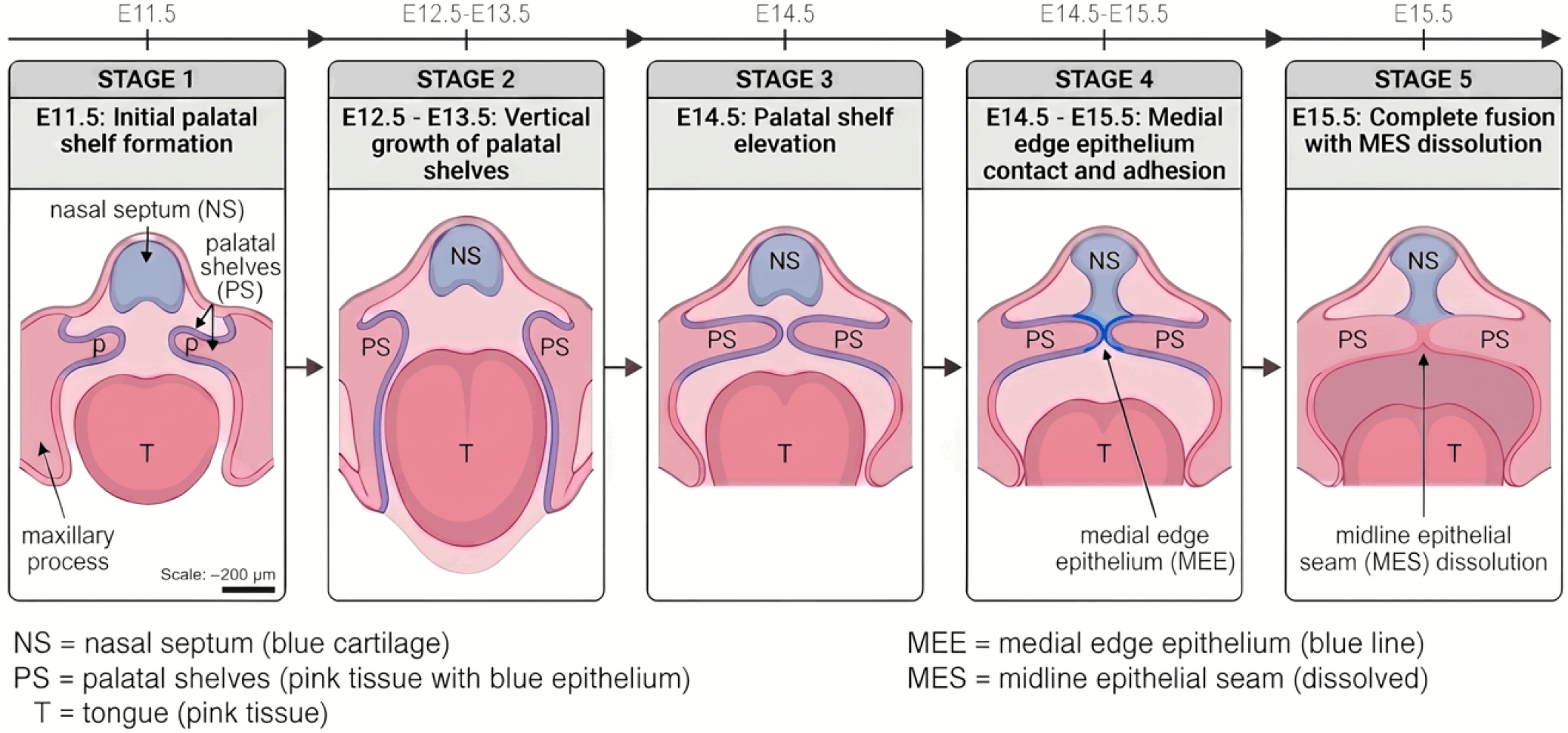
In embryogenesis, the hard palate (anterior bony part) and soft palate (posterior muscular part) are formed to separate the oral cavity from the nasal cavity. In the course of development, the two palatal shelves lift up, contact and unite along the midline to give rise to the intact palate (Fig. 1). The mesenchyme of these palatal shelves is predominantly derived from CNCCs, which function as a pool of multipotent progenitor cells capable of differentiating into the osteoblasts, fibroblasts, and other cell types that constitute the mature palate (5, 11). Development of the upper lip, philtrum, and primary palate arises from the fusion between medial nasal and maxillary processes; disturbance of these fusion events can result in cleft lip with or without cleft palate (CL/P) (12).
Palate development in humans begins around gestation week 6 and completes by week 12 (2). Secondary palate formation begins at approximately embryonic day 11.5 (E11.5) and completes by E15.5. The secondary palate fuses with primary palate anteriorly and nasal septum dorsally to completely separate oral and nasal cavities. Failure of shelf elevation, contact, or fusion results in secondary cleft palate (CP).
Classification of orofacial clefts
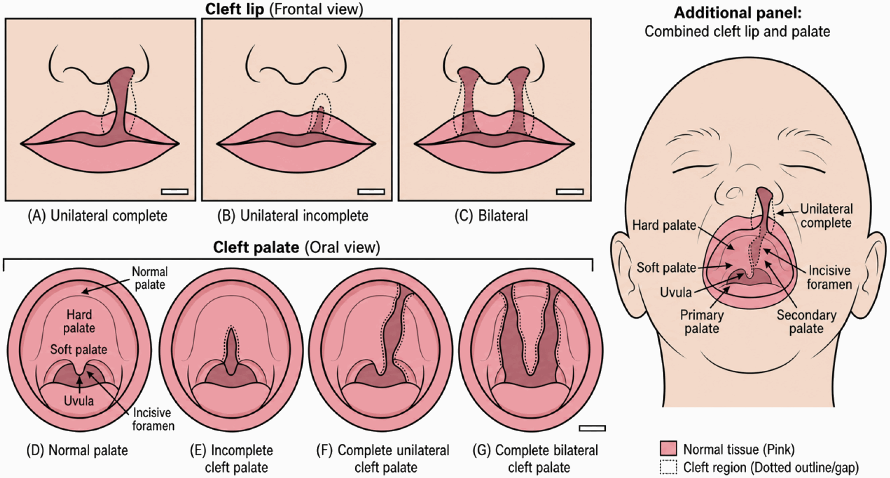
CL/P is distinguished by the location and extent of the cleft. Cleft lip (CL) occurs when tissues of the upper lip fail to fuse properly during fetal development, resulting in a gap on one (unilateral) or both sides (bilateral) (Fig. 2). CP is the failure of maxillary palatal processes to fuse properly, resulting in a complete and/or incomplete palatal defect.
Syndromic and non-syndromic orofacial clefts
OFCs are further classified into syndromic and non-syndromic forms. Non-syndromic cleft lip with or without cleft palate (NSCL/P) makes up approximately 93%∼97% of all cases and has a multifactorial inheritance pattern resulting from interactions between genetic and environmental factors. Remaining cases are considered syndromic forms of OFCs and make up around 5%∼7% of patients with over 200 distinct syndromes associated with OFCs (Table 1).
22q11.2 microdeletion syndrome also known as DiGeorge syndrome or velo-cardio-facial syndrome occurs due to a hemizygous deletion on chromosome 22q11.2. Approximately 55% of patients with this syndrome display craniofacial defects such as CP (13). TBX1 is one gene involved in this deletion and has been studied extensively for its role in palate development. Both overexpression and knockdown of Tbx1 results in CP demonstrating its importance in regulating palatogenesis (14).
Van der Woude syndrome (VWS) is considered the most common form of syndromic clefting representing around 2% of all CL/P cases. Mutations in interferon regulatory factor 6 (IRF6) are considered the primary genetic cause of VWS (10).
Molecular Regulation of Palatal Shelf Growth and Patterning
The cranial neural crest stem cell niche in palatogenesis
The concept of the stem cell niche—a specialized microenvironment that regulates stem cell behavior—is central to understanding palatogenesis. In the developing palate, the CNCC niche is a dynamic milieu composed of both cellular and acellular components that collectively govern CNCC self-renewal, proliferation, migration, and differentiation. Understanding the intricate interplay within this niche is paramount, as disruptions to any of its components—whether by genetic mutation or environmental insult—can lead to the aberrant CNCC behavior that underlies OFCs.
The signaling molecules within the niche include soluble factors such as Wnts, Bmps, Fgfs, and Shh, which are secreted by niche cells and create precise concentration gradients. The spatiotemporal integration of these signals determines whether a CNCC remains in a proliferative, undifferentiated state or commits to a specific lineage, such as osteoblast, chondrocyte, or fibroblast.
The acellular component of the niche is primarily the extracellular matrix (ECM), composed of collagen, fibronectin, laminin, and glycosaminoglycans such as hyaluronic acid. The ECM provides structural support and acts as a reservoir for growth factors, while also conveying mechanical signals through mechanotransduction that influence cell shape, migration, and fate decisions.
The primary cellular component of the palatal niche is the oral epithelium, which secretes critical signaling molecules including Shh and Fgfs. Vascular endothelial cells also contribute by providing nutrients and secreting additional growth factors. Furthermore, the mesenchymal cells themselves engage in autocrine and paracrine signaling, creating a complex feedback network that maintains the homeostasis of the stem cell niche.
The precise orchestration of palatal development is governed by a complex interplay of signaling molecules that regulate the proliferation, differentiation, and patterning of the CNCC-derived mesenchymal stem/progenitor cells. These pathways form a sophisticated signaling network that functions as a stem cell niche, ensuring the shelves grow to the appropriate size, elevate at the correct time, and fuse seamlessly. From a stem cell perspective, these pathways collectively define the microenvironment that controls the balance between progenitor self-renewal and terminal differentiation (3, 15).
Sonic hedgehog signaling pathway
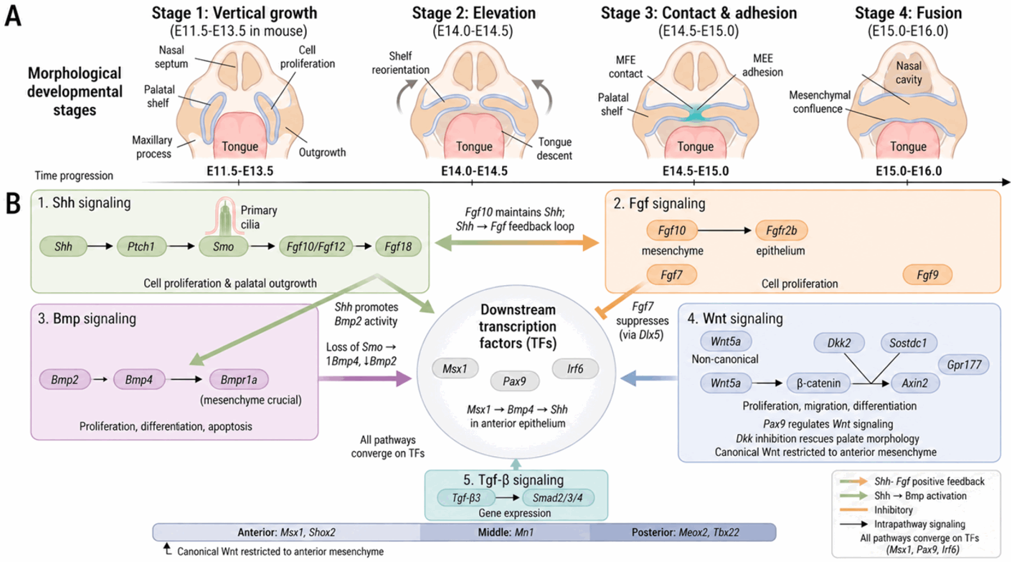
SHH signaling has been shown to play an important role in palate development by regulating cell proliferation and promoting outgrowth of the palatal shelves (Fig. 3). Shh interacts with other signaling pathways including Fgf and Bmp to ensure proper palatogenesis (16). Inactivation of Shh in the epithelium or mesoderm-specific inactivation of smoothened (Smo) impairs palatal cell proliferation and growth.
A key mediator of Shh signaling is the primary cilium, a microtubule-based organelle that acts as a cellular antenna. Primary cilia mediate Shh signaling and there is decreased expression of forkhead box F1 (Foxf1) in the palatal mesenchyme, indicating that primary cilia serve as effectors downstream of Shh signaling (17, 18). Disruption of cilia-related genes, such as Ift88 or Ick, leads to CP, underscoring the essential role of ciliogenesis and cilia-mediated signaling in palatogenesis (17, 18). Recent research has found a complex regulatory network that is comprised of Shh, Foxf1/2 and Fgf18 in palatal shelf development. Disruption of Foxf1 and Foxf2 in mouse embryos leads to palatal outgrowth defects, with altered expression of Fgf18 and Shh (19).
From a stem cell perspective, Shh acts as a critical niche factor for the CNCC-derived mesenchymal progenitors. Shh signaling from the epithelium maintains the proliferative and undifferentiated state of the underlying mesenchymal progenitor cells, and its precise regulation is a prerequisite for their orderly differentiation into osteoblasts and other cell types (15). The interaction between Shh and other pathways is complex and context-dependent. While Shh signaling from the epithelium is known to promote Bmp2 expression in the mesenchyme to stimulate proliferation (20), conditional inactivation of the Shh transducer Smoothened (Smo) in the mesenchyme results in an upregulation of Bmp4 but a downregulation of Bmp2, hinting at a complex regulatory logic that is not fully understood (16). Furthermore, ectopic activation of Hedgehog signaling in the palatal mesenchyme has been shown to cause CP and defective osteogenesis, partly by downregulating Wnt and Bmp pathways (21).
Fibroblast growth factor signaling pathway
Fgf10 is essential for palatal shelf outgrowth, with its absence leading to CP due to impaired proliferation (22). Although Fgf10 is expressed in the mesenchyme, its receptor Fgfr2b is crucial in the epithelium, and epithelial-specific deletion of Fgfr2 causes CP (23). A positive feed-back loop between Shh and Fgf signaling regulates palatal proliferation: FGF10 maintains Shh expression in palatal epithelium, whereas Fgf7 suppresses Shh expression regulated by distal-less homeobox 5 gene (Dlx5) (24).
Fgf9, expressed in both palatal epithelium and mesenchyme during palatogenesis, plays a critical role in craniofacial development (25, 26). Elevated Fgf9 levels induce temporomandibular joint dysplasia, impairing spatial coordination between tongue descent and palatal shelf elevation, thereby exacerbating CP formation (27).
Bone morphogenetic protein signaling pathway
The BMP signaling pathway plays numerous roles during facial morphogenesis by regulating cell proliferation, differentiation, and apoptosis (28). Bmp signaling interacts with Shh signaling pathway; loss of Smo in palatal mesenchyme results in increased Bmp4 expression and decreased Bmp2 levels (16). Shh signaling promotes Bmp2 activity to stimulate cell proliferation in palatal mesenchyme (20).
Bmpr1a, a type I BMP receptor, is essential for palate formation. Deletion of Bmpr1a in maxillary mesenchyme and oral epithelium causes CLP, whereas its loss in oral epithelium alone does not, suggesting that Bmpr1a signaling in mesenchyme is crucial for palatogenesis (29). Loss of Bmpr1a also disrupts Shh expression, indicating that Bmp-Shh interactions regulate palate growth (21).
Wnt signaling pathway
Wnt signaling is crucial for paired box 9 (Pax9)-mediated secondary palate development, regulating cell proliferation, migration, and differentiation (30). Notably, Wnt signaling is a well-established regulator of stem cell self-renewal across multiple tissues. In the context of neural crest biology, combinatorial Wnt and Bmp signaling has been shown to maintain the self-renewal capacity of multipotent neural crest stem cells (3, 31). Thus, the Wnt pathway likely plays a dual role in palatogenesis: maintaining the progenitor pool and directing cell fate decisions. In Pax9−/− mice, decreased Axin2 and β-catenin levels with increased Dickkopf-related protein 2 (Dkk2) expression disrupt Wnt signaling, but pharmacological inhibition of Dkk partially rescues palate morphology (32). Inactivation of Sostdc1 restores Wnt signaling and rescues CP. Pathogenic variants in Wnt pathway genes, such as Wnt3a, are linked to NSCL/P (33).
Anterior-posterior patterning
The growing palatal shelves are regionalized molecularly and morphologically along the A-P axis. Muscle segment homeobox 1 (Msx1) and short stature homeobox 2 (Shox2) are necessary for the proliferation of anterior palatal mesenchymal cells, whereas mesenchyme homeobox 2 and T-box transcription factor 22 are expressed more posteriorly. Msx1 acts via Bmp4 in the mesenchyme to regulate Shh expression in anterior palatal epithelium (20).
Msx1−/− mice show proliferation defects only in the anterior palate, whereas Mn1−/− mice have growth defects in middle and posterior palates. Shox2−/− mice have cleft restricted to the anterior palate, demonstrating regional specificity in palatal development. Canonical Wnt signaling is restricted to anterior palatal mesenchyme and depends on G protein-coupled receptor 177 for Wnt secretion (34).
Genetic Network Controlling Palatal Fusion
The differentiation of multipotent CNCCs into specific craniofacial structures is orchestrated by a hierarchical gene regulatory network of transcription factors. These master regulators interpret upstream signaling cues and execute downstream gene expression programs that progressively restrict the stem cell potential of CNCCs, defining cellular identity and behavior at each stage of palatogenesis (15).
Palatal shelf adhesion and epithelial differentiation
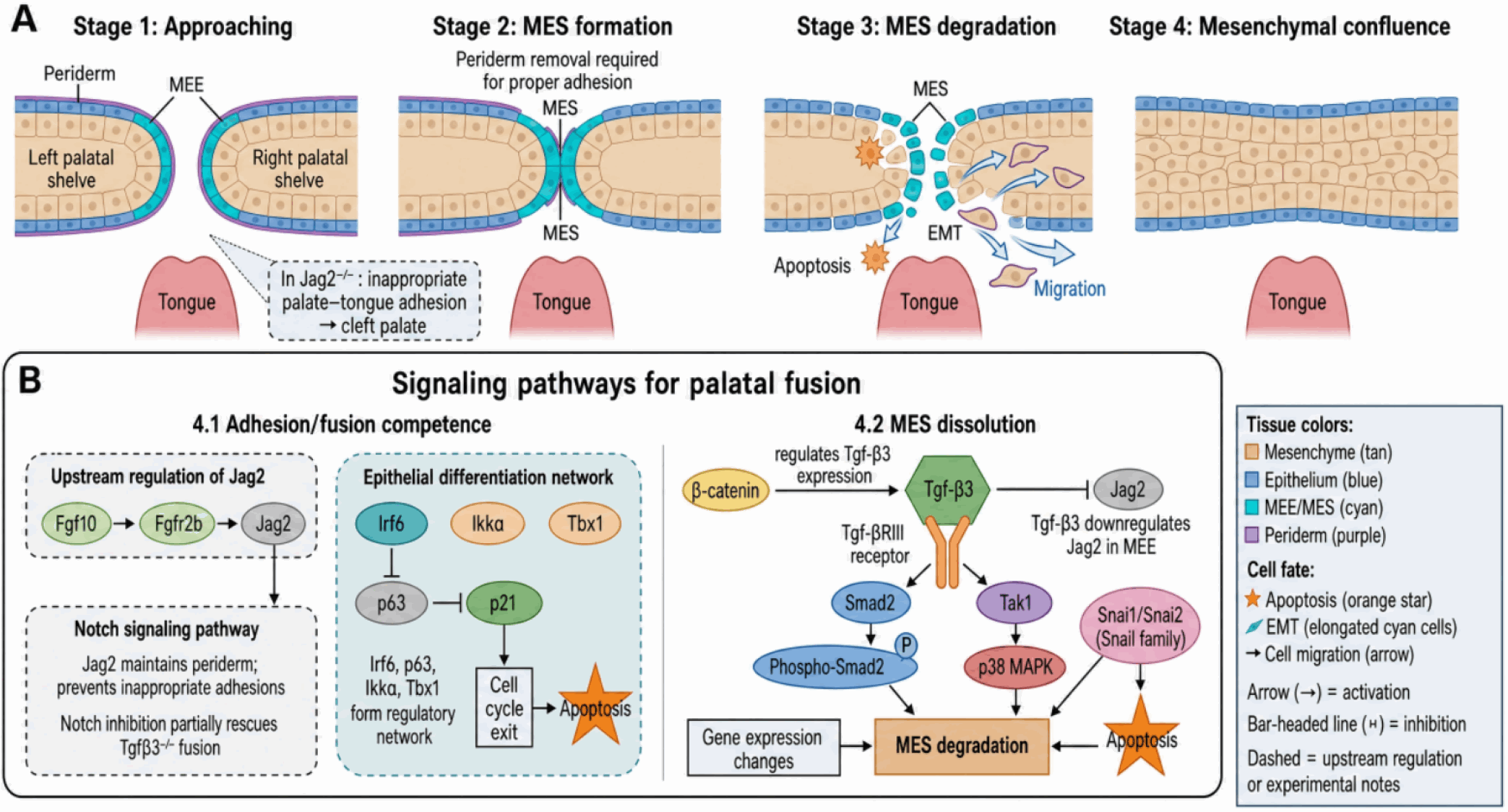
Concomitant with palatal shelf growth, downward and forward movement of the tongue allows palatal shelf elevation, contact, and midline fusion. Before the MES can form and dissolve, the palatal shelves must first adhere. This process requires precise control of epithelial differentiation (Fig. 4). The outer layer of the MEE, the periderm, must be removed to allow the underlying basal epithelial cells to make contact. This process is regulated by a network involving IRF6, p63, and the Jagged2-Notch1 signaling pathway (10, 35). Disruption of Irf6 leads to a failure of periderm removal and results in inappropriate epithelial adhesions and CP. Fgf10 signaling acts upstream of this network, as Fgf10 knockout embryos show reduced Jag2 expression (22).
Mutations in Irf6 causes a complete lack of periderm and results in an undifferentiated hyper-proliferative epidermis. These mice develop CP with inappropriate adhesions of the oral epithelium (10). Irf6 acts together with Jag2 to regulate epidermal differentiation. A genetic network controlling differentiation of palatal epithelium has been defined and includes Irf6, Jag2, p63, Ikk-α, Tbx1, and Fgf10-Fgfr2b signaling.
Medial edge epithelium dissolution
The Tgf-β signaling pathway is arguably the most critical regulator of MES dissolution. Mice lacking Tgf-β3 exhibit a fully penetrant CP phenotype where the MES fails to disappear (2). Tgf-β3 is thought to induce both apoptosis and epithelial-mesenchymal transition (EMT), acting through both SMAD-dependent and SMAD-independent (e.g., p38 MAPK) pathways.
The Tgf-β pathway is required for palatal fusion, which is triggered by signaling mediated through heteromeric type I and type II receptors, followed by Smad2 phosphorylation and changes in gene expression. Smad2 is important for MES degradation, but deletion of Smad4 has no effect on fusion implying participation of alternate pathways such as p38 MAPK. The Tgf-β pathway induces Tak1 and palatal fusion via Smad and p38 MAPK pathways.
The interplay between Tgf-β and other pathways is crucial. For example, Tgf-β3 negatively regulates Jag2 expression in the MEE, and inhibiting Notch signaling can partially rescue the fusion defects in Tgf-β3 knockout mice, suggesting a complex balance between these pathways (36). Furthermore, epithelial β-catenin is required for Tgf-β3 expression, linking WNT signaling directly to MES dissolution (37). The Snail family transcription factors (Snai1, Snai2) are crucial for palatal fusion; fusion fails in Snai1+/−;Snai2+/− compound mutants with reduced MES apoptosis.
Epigenetic Mechanisms in Palatogenesis
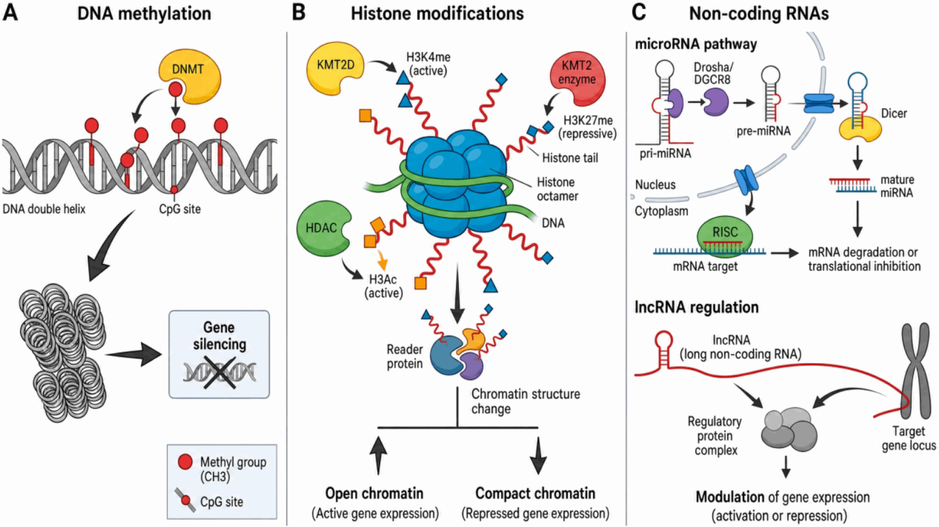
Epigenetic processes regulate gene expression through chromatin remodeling independent of DNA sequence changes (Fig. 5). Epigenetics provides a crucial regulatory layer that translates environmental signals and intrinsic genetic programs into specific stem cell behaviors. In the context of palatogenesis, epigenetic mechanisms are fundamental to establishing and maintaining CNCC identity, controlling their differentiation potential, and ensuring the precise timing of gene expression (38). In embryonic stem cells, many developmental genes are held in a “bivalent” state, carrying both activating (H3K4me3) and repressive (H3K27me3) marks, poising them for rapid activation or stable repression upon receiving differentiation cues (39). It is highly probable that CNCCs utilize a similar mechanism to maintain their multipotency while remaining ready to respond to patterning signals.
DNA methylation
DNA methylation leads to addition of methyl groups to CpG nucleotides (predominantly CpG islands found in promoter regions), that then attract transcription repressing proteins (40). This action is completed by enzymes DNA methyltransferases (DNMTs), which include DNMT1, DNMT3A and DNTM3B. DNA methylation is necessary for cell fate-commitment and tissue specification. DNMT3A was shown to silence neural gene expression such as Sox2 and Sox3 to allow for neural crest specification (9). Methylation at CpG sites was shown to be enriched in palate tissue at E14.5 compared to E13.5 and E18.5. This timepoint represents when the palatal shelves elevate to a position superior to the tongue before formation of the MES.
The causality dilemma: cause or consequence?: Epigenome-wide association studies (EWAS) have revealed many differentially methylated regions (DMRs) associated with OFCs in humans (40). However, a major unresolved question is whether these epigenetic changes are a cause of the clefting or a consequence of the altered developmental state. This is a critical distinction for understanding disease etiology and developing preventative strategies. The challenge is highlighted by studies showing a weak correlation between DNA methylation patterns in easily accessible tissues like blood and those in the relevant craniofacial tissues (41). This discrepancy complicates the interpretation of EWAS results and raises the question: To what extent can peripheral epigenetic marks serve as reliable proxies for developmental processes in the head?
Metastable epialleles and gene-environment interactions: An exciting concept is that of metastable epialleles, which are genomic loci where epigenetic marking is stochastically established early in development and then stably inherited. The Clf1 locus in mice, an intracisternal A-particle retrotransposon insertion near Wnt9b, is a classic example that contributes to incomplete penetrance of cleft lip (9). This raises the possibility that similar loci in humans could explain the variable expressivity of OFCs and act as sensors for environmental insults. Future research should focus on identifying novel human metastable epialleles and investigating how their methylation status is influenced by environmental factors like maternal diet.
Histone modifications
Histone modifications including methylation, acetylation, deacetylation, phosphorylation, and ubiquitination result in condensation or relaxation of chromatin thereby upregulating or downregulating gene transcription. Histone modifications play an essential role in NCC development and must be tightly regulated for normal craniofacial formation (9).
Mutations in genes encoding these enzymes are linked to several syndromic forms of OFCs. For example, mutations in KMT2D (a histone methyltransferase) and KDM6A (a histone demethylase) both cause Kabuki syndrome, which is often associated with CP (42). Interestingly, the conditional deletion of Kdm6a in NCCs revealed a striking sexual dimorphism, with female mice exhibiting a much more severe phenotype. This points to a fascinating, yet poorly understood, X-linked epigenetic regulation in craniofacial development that warrants further investigation. KDM6B is also essential for cranial neural crest development and deletion of KDM6B leads to loss of p53 pathway mediated function, resulting in complete penetrance of CP due to defects in cell proliferation and differentiation. HDAC3 is critical for mouse development; conditional knockout in NCCs causes craniofacial malformations, including CP, by modulating Msx1, Msx2 and Bmp4 (10).
Non-coding RNAs
MicroRNAs (miRNAs) are small (∼22-nucleotide) non-coding RNA molecules that regulate developmental processes by binding to 3'UTRs of target mRNAs (Table 2). Dicer, essential for miRNA processing, is critical for post-migratory NCC survival; NCC-specific Dicer knockout mice exhibit microcephaly, facial hypoplasia, and CP (43).
Specific miRNAs have been shown to regulate key developmental pathways. The miR-17-92 cluster, for instance, directly targets and represses T-box factors like Tbx1 and Tbx3 (44). The miR-200 family plays a crucial role in regulating EMT by targeting zinc finger E-box-binding homeobox 1/2 and is itself regulated by TGF-β signaling during MES fusion (45). In MES, miR-200b expression decreases during palatal fusion in response to palatogenesis; overexpression of the miR-200 family results in CP by repressing TGF-β-induced Smad2 and Snai1 expression (45). miR-22-3p modulates actomyosin contractility to palatal fusion: down-regulation leads to MEE dissolving deficiency and MES persistence (46).
Long non-coding RNAs (lncRNAs) have been shown to act as competing endogenous RNAs or “sponges” that sequester miRNAs, adding another layer of complexity to the regulatory network. LncRNA-NONMMUT100923.1 regulates Cdsn expression by competitively binding to miR-200a-3p during palatogenesis. The MALAT1-miR-124-3p-SP1 and NEAT1-miR-140-3p-CXCR4 ceRNA networks may contribute to non-syndromic orofacial cleft etiology (43).
Environmental Factors in Orofacial Cleft Development
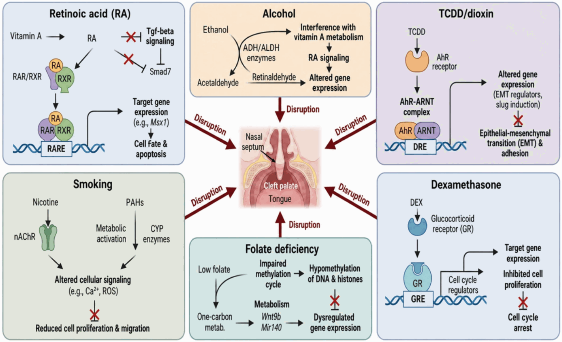
Non-syndromic OFCs are multifactorial birth defects therefore environmental factors and their impact on genetic susceptibility are thought to play a large role (Fig. 6). On one hand, the incredible plasticity that allows CNCCs to differentiate into any cell lineage within the derivative structures also makes them susceptible to environmental insults. Teratogens can alter homeostasis within the CNCC stem cell niche and lead to abnormal apoptosis, decreased proliferation, or accelerated differentiation of these cells (5). For many risk factors, there are unknown mechanisms behind this association.
Retinoic acid
RA, a metabolic derivative of vitamin A, is an important signaling molecule for morphogenesis and differentiation during embryonic development, including craniofacial morphogenesis. Both deficiency and excess of retinoids induce CL/P in rodents (10). Mutations in RARs (α, γ) result in CP and midfacial clefts. Cyp26 genes (Cyp26a1, Cyp26b1, Cyp26c1) that metabolize RA are dysregulated in Tbx1 mutants, leading to RA accumulation and reduced expression of Bmp2 and Fgf10, resulting in CP (10). RA suppresses Tgf-β signaling through upregulation of Smad7 and inhibition of phosphorylated Smad2. All-trans retinoic acid signals through Notch in palatal cells to induce apoptosis and cell proliferation. RA-treated CP is associated with upregulation of miR-181a-5p, miR-410-3p, miR-124-3p and miR4680–3p (43).
Alcohol
Regular alcohol consumption correlates with increased risk of OFC formation among numerous demographics. The mechanisms by which alcohol increases OFC risk are still under investigation; proposed theories include RA signaling inhibition, disruption of epigenetic regulation, and oxidative stress (10). Acetaldehyde, the metabolite of ethanol, competes with retinaldehyde for aldehyde dehydrogenase (ALDH) enzymes required for RA synthesis. Ethanol treatment prevents Aldh1a2 activity thereby inhibiting synthesis of RA. Additionally, ethanol alters the epigenetic landscape of neural stem cells by altering methylation patterns of cell cycle regulating genes. Oxidative stress from metabolism of ethanol to acetaldehyde by Cyp2e1 leads to apoptosis and activation of signaling pathways involved in orofacial development (10).
2,3,7,8-Tetrachlorodibenzo-p-dioxin/dioxin
2,3,7,8-Tetrachlorodibenzo-p-dioxin (TCDD), the most teratogenic dioxin compound, disrupts gene expression within epithelial and mesenchymal cells involved in multiple stages of palatogenesis including growth and shelf fusion (10). The aryl hydrocarbon receptor (AhR) is crucial for teratogenic effects of TCDD; AhR is highly active in palatal shelf epithelial cells. In TCDD-exposed fetuses, MEE is covered by thinner epithelial monolayer with fewer filopodia, impairing proper palatal fusion. TCDD promotes EMT inappropriately through Slug induction, a repressor of E-cadherin containing dioxin response elements in its promoter modulated by AhR (43).
Dexamethasone
Dexamethasone (DEX) is a synthetic glucocorticoid prescribed to treat various conditions yet displays teratogenic side effects. CL/P risk increased 2-9 fold if the mother was exposed to oral or systemic corticosteroids during pregnancy (10). DEX crosses the blood-placental barrier and binds to cytoplasmic glucocorticoid receptors, inducing CP by inhibiting cell proliferation in palatal mesenchyme. Analysis of miRNA expression in DEX-exposed developing palatal shelves revealed that the miR-449 family shows increased expression at E14.5, whereas miR-19a-3p, miR-130a-3p, and miR-301a-3p show decreased expression (43).
Folate deficiency
Folate deficiency and metabolism play an important role during embryogenesis. Despite numerous studies, systematic reviews have shown inconsistent but inverse associations between folic acid use and OFCs. The meta-analysis found that if folic acid is taken periconceptionally there is a reduced risk (odds ratio [OR] = 0.64) but if taken during pregnancy there is little effect (OR = 0.90) (10). Folate is critical for DNA methylation because the folate cycle provides methyl groups to S-adenosylmethionine. The methylenetetrahydrofolate reductase (MTHFR) c.677C>T polymorphism decreases enzymatic activity and increases CL/P risk, particularly combined with low maternal folic acid intake. CL/P cases demonstrated significant epigenome-wide hypomethylation compared to controls. CL/P-associated DMRs include genes involved in palate development such as Wnt9b, Mir140, and LIM homeobox 8 (9).
Maternal smoking
Maternal cigarette smoking is an established risk factor for OFCs. One meta-analysis found relationships of maternal smoking with CL/P (risk ratio [RR] = 1.34) or CP (RR = 1.22) (10). Gene polymorphisms of ligands in developmental signaling (Tgf-α, Tgf-β3, Bmp4) have been found associated with OFCs in the context of maternal smoke exposure. As nicotine is a potent vasoconstrictor, maternal smoking causes impaired vascular function in the uterus resulting in reduced oxygen delivery to fetus. Components of tobacco smoke include PAHs, polycyclic aromatic hydrocarbons (PAHs), dioxins, carbon monoxide, and metals such as cadmium which are potential teratogens (10).
Several key questions in gene-environment interactions remain unresolved. First, it is unclear whether folic acid prevents OFCs primarily by supporting the DNA methylation cycle or through other mechanisms such as nucleotide synthesis; the inconsistent association across different studies and populations, together with its interaction with polymorphisms in genes like MTHFR, suggests a complex relationship that is far from resolved (10). Second, for maternal smoking and alcohol consumption, the specific teratogenic agents and their precise molecular targets have yet to be fully elucidated. For smoking, it remains uncertain whether the primary culprit is nicotine, PAHs, or hypoxia induced by carbon monoxide; for alcohol, the precise mechanisms by which it disrupts RA signaling and induces oxidative stress in the developing craniofacial tissues require further investigation (10). Third, defining the critical windows of exposure and the dose-response relationships for these teratogens represents a crucial challenge. Furthermore, understanding why only a fraction of exposed embryos develop clefts requires a deep appreciation of how an individual’s genetic background, such as polymorphisms in metabolic enzymes like CYPs or ALDHs, confers susceptibility or resilience to environmental insults.
Unresolved Questions and Future Perspectives
While significant progress has been made, the field of palatogenesis is ripe with fundamental questions that are now becoming tractable with emerging technologies. Acknowledging these gaps is essential for guiding the next decade of research.
While this review focuses on CNCCs, it is conceptually valuable to consider that similar regulatory mechanisms govern non-cranial neural crest populations, albeit resulting in distinct regional phenotypes upon dysregulation. For instance, perturbations in Bmp and Wnt signaling within the vagal and sacral neural crest lead to migration defects characteristic of Hirschsprung disease, an enteric neuropathy. Similarly, disruptions in cardiac neural crest migration and differentiation—often involving Tgf-β and Fgf pathways—result in congenital outflow tract anomalies such as persistent truncus arteriosus. Comparing these distinct region-specific outcomes highlights the shared multiscale regulatory logic of neural crest biology, while underscoring the unique vulnerability of the palatal shelves to specific environmental and genetic insults (47).
Key unresolved questions
A fundamental question concerns CNCC heterogeneity, plasticity, and fate decisions: whether the mesenchymal and epithelial cells of the palate constitute a homogenous population or consist of functionally distinct subpopulations remains to be determined. Given that CNCCs transiently reactivate pluripotency programs (6), the degree of plasticity retained once they have colonized the palatal shelves, and the signals that irreversibly commit them to specific fates, are critical unknowns. Equally important is the integration of signaling and mechanics: how molecular signals from canonical pathways translate into the physical, mechanical forces that drive shelf elevation and fusion, and how the ECM, as a key component of the stem cell niche, regulates CNCC behavior through mechanotransduction, remain poorly understood.
At the epigenetic-genetic interface, it is essential to elucidate how environmental signals write, read, and erase epigenetic marks at specific gene loci, and whether we can identify the causal chain of events from an environmental exposure to an altered gene expression program and, ultimately, a structural birth defect. Finally, the extent to which mouse models can recapitulate human palatogenesis and the key molecular differences that may explain species-specific aspects of craniofacial development constitute important unresolved questions.
Future directions: a technological roadmap
Addressing these questions requires a move beyond traditional approaches and an embrace of cutting-edge technologies. The integration of single-cell RNA-seq, ATAC-seq for chromatin accessibility, and spatial transcriptomics is creating unprecedented opportunities to dissect cellular heterogeneity and regulatory networks in their native tissue context (48-51), enabling the construction of comprehensive, spatiotemporal atlases of palate development. Furthermore, the development of cranial neural crest-derived craniofacial organoids from human pluripotent stem cells offers a powerful in vitro system to study human-specific developmental mechanisms and screen for potential teratogens or therapeutic compounds (52). Patient-derived induced pluripotent stem cell (iPSC) can be differentiated into neural crest cells and assembled into organoids that recapitulate key aspects of craniofacial development, providing an unprecedented platform for personalized disease modeling (53). CRISPR-based epigenome editing, using catalytically dead Cas9 fused to methyltransferases or acetyltransferases in cellular and animal models, will allow researchers to move from correlation to causation by directly testing the functional consequences of specific epigenetic modifications. Additionally, computational approaches such as deep learning-based frameworks can predict the functional impact of non-coding genetic variants identified in genome-wide association study and integrate vast multi-omics datasets to uncover novel regulatory networks (54).
Future directions in translational research
Ultimately, this fundamental research should pave the way for novel therapeutic and preventative strategies. A deeper understanding of signaling crosstalk may reveal new targets for pharmacological intervention; for example, the successful rescue of CP in Ick mutant mice using a Smoothened agonist provides a compelling proof-of-concept for prenatal therapeutic strategies (17). Recent advances in human iPSC technologies have provided unprecedented opportunities to model CNCC-related craniofacial disorders in vitro (55). For example, iPSC-derived CNCC models of Treacher Collins syndrome have revealed specific defects in neural crest survival and migration associated with treacle ribosome biogenesis factor 1 mutations. Similarly, iPSC models of DiGeorge (22q11.2 deletion) syndrome have elucidated how haploinsufficiency alters the early specification and migration trajectory of cranial neural crest populations. These human-specific, patient-derived models offer a powerful platform for discovering novel therapeutic targets and testing pharmacological interventions to prevent or ameliorate cleft-related phenotypes. In the realm of stem cell-based regenerative medicine, the discovery of adult pNC-SCs within the palate itself, expressing pluripotency markers such as Sox2, Oct4, Klf4, and c-Myc, has opened a new frontier for autologous regenerative therapies (7, 56). Current clinical research is actively exploring the use of various mesenchymal stem cells, including those derived from bone marrow and adipose tissue, to enhance alveolar bone regeneration in cleft patients, with results comparable to traditional iliac bone grafting (56-59). The integration of these stem cells with advanced biomaterials and 3D bioprinting technologies holds immense promise for reconstructing complex orofacial tissues (31). In the long term, identifying high-risk individuals based on their genetic and epigenetic profiles could allow for targeted preventative measures, such as personalized nutritional supplementation or avoidance of specific environmental triggers.
Conclusions
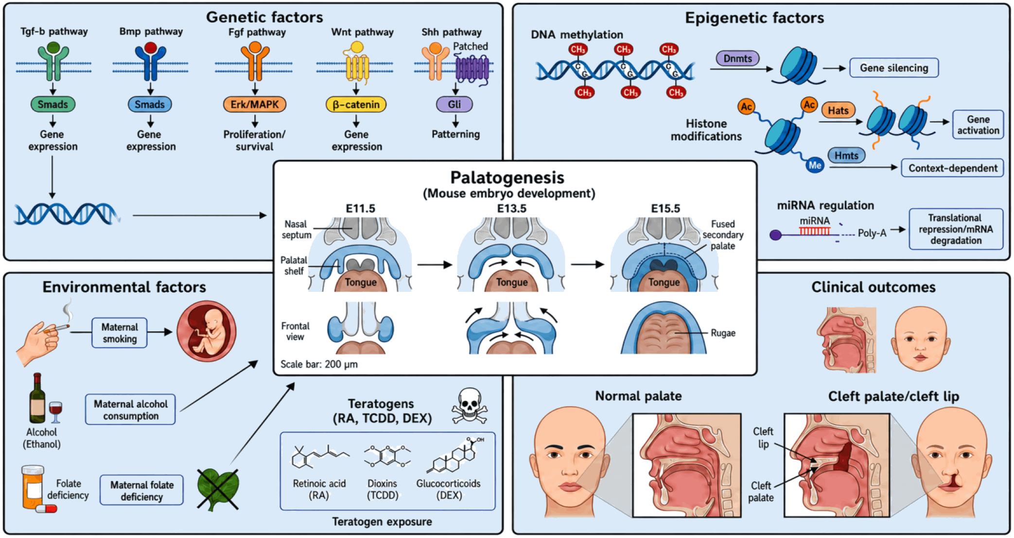
To synthesize the complex interplay of these mechanisms, we propose an integrative conceptual model (Fig. 7). In this model, the morphological progression of palatogenesis—from initial palatal shelf formation (E11.5) through vertical growth, elevation, and fusion (E15.5)—occupies the central core, governed by a multiscale network of genetic factors (Tgf-β, Bmp, Fgf, Wnt, and Shh signaling pathways) and epigenetic factors (DNA methylation, histone modifications, and non-coding RNA regulation) discussed in previous sections. Crucially, environmental insults—including maternal smoking, alcohol consumption, folate deficiency, and teratogen exposure (RA, TCDD, DEX)—act as external disruptors capable of penetrating and destabilizing this regulatory architecture, ultimately leading to clinical outcomes such as CL/P.
This review highlights the complex interplay of genetic, epigenetic and environmental risk factors in palatogenesis and CL/P pathogenesis. By framing these processes through a stem cell-centric lens, we have emphasized that palatogenesis is fundamentally a story of how a multipotent stem cell population—the cranial neural crest—builds complex anatomical structures under the precise guidance of its signaling niche. We have moved beyond a simple summary of established pathways to critically examine the ongoing debates, unresolved questions, and exciting future directions that are shaping the field. Many of the signaling pathways such as Tgf-β, Bmp, Fgf, Wnt and Shh in association with epigenetic mechanisms (DNA methylation, histone modifications and noncoding RNAs) are vitally involved in the formation of craniofacial structures. These pathways collectively define the stem cell niche that controls the balance between CNCC self-renewal and differentiation, and disruptions at any level of this hierarchy can lead to OFCs. The urgent need for the tight regulation of different phases in development such as proliferation, migration, differentiation and apoptosis has been highlighted by recent findings (2).
Environments, such as maternal tobacco cigarette use, alcohol exposure, folate deficiency and teratogens (RA and TCDD) all interact with genetic and epigenetic factors to interfere with normal development and cause birth defects like CL/P (10). The journey from understanding the fundamental biology of a single epithelial seam to preventing a common and distressing birth defect is long, but the technological and conceptual advances discussed herein provide a clear and promising roadmap for the future. The application of powerful new technologies, from single-cell omics to patient-derived neural crest organoids, promises to unravel the remaining mysteries of this intricate process and unlock the full regenerative potential of craniofacial stem cells, bringing us closer to a future where we can not only repair but also prevent OFCs. An integrated, multidisciplinary approach will be paramount in translating these scientific discoveries into tangible clinical benefits.
 XML Download