



 PDF
 Citation
 Print


I. Introduction
The integration of dental implants has become a central strategy in modern oral rehabilitation. However, clinicians often encounter significant challenges when treating patients with complex factors such as systemic disease, severe alveolar atrophy, or chronic infection. Notably, chronic odontogenic maxillary sinusitis leading to extensive bone loss frequently precludes straightforward implant therapy. Such scenarios demand a highly coordinated, sequential approach. Initially, the clinician must resolve the sinus infection to eliminate chronic inflammation and restore a healthy mucosal environment. Once stabilized, reconstruction of the deficient maxilla is essential, often requiring autogenous bone grafts—such as those harvested from the mandibular ramus—combined with xenografts to provide volumetric stability. While conventional bone block grafting typically requires metallic screw fixation, the use of a press-fit technique offers an alternative that achieves primary stability through mechanical interlocking, potentially reducing surgical morbidity and the need for hardware removal. Following a sufficient healing period to ensure graft maturation and adequate bone density, stable implant placement and subsequent functional restoration can be achieved. This multidisciplinary process requires not only precise surgical execution but also high patient motivation and compliance throughout the extended rehabilitation period.
II. Case Report
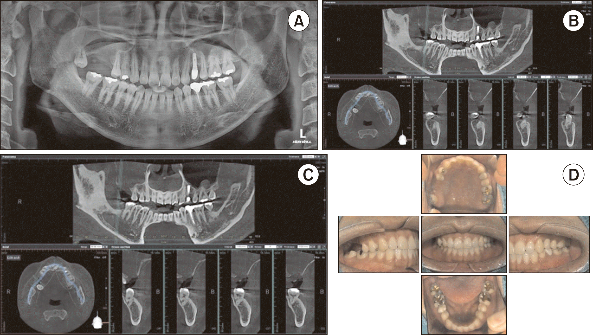
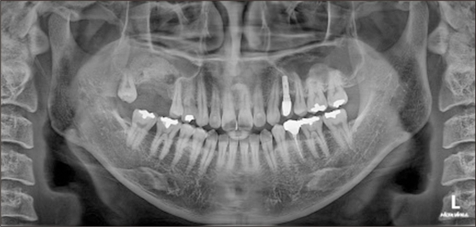
A 37-year-old male presented with impaired masticatory function and persistent malodor in the right maxillary posterior region. The clinical history indicated that symptoms had progressed for eight months following the extraction of the maxillary right first and second molars (#16, #17). Clinical examination and cone-beam computed tomography (CBCT) revealed significant nasal congestion, rhinorrhea, and extensive alveolar bone loss with a complete absence of the sinus floor, accompanied by severe opacification of the right maxillary sinus consistent with odontogenic sinusitis.(Fig. 1)
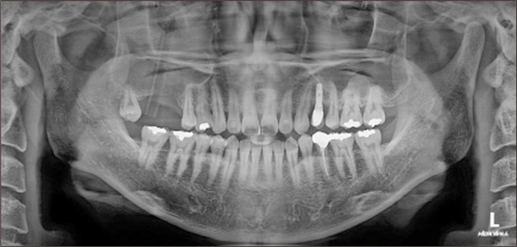
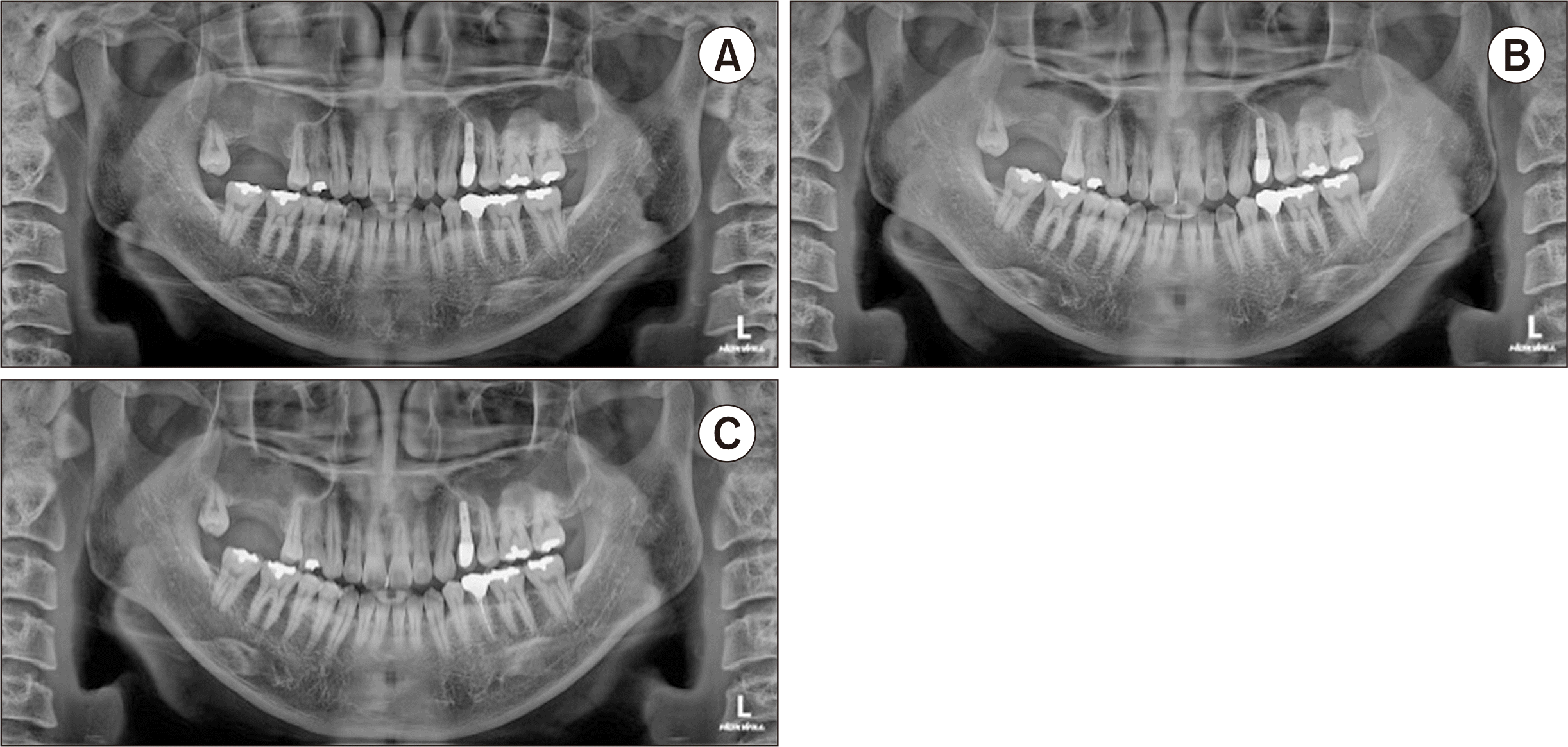
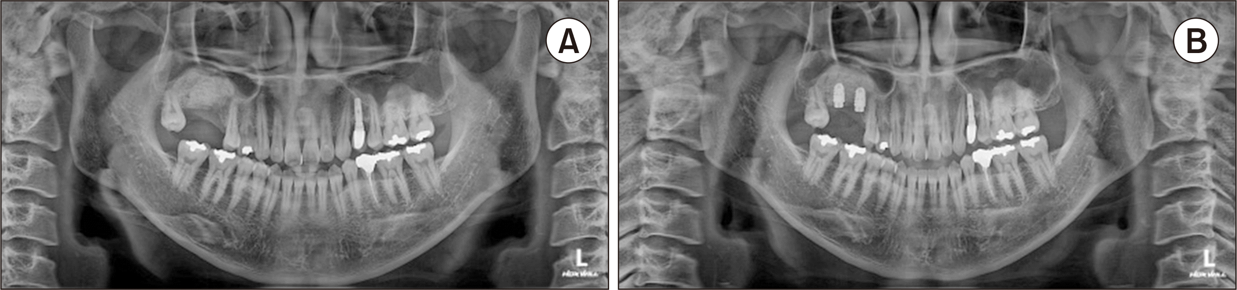
Given the severity of the sinus infection, a staged therapeutic approach was initiated. Phase I focused on the resolution of sinusitis through a lateral surgical approach. Following a buccal incision, the sinus was accessed and subjected to thorough saline irrigation until all purulent exudate was eliminated. A silicone Penrose drain was stabilized at the site and the wound was closed.(Fig. 2) The patient was prescribed a rigorous 14-day pharmacological regimen, including amoxicillin/clavulanate (Croanan 625 mg; Hanmi Pharm), loxoprofen, almagate, and betahistine (Beartilin; Daewon Pharm). Systemic methylprednisolone (Beasolone; Daewoong Bio) was administered as a short-term adjunctive therapy with tapering to reduce mucosal edema and inflammatory exudate. Clinical resolution of the mucosal inflammation and sinonasal symptoms was confirmed at the 2-week follow-up.(Fig. 3. A) However, a subsequent 3-month and 5-month radiographic re-evaluation indicated that the bone deficiency remained inadequate for functional rehabilitation, necessitating an advanced reconstructive procedure.(Fig. 3. B, 3. C)
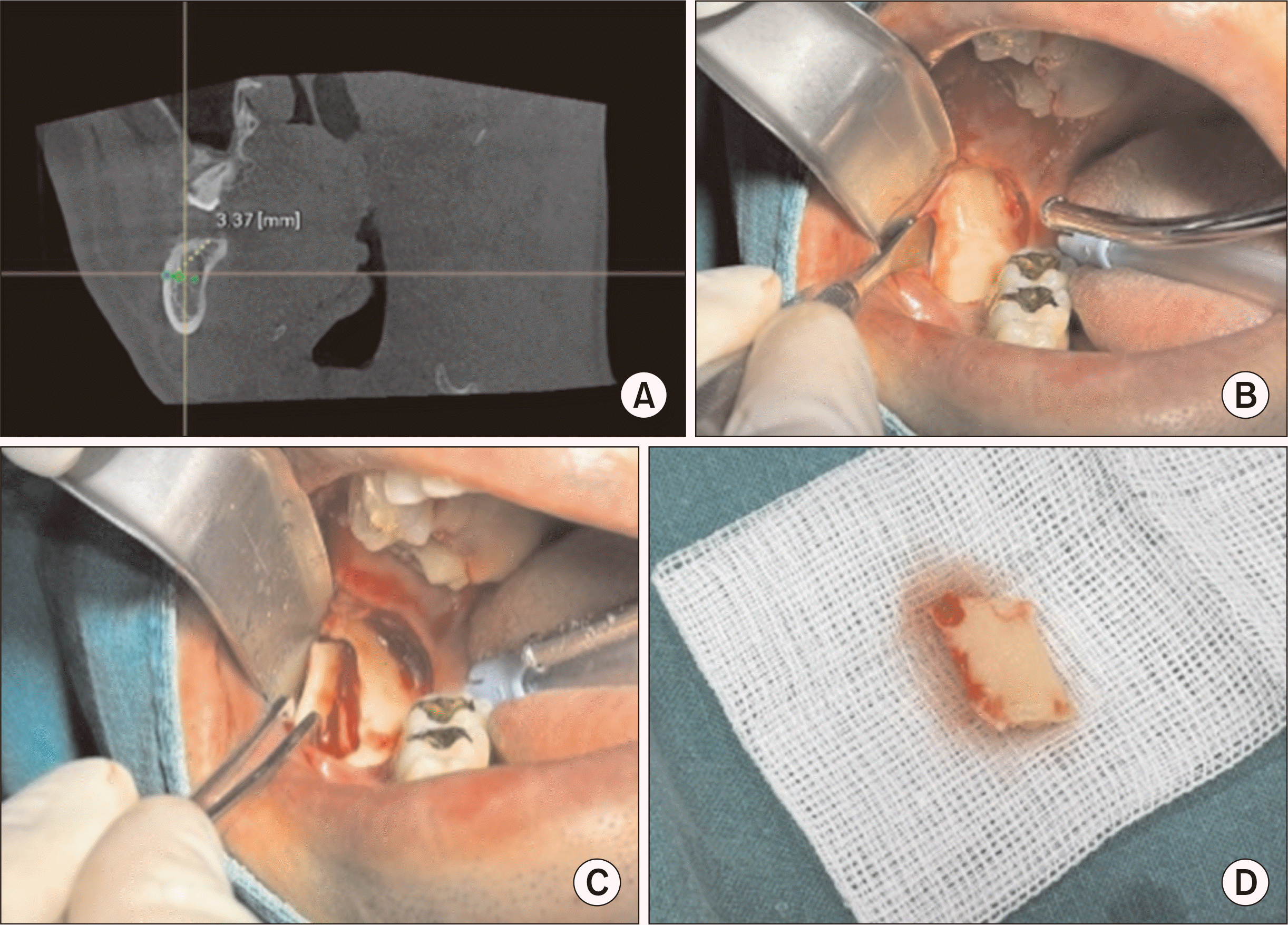
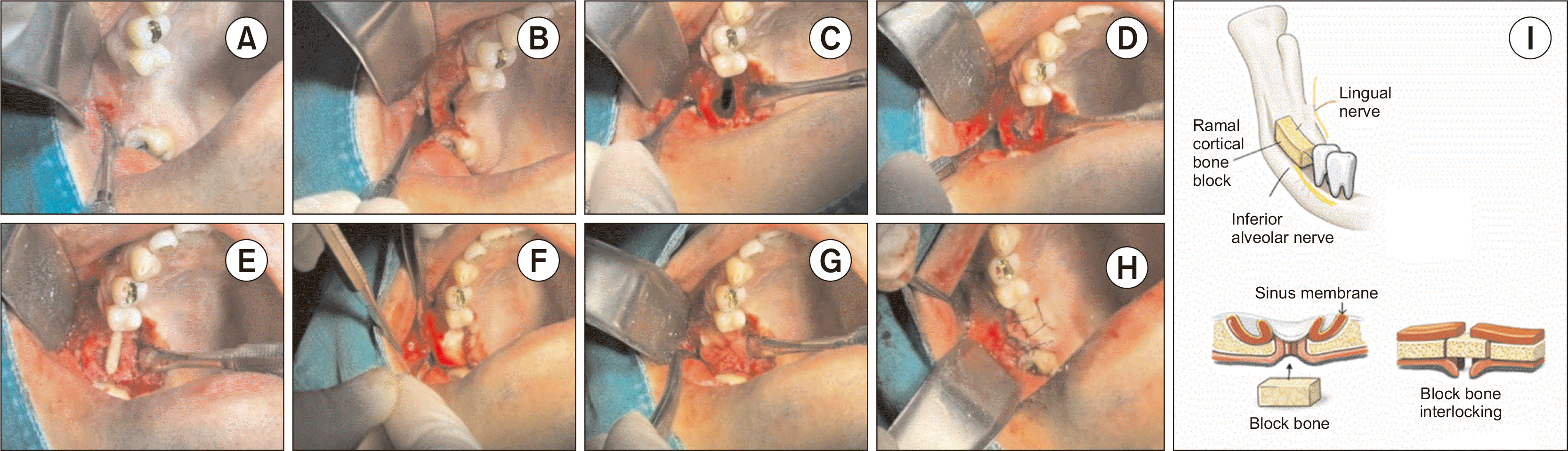
The secondary surgical phase involved a combined autogenous-xenogenic bone grafting procedure. Preoperative CBCT was utilized to plan the harvest of a cortical bone block from the right mandibular ramus, ensuring a safe clearance from the inferior alveolar nerve (IAN) canal.(Fig. 4) The harvesting was performed using a low-speed rotary instrument (RB-018; Hudens Bio). The harvested ramus block was precisely contoured to be slightly larger than the recipient defect to facilitate a screwless fixation. This block was then secured using a press-fit technique, achieving immediate mechanical stability through frictional force and mechanical interlocking between the graft and the host bone.(Fig. 5) By eliminating the need for fixation screws, the potential for hardware-related complications was minimized while maintaining a stable environment for graft maturation. This was supplemented by a lateral window sinus augmentation using a composite of xenografts, comprising 0.25 cc of SureOss (HansBioMed) and 0.5 g of Ovis XENO-P (Purgo Biologics). The entire graft complex was protected with two layers of collagen membrane (Lyoplant; Aesculap) and two units of platelet-rich fibrin to enhance soft tissue healing and graft maturation. Finally, tension-free primary closure was achieved using eversion sutures to prevent marginal inversion and promote stable wound healing1.
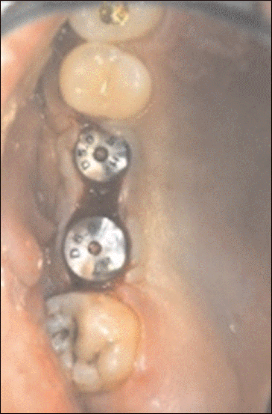
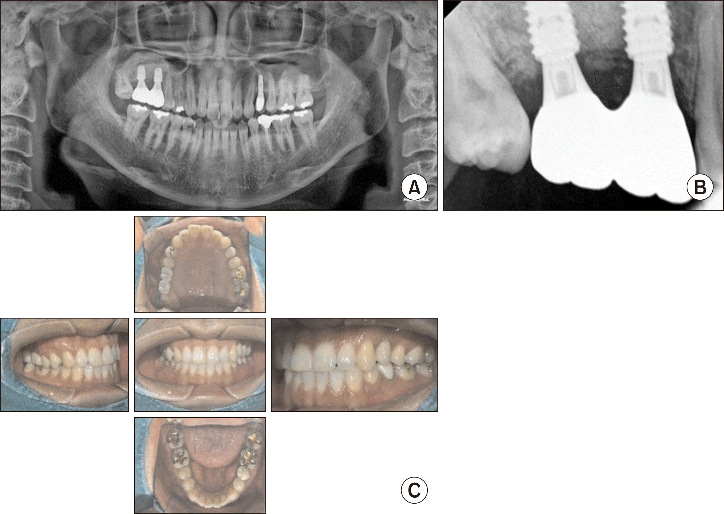
Following a 5-month healing period, radiographic imaging demonstrated successful integration of the graft and sufficient bone volume for stable implant support. Two Osstem BA fixtures (Osstem) were placed at the #16 and #17 positions.(Fig. 6, 7) After three months of undisturbed osseointegration, second-stage surgery was performed to install healing abutments. The stability of the implants was verified using resonance frequency analysis with an Osstell Beacon (Osstell), yielding Implant Stability Quotient (ISQ) values of 76 and 77 for the respective sites.(Fig. 8) Final monolithic zirconia prostheses were delivered following one week of peri-implant soft tissue conditioning. Post-prosthetic radiographic and clinical evaluations confirmed the successful restoration of normal occlusion and masticatory function.(Fig. 9)
III. Discussion
This case illustrates the efficacy of a staged multidisciplinary protocol for reconstructing extensive maxillary defects. The treatment was organized into three distinct clinical pillars: (1) resolution of odontogenic sinusitis, (2) ramal bone grafting, and (3) implant rehabilitation.
The healthy maxillary sinus microbiome is typically dominated by Corynebacterium and Staphylococcus2. In chronic sinusitis, this shifts toward pathogenic biofilms of gram-negative anaerobic rods, such as Prevotella and Fusobacterium, with a significantly higher prevalence of Staphylococcus aureus3. Therefore, the therapeutic objective is the restoration of a physiological microbial environment rather than total eradication.
In this case, the resolution of infection was prioritized prior to any reconstructive efforts. Various surgical techniques exist for the closure of an oro-antral fistula, including palatal flaps or buccal fat pads; however, a buccal flap was selected here for its reliability and excellent surgical accessibility4,5. To reduce the microbial load, incision and drainage were performed in a manner similar to the lateral sinus window technique. This surgical decompression with saline irrigation effectively reduces microbial load and purulent exudate, restoring mucociliary function and capillary perfusion to ischemic mucosa6. Such revitalization of oxygen and immune cell delivery is essential to suppress over-activated pro-inflammatory cytokines, such as IL-1, IL-6, and TNF-α, while inducing anti-inflammatory mediators necessary for clinical healing7.
The success of this initial phase was further ensured by a targeted pharmacological strategy. Following the American Academy of Otolaryngology–Head and Neck Surgery (AAO-HNS) guidelines, amoxicillin/clavulanate was prescribed to provide broad-spectrum coverage against both aerobic and anaerobic pathogens8. The inclusion of short-term systemic methylprednisolone was instrumental in alleviating mucosal swelling and reducing inflammatory exudate, a protocol supported by evidence showing significant symptomatic improvement in chronic sinusitis cases9,10. This integrated medical and surgical management successfully converted a contaminated environment into one suitable for subsequent bone augmentation. In cases of odontogenic sinusitis accompanied by purulent discharge and extensive bone destruction, a prolonged antibiotic regimen may be required following adequate surgical drainage. Short-term systemic corticosteroids may be used as an adjunct to reduce mucosal edema and facilitate restoration of sinus ventilation prior to reconstruction.
For reconstruction, autogenous bone was harvested from the mandibular ramus. Despite the theoretical risk of IAN injury associated with rotary instruments, meticulous CT analysis allowed for a safe and efficient harvest11. The ramus is often preferred over the symphysis due to its superior safety profile regarding IAN preservation and higher subjective patient satisfaction12. In the present case, mechanical stabilization of the ramal block was achieved through press-fit fixation rather than conventional screw osteosynthesis. By contouring the autograft to be slightly larger than the recipient defect, primary stability was ensured via compressive friction and mechanical interlocking between the graft and the host bone13. This approach not only prevents micro-motion during the initial healing phase but also eliminates the need for a secondary procedure to remove fixation screws, thereby minimizing surgical morbidity. Adequate graft stability and intimate adaptation to the recipient site are essential prerequisites for successful graft integration, regardless of fixation modality in selected clinical situations. When sufficient primary stability is achieved, press-fit fixation may represent a viable alternative to conventional screw fixation.
The biological success of such grafts relies on the tripartite potential of autogenous bone: osteogenesis, osteoinduction, and osteoconduction14. The ramal particles facilitate “creeping substitution,” where simultaneous resorption and re-formation generate a continuous structure integrated with the host bone, regulated by the RANKL/OPG ratio and BMP-215. However, because autogenous bone is prone to high early resorption in the sinus environment16, a composite graft with xenogenic substitutes was utilized to ensure volumetric stability17. This synergy leverages the rapid vitality of the autograft while employing the xenograft as a slow-resorbing scaffold17.
In the present case, two layers of collagen membrane (Lyoplant; Aesculap) were used to cover the grafted area to prolong the barrier function and provide additional mechanical protection in the maxillary sinus environment. Previous studies have shown that collagen membranes used in sinus augmentation may exhibit accelerated biodegradation due to continuous mucosal moisture and sinus dynamics; therefore, a layered membrane technique was selected to maintain graft stability during early healing18.
Finally, monolithic zirconia was selected for the final prosthesis due to its exceptional mechanical strength and low plaque affinity, which is paramount for maintaining peri-implant health in patients with a history of sinus pathology19. Although the follow-up period was relatively short, the 10-year implant survival rate of 100% using ramus autogenous grafts, as reported by Nissen and Starch-Jensen20, supports the long-term predictability of this staged approach. The follow-up period after final prosthesis delivery was 5 months, which is insufficient to fully evaluate long-term volumetric stability. This represents a limitation of the present case report.

 XML Download