



 PDF
 Citation
 Print


INTRODUCTION
Mixed martial arts (MMA) training and competition may cause significant muscle damage and physiological stress [1]. Studies have shown elevated levels of muscle damage markers like creatine kinase (CK) and myoglobin following MMA matches, with peaks occurring 24 hours and 2 hours post-combat, respectively [1]. Fatigue and muscle damage persist for at least 24 hours after competition, as evidenced by increased lactate dehydrogenase levels and decreased countermovement jump performance [2]. However, in a simulated Brazilian jiu-jitsu fight involving athletes with disabilities, muscle damage and functional impairments were found to be temporary, with full recovery occurring within 72 hours [3]. These findings suggest that while muscle damage from MMA is significant, it is also transient. Therefore, implementing recovery strategies could help accelerate the restoration of muscle function and readiness for training, ultimately supporting athletic performance.
Some studies show intermittent pneumatic compression may provide immediate subjective benefits, such as reduced muscle pain, soreness, and fatigue compared to rest [4]. However, these effects appear short-lived, with no significant differences observed 1–7 days post-treatment [4]. A recent meta-analysis suggests intermittent pneumatic compression may have trivial to small benefits for muscular function and pain reduction, with highly variable effects on muscle damage markers [5]. On the other hand, recent studies have investigated the effects of cold and heat therapy on recovery in MMA athletes in combination with compression. The Game Ready device, which provides localized cold and heat compression, has showed positive effects on muscle biomechanics, pain threshold, and tissue perfusion in MMA athletes [6]. A 10-minute application was found to be sufficient for achieving beneficial changes in muscle tone, stiffness, and strength [6].
An increasingly prominent area of recovery research is the use of ischemic preconditioning (IPC). IPC involves brief, repeated episodes of restricted blood flow to skeletal muscles, followed by reperfusion. This protocol has been shown to reduce muscle damage following eccentric exercise-induced or ischemia–reperfusion injury, increase blood flow and enhance endothelial function, improve tissue oxygenation in animal models, and activate protective cellular pathways that confer resilience against ischemic insults [7]. Both acute and repeated IPC protocols have shown potential benefits, with repeated applications resulting in faster recovery of maximal voluntary isometric contraction and reduced muscle swelling compared to acute IPC or placebo treatments [8]. However, the efficacy of IPC varies across different exercise types and individual characteristics. Current research aims to optimize IPC protocols, including investigating the effects of different occlusion pressures on muscle damage reduction [9]. Despite promising results, more research is needed to fully elucidate the mechanisms and determine the most effective IPC protocols for various sports and exercise modalities.
Both cold compression (CC) and ischemic preconditioning with ice (IPCice) are commonly used recovery modalities in combat sports; however, their mechanisms differ markedly, and a direct comparison of their efficacy remains underexplored. CC combines cryotherapy with static compression—typically applied to the calf at pressures between 25–75 mmHg—to reduce swelling and enhance venous return through mechanical support and cryovascular effects [10]. By contrast, IPCice involves high-pressure occlusion (e.g., ~200 mmHg) at the thigh, followed by reperfusion combined with localized ice application to the muscle, aiming to activate protective ischemia–reperfusion pathways and analgesic responses [11]. Although these protocols differ in pressure magnitude, anatomical site, and ice delivery method, both are widely adopted by athletes and clinicians as practical means to accelerate post-exercise recovery. From a mechanistic standpoint, comparing IPC with IPCice would indeed help isolate the incremental contribution of cryotherapy; however, our study was designed to address a more applied and clinically relevant question: whether IPCice provides superior recovery benefits compared with CC, which represents one of the most established and accessible cryo-compression modalities in combat sports practice. Therefore, a head-to-head comparison between CC and IPCice is both clinically meaningful and necessary to guide evidence-based recovery strategies—even as we acknowledge their mechanistic disparities.
Additionally, existing studies that compare these techniques are few and often lack comprehensive analysis across multiple recovery parameters. This gap in the literature presents an opportunity to provide valuable insights for both researchers and practitioners seeking to determine the most effective recovery strategies for combat sports athletes. Specifically, there is a need for clarity on when and how these techniques should be applied for optimal recovery.
To align our measurements with a rehabilitation view of recovery as multidimensional functioning, we selected outcomes spanning body functions, activity, and perception, consistent with the International Classification of Functioning, Disability and Health (ICF) model [12]. We assessed muscle stiffness (MS) with ultrasound shear-wave elastography because it quantifies mechanical properties of muscle and is sensitive to damage-related changes in tissue stiffness [13]. Pressure pain threshold (PPT) via digital algometry was included to index mechanical hyperalgesia after eccentric exercise and because PPT demonstrates acceptable reliability for monitoring change [14]. We measured microvascular perfusion as a mechanistic marker relevant to nutrient delivery and repair, using established noninvasive imaging approaches, and because post-exercise perfusion differences relate to muscle protein synthesis and are modifiable by compression [15]. To capture activity-level functioning, we used the reactive strength index (RSI), which is reliable and associated with stretch–shortening cycle performance and readiness in return-to-sport decision contexts [16]. CK was included as a classical but indirect marker of muscle damage, interpreted cautiously given its substantial inter- and intra-individual variability and inconsistent correspondence with functional recovery [17]. Finally, we captured perceived recovery using the total quality recovery (TQR) scale to reflect the psychophysiological dimension that complements objective tests and helps guide day-to-day progression in rehabilitation and sport settings [18].
Therefore, the primary objective of this study is to evaluate and compare the immediate recovery effects of CC and IPCice on outcomes including PPT, tissue congestion, muscle strength, CK levels, and stiffness of the gastrocnemius muscles, following an intensive fatigue-inducing jump rope test in MMA athletes.
METHODS
Study design and experimental approach
This study employed a single-blind, parallel-group, randomized controlled trial design. Participants were randomly allocated in a 1:1:1 ratio to one of three intervention groups. The allocation sequence was concealed from both participants and the assessor performing the measurements.
The three experimental groups were: (i) group CC: Game Ready CC, in which this group received treatment using the Game Ready system (Game Ready Cold & Compression Therapy System; Game Ready®), which combines cold therapy with pneumatic compression; (ii) group IPCice: passive ice pack application combined with occlusion, in which participants in this group underwent application of ice packs to the gastrocnemius concurrently with vascular occlusion; and (iii) control group: the control group received no active intervention during the study period. It is important to note that the compression/occlusion site differed between interventions (CC: calf at 25–75 mmHg; IPCice: proximal thigh at 200 mmHg), whereas the cooling target was identical in both protocols (gastrocnemius); cooling was delivered via an integrated cryo-compression cuff in CC vs. manual ice bags in IPCice.
Baseline (rest) measurements were collected from all participants prior to randomization to ensure comparability between groups. Follow-up assessments were conducted at four standardized time points: (i) immediately after the fatigue program (1–5 minutes post-fatigue, PoExe); (ii) immediately after the first treatment session (approximately 30 minutes post-fatigue, Po30min); (iii) 48 hours after the end of the fatigue program, following completion of the final (fourth) treatment session (Po48h). Participants received four treatment sessions over a 48-hour period—two per day, one in the morning and one in the afternoon—but outcomes were consistently analyzed at these four main time points.
Randomization and blinding procedures
Randomization was performed using an electronic, computer-generated random number sequence to ensure allocation concealment (Randomizer software, Randomizer.org; Urbaniak & Plous). A researcher, who was not involved in participant recruitment or assessment, managed the randomization process. Participants were blinded to their group assignment. Due to the nature of the interventions, it was not feasible to blind the therapists administering the treatments. However, the assessor responsible for all outcome measurements was blinded to the participants' group allocation to minimize observer bias.
Ethical considerations and registration
This study received ethical approval from the ethics committee of the National Council of Physiotherapists (approval number 26/2022 dated January 12, 2023) and was registered as a clinical trial (ISRCTN10378682). The study was conducted in accordance with the principles outlined in the Declaration of Helsinki. All participants provided written informed consent prior to enrollment, after receiving a thorough explanation of the study procedures, potential risks, and benefits. Participants were informed of their right to withdraw from the study at any time without consequence.
Participants
For the a priori sample size estimation, G*Power software (version 3.1.9.7) was utilized. To determine the necessary sample size for detecting a significant interaction effect in the repeated measures ANOVA (within-between interaction), the following parameters were specified: effect size f=1.248 estimated from a partial eta squared of 0.609 observed in a previous study using compression contrast therapy in athletes for perfusion unit [19], α error probability=0.05, power (1-β error probability)=0.80, number of groups=3, and number of measurements=4 (corresponding to the four time points). Based on these parameters and employing the “ANOVA: Repeated measures, within-between interaction” test in G*Power, the estimated total sample size required to achieve adequate statistical power was calculated to be N=6.
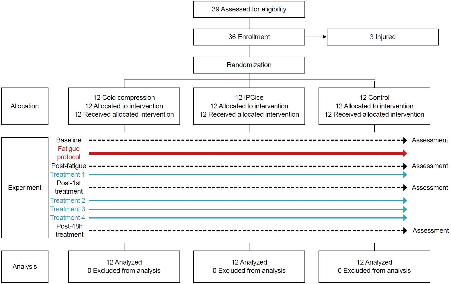
The recruitment process included inviting MMA clubs in the region through individual communication with the coach and administration and advertising the study to athletes associated with these clubs (Fig. 1). A total of 36 healthy amateur MMA athletes participated in this study, comprising 11 female and 25 male (Table 1). Inclusion criteria mandated participants to be aged 18–40 years, possess a minimum of 3 years of training experience, and train at least three times per week being at least tier 2 (trained/developmental) based on McKay’s participant classification [20]. Exclusion criteria included: resting blood pressure exceeding 140/90 mm Hg, any musculoskeletal injuries in the upper or lower limbs within the preceding 6 months, skin lesions or tattoos near the intended measurement sites (preventing accurate tissue perfusion assessment), fever, infection, pregnancy, and extreme fatigue. To minimize potential confounding factors, participants were instructed to abstain from ergogenic drinks for 6 hours prior to the study and to refrain from training for 24 hours before and throughout the study duration (until 48 hours post-study), as these could influence blood perfusion (PU) and muscle strength measurements.
Fatigue program
The fatigue program consisted of a standardized 8–10 minutes preparatory phase (brief jog, dynamic movements, and stretches) followed immediately by intermittent rope jumping designed to induce gastrocnemius fatigue. Rope jumping is a demanding activity frequently used in combat sports warm-ups [21] and has a metabolic equivalent ranging from 11.7 to 12.5, depending on the jumping rate [22]. Given that rope jumping engages both aerobic and anaerobic systems, similar to the demands of combat sports, it was included in the fatigue program. In this study, participants performed intermittent rope jumping for a total of 30 minutes, consisting of 5-minute bouts of jumping followed by 1-minute rest periods, repeated five times. This intermittent pattern was designed to mimic the calf muscle exertion experienced during combat sports rounds (3 or 5 minutes). The jumping pace was maintained between 135 and 145 jumps per minute, monitored by an assistant using a wireless counter in the Rival Jump Rope (Rival Boxing Gear).
Recovery interventions
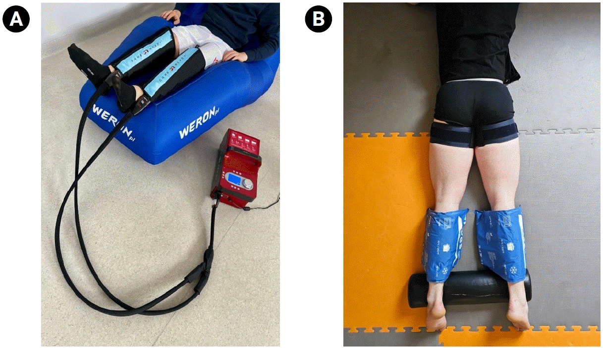
Following the post-exercise measurements, the CC group underwent a 20-minute recovery intervention targeting the gastrocnemius muscle (Fig. 2A). Cryo-compression cuffs were applied at alternating sub-diastolic pressures of 25–75 mmHg, cycling every two minutes, while maintaining a constant cuff temperature of approximately 1°C–3°C. These pressures are insufficient to occlude arterial inflow and are intended to enhance venous return, limit interstitial edema, and deliver localized cryotherapy to the gastrocnemius.
The IPCice group received recovery intervention involving passive ice pack application combined with occlusion (Fig. 2B). Compression cuffs were applied at the proximal thigh and inflated to 200 mmHg (~100% arterial occlusion pressure) for 2 minutes, followed by 2 minutes of reperfusion, during which ice bags were applied directly to the gastrocnemius (same cooling target as CC). This 2-minute on/2-minute off cycle was repeated for 20 minutes. Although both groups cooled the gastrocnemius, CC provided standardized circumferential cryo-compression at 25–75 mmHg, whereas IPCice used manual ice packs (no controlled cuff pressure at the calf); the only high-pressure component in IPCice was the proximal thigh occlusion.
The control group received no active intervention and engaged in passive rest. Both the CC and IPCice groups received a total of four recovery interventions within a 48-hour period following the initial fatigue program.
Measurements and outcomes
During this 48-hour recovery window, participants completed four recovery sessions (two per day), but outcome assessments were consistently anchored to these four time points. All examinations were conducted between 9:00 AM and 12:00 PM at the Provita Medical Center in Poland.
Given the extensive number of measurements, a single measurement site on the gastrocnemius muscle of each limb was chosen based on scientific literature demonstrating high measurement repeatability at this location. Reference lines were marked using a flexible ruler, extending between the medial epicondyle of the femur and the lateral malleolus of the fibula. A measurement marker was then placed at a point corresponding to 15% of this measured length.
Blood perfusion
Microvascular perfusion was assessed in vivo through laser Doppler flowmetry (LDF) using the PeriFlux 6000 Combined System (Perimed AB). The methodology for blood flow measurement adhered to methodological recommendations [23], involving the placement of a probe on the skin’s surface at the designated measurement point. Data were collected from a tissue depth of 2.5 millimeters within a measurement volume of one cubic millimeter over a two-minute interval. This non-invasive LDF method is valued for its consistent and responsive evaluation of microcirculatory dynamics, both at baseline and during physiological provocation [24]. Considered a robust approach for quantifying the function of the microvasculature, LDF offers precision and reliability in its measurements. The resulting microperfusion data were quantified and presented in perfusion units.
MS
The mechanical characteristics were assessed using a MyotonPRO AS myotonometer (Myoton Ltd.), a digital instrument comprising a main unit and a 3 mm diameter depth probe. During measurement, an initial preload of 0.18 N was applied to the skin surface, gently compressing the underlying tissue. Subsequently, a brief mechanical impulse (0.4 N, 15 ms) was delivered by the device, causing a transient deformation of the tissue. Myotonometry is a dependable measurement technique capable of discerning variations in the physical characteristics of muscle fibers, including stretched fibers. Measurements were conducted with participants in a standardized, relaxed supine position on a treatment couch, focusing on the rectus femoris muscle. Three distinct points along the muscle belly were assessed, with these locations identified by dividing the muscle into thirds after marking reference points (a line drawn between the anterior superior iliac spine and the center of the patella). The reported data represent the average of these three measurements taken on both the right and left rectus femoris muscles. The final outcome, MS, was reported as the average of these three measurements taken on both the right and left rectus femoris muscles, expressed in Newtons per meter (N/m).
RSI
Lower limb reactive strength was evaluated using the RSI. The reliability of RSI and related kinematic variables derived from the countermovement jump has been previously established. Data acquisition was performed using a Force Decks ground reaction force plate (VALD Performance). RSI quantifies an individual’s capacity for rapid transition between eccentric and concentric muscle actions, serving as an indicator of reactive strength. The testing protocol involved participants stepping off a 50 cm box and immediately performing a maximal vertical jump upon landing on the force plate. Prior to testing, participants completed a one-minute bout of passive lower limb stretching. A research assistant provided standardized instruction on the jump technique, and each participant performed three familiarization trials before data collection. The highest RSI value obtained across the three trials was used for subsequent analysis. RSI was calculated using the formula:
RSI =jump height/contact time [m/s-1].
CK
For serum enzyme activity assessment, a total of 8 ml of blood was collected via venipuncture from the antecubital vein of participants in a seated position, using Innmedis clotting tubes and performed by qualified personnel. We collected a single 8 mL venous sample solely to quantify the primary outcome (serum CK) and to retain backup aliquots for quality-control re-analysis.
The collected tubes were promptly transported to the laboratory, where they underwent centrifugation at 3,000 g for 10 minutes at 4°C using a Mechanika Precyzyjna Warszawa -54 centrifuge. The separated serum was then aliquoted and stored at -80°C until analysis. To mitigate inter-assay variability, all samples were analyzed by a certified laboratory diagnostician. Enzyme activity was quantified in triplicate using a spectrophotometer. Specifically, dehydrogenase activity in the serum was determined through colorimetric spectrophotometry at a wavelength of 340 nm and a temperature of 37°C, employing ready-to-use diagnostic reagents (Alpha Diagnostic) on an automated biochemical analyzer A15 (Randox).
The TQR
The TQR scale, a subjective measure of an athlete’s recuperation, ranges from 0 to 20. Participants provided a rating of their recovery, where scores between 0 and 4 were indicative of poor recovery, 5 to 8 suggested moderate recovery, 9 to 12 indicated good recovery, and 13 to 20 reflected excellent recovery. The scale was administered on an individual basis, and all participants possessed prior familiarity with its use as part of their standard club procedures. The recorded measurements were expressed in arbitrary units (A.U.).
Statistical procedures
Statistical analyses were performed using SPSS version 29 (IBM Corp.). Prior to conducting the repeated measures ANOVA with Group (3 levels: CC, IPCice, control) as the between-subjects factor and Time (4 levels: Rest, PoExe, Po30min, Po48h) as the within-subjects factor, the assumptions of sphericity (assessed using Mauchly’s test) and normality of residuals (assessed visually via histograms and Q-Q plots, and statistically using Shapiro–Wilk tests) were examined for each dependent variable. In cases where sphericity was violated, the Greenhouse-Geisser correction was applied to adjust the degrees of freedom. The primary analysis involved a two-way repeated measures ANOVA to determine the main effects of Group and Time, as well as their interaction effect on each outcome measure. Significant main or interaction effects were followed up with Bonferroni post hoc tests to identify specific pairwise differences between groups and time points. The magnitude of the effects was quantified using partial eta squared (ηp²) for the ANOVA (with thresholds of 0.01, 0.06, and 0.14 indicating small, medium, and large effects, respectively). An alpha level of p<0.05 was set for statistical significance for all analyses.
RESULTS
Table 2 presents the descriptive statistics for the three groups across different outcomes at the four time point assessments. Significant interactions were observed between the time points of assessment and the three groups for perfusion in the right (F=36.982; p<0.001; ηp²=0.691) and left legs (F=23.631; p<0.001; ηp²=0.589), MS in the right (F=27.352; p<0.001; ηp²=0.624) and left legs (F=30.258; p<0.001; ηp²=0.647), PPT in the right (F=6.129; p<0.001; ηp²=0.271) and left legs (F=20.096; p<0.001; ηp²=0.549), RSI (F=4.295; p=0.007; ηp²=0.207), and TQR (F=20.889; p<0.001; ηp²=0.559). No significant interaction was found for CK (F=1.718; p=0.186; ηp²=0.094).
No significant differences between groups at rest were observed for any of the outcomes (p>0.05), as shown in Table 2. Moreover, immediately post-exercise, significant differences were only found for CK (p=0.009; ηp²=0.248), with the control group exhibiting significantly higher levels than the CC group (p=0.007). No other significant differences were observed between groups for the remaining outcomes (p>0.05).
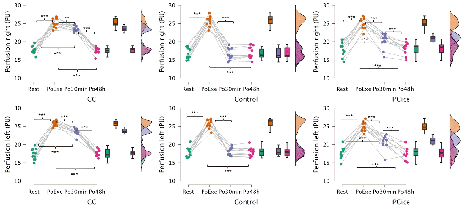
Fig. 3 shows the variations in muscle perfusion across the time points and the within-group inferential analysis. Significant differences between groups were observed Po30min (p<0.05), but not after Po48h for either leg (p>0.05). Specifically, the control group exhibited significantly lower perfusion in the right and left legs compared to the IPCice (p<0.001) and CC (p<0.001) groups in Po30min. Additionally, CC showed significantly greater perfusion than IPCice Po30min in both the legs (p<0.001).
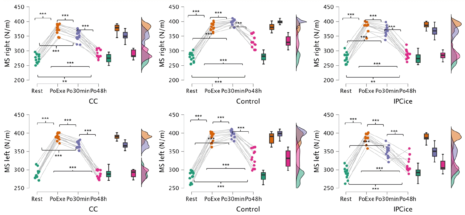
Fig. 4 illustrates the variations in MS across the time points, along with the within-group inferential analysis. Significant differences between groups were observed both Po30min (p<0.05) and Po48h in both legs (p<0.05). Po30min, the CC group showed significantly lower MS in the right leg compared to IPCice (p=0.020) and control (p<0.001), while IPCice also had significantly lower MS than control (p<0.001). At Po48h, the control group exhibited significantly higher MS in the right leg compared to both IPCice (p<0.001) and CC (p<0.001). Similarly, in the left leg Po30min, the CC group showed the lowest MS values, significantly lower than IPCice (p=0.003) and control (p<0.001). IPCice also showed significantly lower MS than control (p<0.001). However, at Po48h, the control group presented significantly lower MS in the left leg compared to IPCice (p=0.013) and CC (p<0.001).
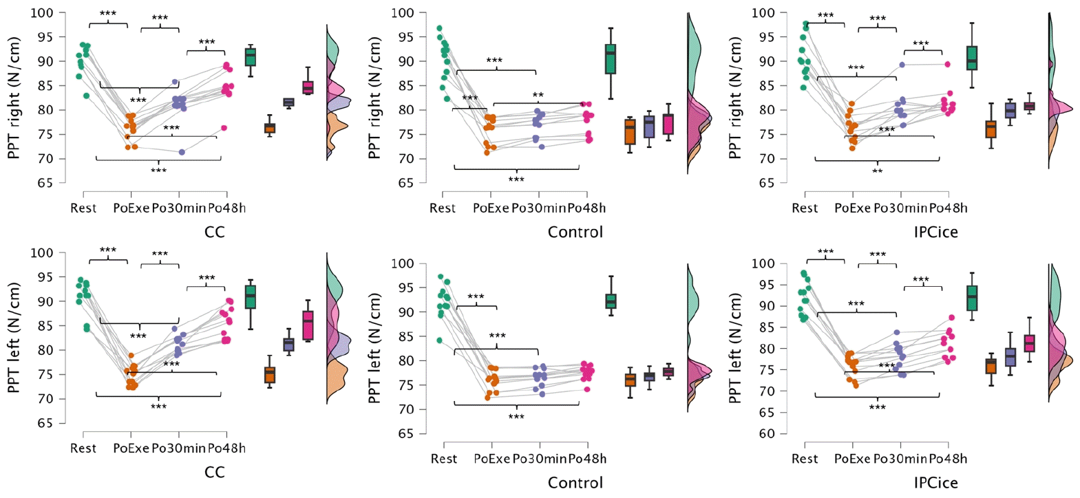
Fig. 5 shows the variations in PPT across the time points and the within-group inferential analysis. Significant differences between groups were observed at Po30min (p<0.05), and at Po48h for either leg (p<0.05). Specifically, in Po30min, the control group showed significantly lower PPT in the right leg compared to both the IPCice (p=0.033) and CC (p=0.006) groups. Interestingly, in the left leg, the CC group demonstrated significantly higher PPT than both IPCice (p=0.004) and control (p<0.001). At Po48h, the control group continued to show significantly lower PPT than both IPCice (right leg: p=0.010; left leg: p=0.005) and CC (right leg: p<0.010; left leg: p<0.001) in both legs. Additionally, the CC group exhibited significantly greater PPT than IPCice in both the right (p=0.047) and left legs (p=0.001).
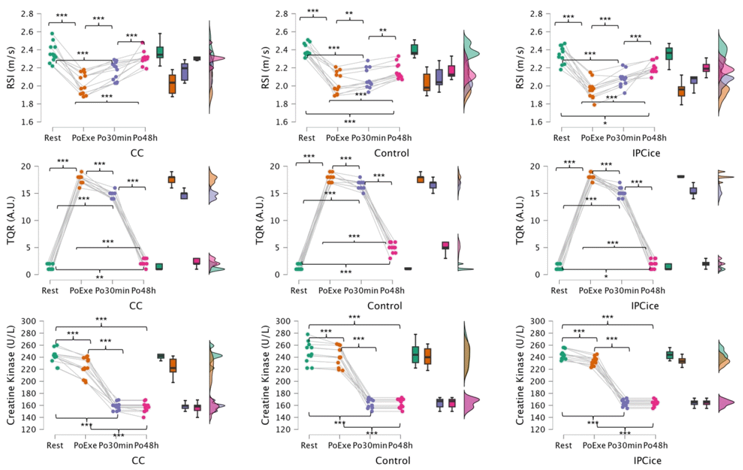
Fig. 6 presents the variations in RSI, CK, and TQR across the time points, along with the within-group inferential analysis. For RSI and CK, significant differences between groups were found only at Po48h (p<0.05), with no significant differences observed immediately after the Po30min (p>0.05). In contrast, TQR showed significant differences between groups both Po30min (p<0.05) and at 48 hours (p<0.05). At Po48h, the CC group exhibited significantly higher RSI compared to both IPCice (p=0.005) and control (p<0.001), with no significant differences observed between IPCice and control (p=0.528). Regarding CK, at Po48h, CC showed significantly lower values than IPCice (p=0.050). For TQR, at Po30min, the control group presented significantly higher scores compared to both IPCice (p<0.001) and CC (p<0.001). Similarly, at Po48h, TQR was significantly higher in the control group than in both IPCice (p<0.001) and CC (p<0.001).
DISCUSSION
The main findings of this study suggest that both IPCice and CC recovery interventions were more effective than passive recovery in facilitating post-exercise recovery, with the CC intervention exhibiting the most consistent and pronounced benefits across outcomes. Specifically, both IPCice and CC significantly improved muscle perfusion, reduced MS, and increased PPT compared to the control group, particularly in the Po30min period. These benefits were largely maintained at the Po48h mark, especially for CC. Interestingly, CC was better than IPCice in measures of perfusion, MS, and RSI, highlighting its superior effectiveness in some time points. However, IPCice still provided measurable recovery advantages over control, particularly in MS and PPT, suggesting it is a viable recovery strategy.
The results of our study revealed that both CC and IPCice were significantly effective in enhancing short-term perfusion following the PoExe. Both interventions outperformed the control group. Interestingly, CC was significantly more effective than IPCice. This finding is partially consistent with previous research: while some studies suggest that post-exercise IPC can accelerate recovery and reduce muscle damage [25], others report no improvement in acute recovery among amateur rugby players [26]. In contrast, previous research involving MMA fighters found that CC significantly enhances tissue perfusion [27]. CC likely promotes vasoconstriction followed by reactive hyperemia upon removal, enhancing blood flow to the tissues [28]. Additionally, external pressure may assist in venous return and reduce interstitial edema, facilitating nutrient delivery and waste removal. IPC, on the other hand, is thought to stimulate endothelial shear stress, promoting vasodilation and improving microvascular function [29]. The greater effectiveness of CC over IPC with ice may be due to the synergistic impact of continuous mechanical pressure and localized cooling, which more effectively reduces tissue inflammation and facilitates reactive hyperemia.
Regarding PPT, our study found that both recovery strategies were effective in enhancing recovery at Po30min, with effects lasting up to 48 hours compared to the control group. Interestingly, CC was also significantly more effective than IPCice both PoExe and at Po48h. Previous research has shown that IPC applied at 110% and 150% of arterial occlusion pressure can significantly increase PPT [30]. Similarly, DuPont et al. [31] reported that CC therapy following resistance exercise enhanced recovery by reducing muscle damage, soreness, and pain compared to controls. CC’s effectiveness can be attributed to vasoconstriction, reducing blood flow to the area. This may minimizes swelling, inflammation, and pain by decreasing the delivery of inflammatory mediators and numbing nerve endings [32]. The compression component further aids in promoting fluid drainage [32]. In contrast, IPCice operates by inducing brief periods of ischemia followed by reperfusion, which can activate endogenous protective pathways and improve vascular function. However, its analgesic effects may be less pronounced compared to CC, possibly due to the intermittent nature of the stimulus and the absence of continuous mechanical pressure, which is crucial for sustained activation of pain inhibitory mechanisms.
Regarding MS, both recovery techniques produced beneficial effects starting at Po30min, with improvements lasting up to Po48h and showing significantly better results than the control condition. Interestingly, CC also showed superior outcomes compared to IPCice. The application of cold induces vasoconstriction, which is the narrowing of blood vessels. This likely reduces blood flow to the affected muscle tissue, helping to minimize swelling and inflammation – major contributors to post-exercise MS. The decreased blood flow also reduces the delivery of inflammatory mediators that can exacerbate stiffness [33]. IPCice was likely not as effective as CC in recovering MS post-exercise because while ice can reduce inflammation and potentially provide some pain relief by decreasing nerve conduction, and blood flow restriction might theoretically alter metabolic byproduct removal [34], the lack of consistent, circumferential compression in ice packs combined with BFR may not have adequately addressed tissue mobility as effectively as the sustained, uniform pressure provided by CC garments, which simultaneously manage swelling and reduce inflammation.
RSI also improved in the CC group, with significantly better results than both IPCice and the control group at Po48h. In contrast, IPCice did not produce a significant difference compared to the control. Studies indicate that CC effectively reduces localized inflammation, potentially leading to faster restoration of neuromuscular function and power output [31], which are key components of RSI. The sustained compression may also enhance proprioceptive feedback. In contrast, while IPCice might offer some analgesic or systemic benefits, the lack of significant improvement in RSI compared to the control group suggests that its mechanisms, possibly related to transient alterations in blood flow and cellular stress responses, do not as effectively facilitate the recovery of explosive strength qualities measured by RSI in this context.
Regarding TQR, both experimental groups showed similarly improved scores compared to the control group, both Po30min and at Po48h. In contrast, CK levels were lower in the CC group than in the IPCice group at Po48h, although no significant difference was found between either group and the control. The higher TQR in the control group despite CC’s advantages on physiological and semi-objective measures underscores that recovery is multidimensional and that subjective and objective indicators often index complementary constructs rather than the same signal [35]. Large-scale syntheses and in-season tracking studies show that athlete-reported wellness and perceived recovery frequently diverge from CK, HRV, and jump-based neuromuscular tests, yet remain sensitive to recent load and fatigue status [35,36]. Perceived recovery also has practical relevance for readiness, adherence, and load adjustment decisions, supporting its integration alongside mechanistic and performance outcomes in rehabilitation and sport practice [37,38]. Consistent with this view, TQR has been used as a responsive psychometric tool to track recovery across microcycles and to inform day-to-day programming in team-sport cohorts [39,40]. We therefore interpret the TQR–physiology mismatch in our data as complementary information about the psychological facet of recovery rather than a methodological artifact, and recommend monitoring both trajectories to guide progression and participation decisions in rehabilitation-oriented contexts [35,36].
Recovery in the context of rehabilitation
Recovery in rehabilitation is best understood as the optimization of functioning and participation, not merely biological restitution, consistent with the ICF-based view of rehabilitation as a strategy that aims to optimize human functioning across body functions, activities, and participation [12]. Within this frame, our Po30min improvements in microvascular perfusion, reductions in MS, and increases in PPTs with both IPCice and CC—most pronounced with CC—reflect early restoration of body functions that enable activity, rather than isolated symptom change [33]. These objective gains were sustained at 48 h for several outcomes, with CC outperforming IPCice and producing higher RSI—a functional indicator linked to stretch–shortening cycle performance and readiness used in rehabilitation decision-making—thereby connecting physiological recovery to activity-level capacity [41]. In ICF terms, perfusion, pain sensitivity, and tissue mechanics map to body functions, whereas RSI captures activity-level capabilities relevant to locomotor tasks, which clarifies how the measured changes translate to function in a rehabilitation context. The superiority of CC over IPCice aligns with evidence that adding pneumatic compression to cryotherapy more effectively reduces pain/edema and expedites functional milestones than cryotherapy alone in postoperative and musculoskeletal rehabilitation settings, supporting its positioning as a pragmatic early-phase modality [42]. Importantly, CK showed only modest, group-dependent differences and did not mirror functional and perceptual changes, consistent with meta-analytic and narrative evidence that CK responses are highly variable and often poorly coupled to recovery of performance, cautioning against over-reliance on damage biomarkers when judging rehabilitation progress [43]. In contrast, the higher TQR values observed in the control arm at Po30min and Po48h illustrate that subjective recovery can diverge from objective functional restoration—an acknowledged “mismatch” in rehabilitation outcomes—underscoring the need to monitor psychometric and performance measures in parallel. Practically, these findings support using CC to optimize body-function targets (perfusion, tissue mechanics, pain thresholds) that scaffold activity and participation goals, while integrating patient-reported recovery and individualized goal setting to guide progression across the rehabilitation continuum. By explicitly interpreting our results as gains in functioning within an ICF-based framework, we enhance clinical relevance for rehabilitation programs that must balance biological restoration with activity and participation outcomes across early and later recovery phases.
Study limitations, future research and practical applications
While the current study offers interesting findings regarding the comparative effectiveness of IPCice and CC recovery strategies, several limitations should be acknowledged. First, the study focused exclusively on the gastrocnemius muscle; therefore, further research is needed to assess whether similar effects are observed in other muscle groups, particularly those with different vascularization patterns or muscle fiber distributions. Additionally, the study examined only short-term recovery (up to 48 hours post-exercise), leaving the long-term effects unaddressed. Another limitation is that, although both protocols cooled the same target muscle (gastrocnemius), the mechanical characteristics differed: CC applied standardized sub-diastolic cryo-compression at the calf (25–75 mmHg), whereas IPCice combined suprasystolic occlusion at the proximal thigh (200 mmHg) with manual ice packs at the calf. These methodological distinctions may have contributed to differences in outcomes and complicate direct mechanistic comparisons. It should also be noted that a direct comparison between IPC and IPCice could have provided clearer mechanistic insight into the incremental contribution of cryotherapy; however, our study prioritized the more clinically relevant comparison between IPCice and CC, which are both widely used recovery strategies in combat sports. Furthermore, the IPC parameters—such as occlusion pressure and duration—were standardized and may not represent the most effective or individualized protocols. Future research should explore the potential benefits of combining different recovery modalities or applying them in varied sequences to optimize outcomes.
Despite the limitations, the findings of this study suggest that CC is an effective recovery strategy, offering consistent benefits in muscle perfusion, stiffness reduction, and performance restoration up to 48 hours post-exercise. Practitioners and athletes seeking efficient recovery tools may consider prioritizing CC, especially when rapid recovery between sessions is critical. IPCice also showed advantages over passive recovery, making it a viable alternative when CC is unavailable.
Conclusions
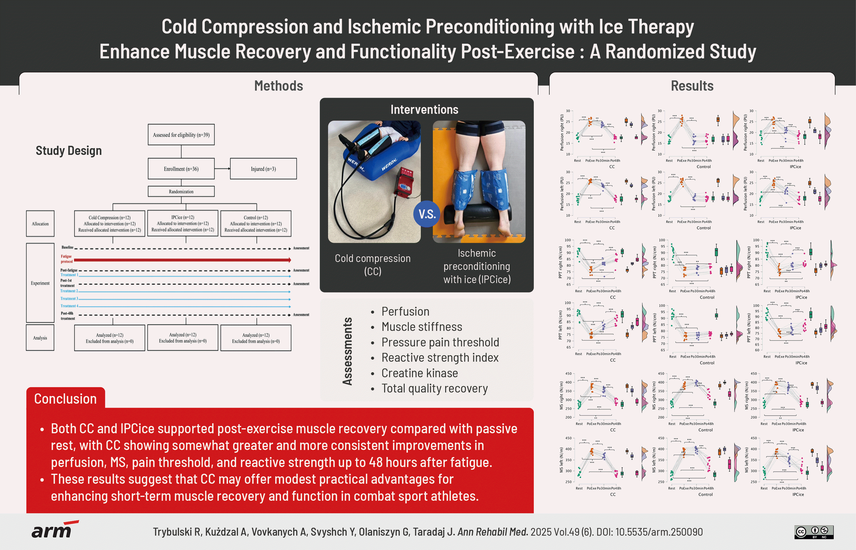
Both CC and IPCice supported post-exercise muscle recovery compared with passive rest, with CC showing somewhat greater and more consistent improvements in perfusion, MS, pain threshold, and reactive strength up to 48 hours after fatigue. These results suggest that CC may offer modest practical advantages for enhancing short-term muscle recovery and function in combat sport athletes. However, given the study’s limited sample size, focus on a single muscle group, and short follow-up, the findings should be interpreted cautiously. Further research is warranted to confirm these effects across different populations, recovery durations, and exercise contexts before firm clinical recommendations can be made.

 XML Download