



 PDF
 Citation
 Print


INTRODUCTION
The liver frequently harbors tumor metastases, although the role of resection for metastases from non-colorectal, non-neuroendocrine carcinomas remains unclear. Adam et al. [1] reported the largest cohort of hepatic resections for metastases deriving from tumors outside the abdominal region, noting that metastasis primarily occurs through systemic circulation rather than lymphatic pathways.
Uveal melanoma (UM) is a rare malignancy and represents the fourth most common primary site, with up to 50% of patients ultimately developing metastatic disease, most frequently in the liver (90%) [1,2]. Due to the lack of lymphatic drainage in the eye, UM primarily spreads hematogenously, unlike cutaneous melanoma. The development of liver metastases is a key clinical factor influencing overall survival (OS) in UM. Treating uveal melanoma liver metastases (UMLV) remains challenging because standard systemic therapies infrequently yield durable responses or significant survival improvement [3]. Median OS in this patient population is approximately 1 year [3,4]. Therapeutic strategies include both systemic treatment and liver-specific targeted therapy such as surgical resection, ablation, isolated hepatic perfusion (IHP), hepatic artery infusion (HAI), transarterial chemoembolization (TACE), selective internal radiation therapy (SIRT), and immunoembolization (IE) [5]. However, a consensus regarding the optimal management of liver metastases from melanoma has not yet been established, and available therapeutic modalities remain limited. Currently, liver resection is regarded as the most effective option for improving the prognosis in those with potentially resectable metastases. In a comparison of patients urdergoing surgery with curative intent versus those receiving palliative care, the median OS among surgical patients was 27 months (range, 14–90 months), compared to 8 months (range, 1–30 months) in the palliative group. The 1-, 3-, and 5-year OS rates were 100%, 41.4%, and 41.4%, respectively, in patients undergoing surgery; by contrast, the 1- and 3-year OS rates in the palliative group were 31.8% and 7.4%, respectively (p < 0.001) [6]. Notably, tumor debulking strategies in selected cases have demonstrated efficacy in delaying hepatic disease progression [7,8]. This paper presents our surgical approach to managing bilobar liver metastases from UM infiltrating the hepatic veins, focusing on surgical strategy and clinical outcomes.
CASES
Case 1
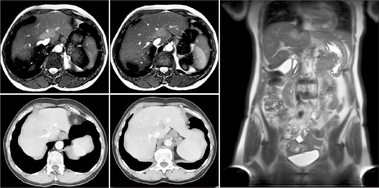
A 55-year-old female patient with a history of fibromyalgia and gastric ulcer was referred to our center for the assessment of metachronous liver metastases detected 8 years after enucleation of UM. During surveillance, CT and magnetic resonance imaging (MRI) revealed two confluent lesions located in segments 1, 4, and 8, encasing the left and middle hepatic veins and abutting the anterior aspect of the right hepatic vein (Fig. 1). No evidence of extrahepatic disease was identified. After multidisciplinary evaluation, the lesions were considered potentially resectable, requiring a left hepatectomy extended to segment 8 with reconstruction of the right hepatic vein. This procedure could necessitate total vascular exclusion, use of a veno-venous bypass, and potentially “in situ” hypothermic perfusion of the right liver.
Surgical technique
In the supine position, with a wide operative field including the left axillary fossa and Scarpa’s triangles, the patient underwent a bilateral subcostal laparotomy with a median incision extending to the xiphoid process. Upon opening the peritoneum, neither peritoneal carcinomatosis nor adhesions were detected. Intraoperative exploration confirmed the absence of extrahepatic disease. The liver exhibited a normal shape, size, color, and texture. The tumor mass was located in the upper region of the liver, involving segments 1, 8, and 4, and intraoperative ultrasound confirmed that it encased the left and middle hepatic veins by several centimeters and was in contact with the anterior surface of the right hepatic vein. Macroscopically, the lesion displayed the characteristic pigmented appearance of melanoma.
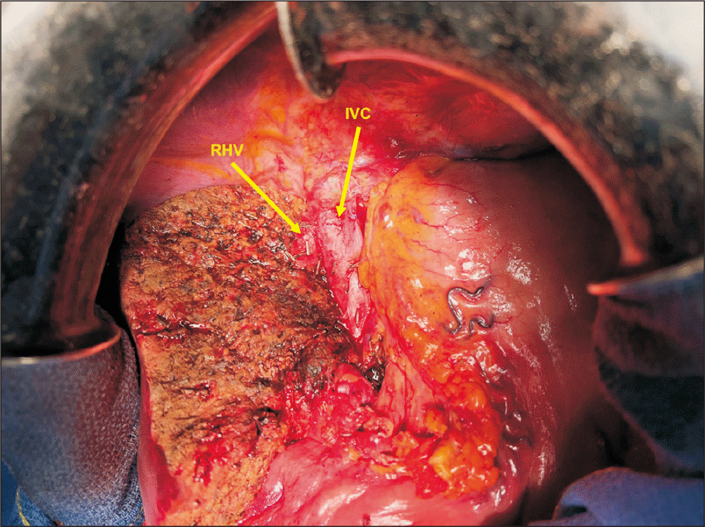
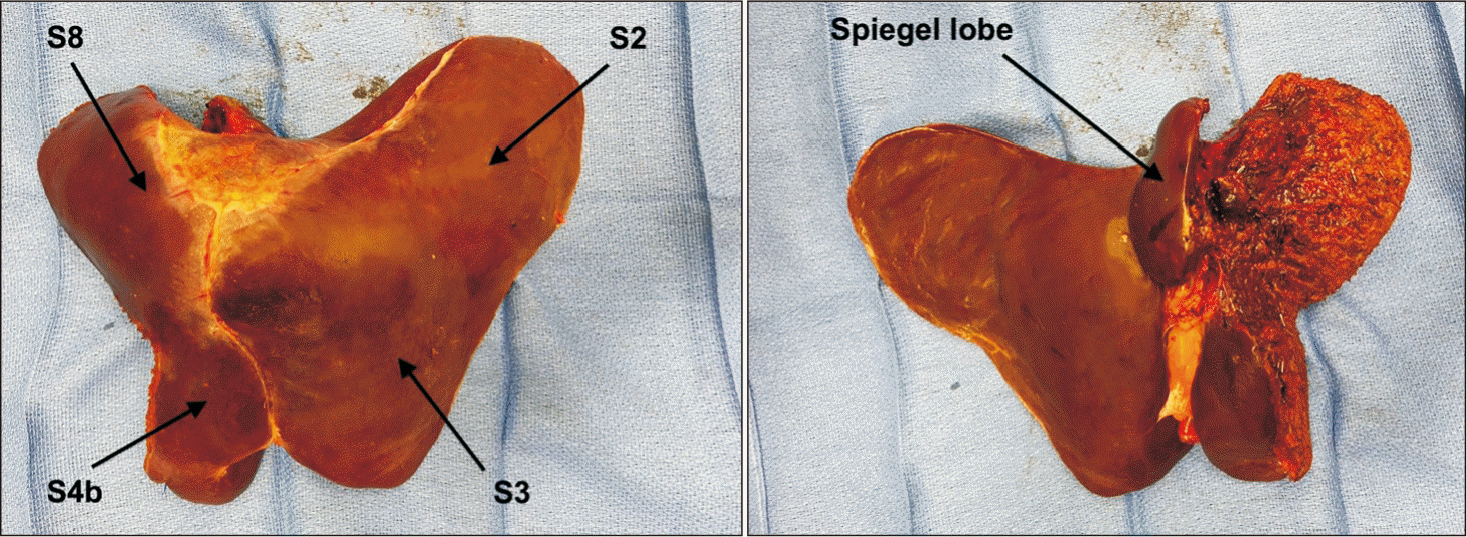
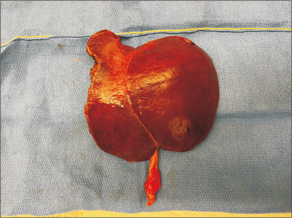
Additionally, other pigmented subcapsular (miliary) lesions ranging from 1 to 3 mm were identified in the remaining three segments of the right liver. Seven miliary lesions were excised using electrocauterization. Additional small miliary lesions were present, but these were located in the left-side of the liver and were removed during the left liver resection. Following complete mobilization of the liver, both the infra-hepatic and supra-hepatic inferior vena cava (IVC) were dissected and taped. The Spiegel lobe was separated from the IVC. The hepatocaval ligament was divided, and the right superior hepatic vein was then dissected and taped. Subsequently, the common trunk of the left and middle hepatic veins was isolated and taped. During this step, a suspicious dark lymphadenopathy, likely containing melanin, was detected near the left branch of the hepatic artery in the hepatic peduncle. As a result, lymphadenectomy of the hepatic artery was performed. The left portal vein and the left branch of the hepatic artery were isolated, ligated, and divided. After verification of the permeability of the right branches of the hepatic artery and portal vein using ultrasound, an antero-posterior parenchymal transection was performed, thereby achieving a left hepatectomy extended to segment 8, with the transection plane located immediately anterior to the right hepatic vein. A “vascular R1” resection was selected over a complex vascular reconstruction due to the extent of liver metastases identified intraoperatively (Fig. 2, 3). At the conclusion of the procedure, a further doppler ultrasound was conducted to confirm the patency of the portal vein, right branch of the hepatic artery, and right superior hepatic vein. The operative duration was 339 minutes. The estimated blood loss was approximately 300 mL, with no transfusion required. Normothermic ischemia lasted 40 minutes. The postoperative course was without complications, and the patient was discharged 9 days after surgery. Histopathological analysis confirmed metastatic UM in the left hepatic lobe, with twelve additional subcapsular metastases detected in the left liver, all of which were BRAF and NRAS wild-type, and no lymph node involvement was found. All seven miliary lesions resected from the right lobe were confirmed as metastases, and resection margins were negative. Imaging at 6 months follow-up revealed no recurrence of disease.
Case 2
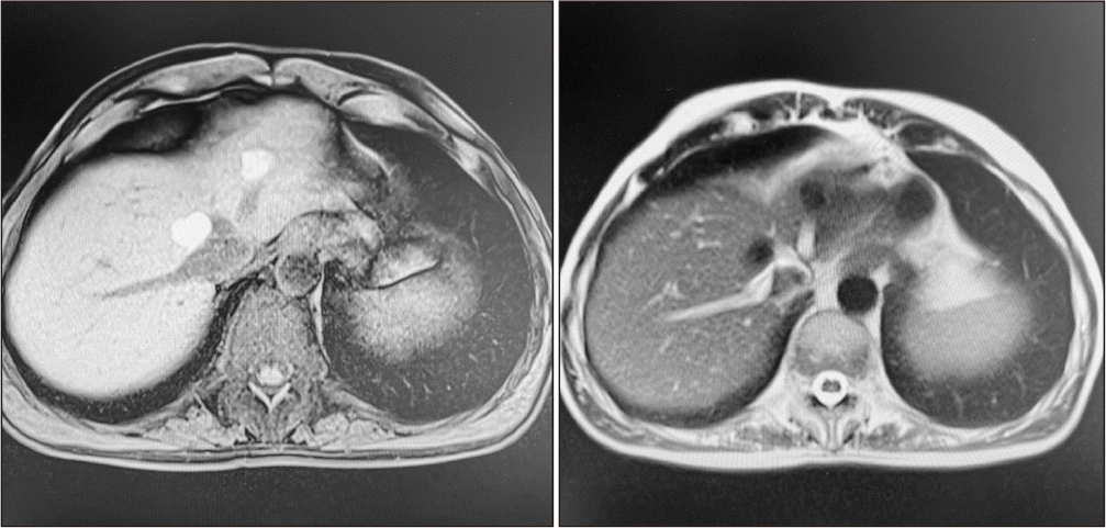
A 50-year-old male patient was referred to our center for evaluation of liver metastases 11-years after treatment for UM. Surveillance MRI identified two nodules measuring 15 and 20 mm; the first was located in segment 2, adjacent to the left hepatic vein, and the second in segment 8, closely associated with the middle hepatic vein and the anterior surface of the IVC. Both nodules appeared hyperintense on T1 sequence, presumably reflecting their pigmentary composition (Fig. 4). Following a multidisciplinary team discussion, these lesions were deemed suitable for resection via a left hepatectomy extended to segment 8 (H2348), including excision of the anterior wall of the IVC and reconstruction employing a peritoneal patch, necessitating vascular exclusion of the liver.
Surgical technique
A bilateral subcostal laparotomy with a median incision extending to the xiphoid process was performed. Upon opening the abdomen, there was no evidence of peritoneal carcinomatosis and no signs of extrahepatic disease during surgical inspection.
The liver exhibited normal morphology, size, texture, and color. The tumors were not visible but could be palpated, and were confirmed by intraoperative ultrasound, which demonstrated vascular involvement consistent with preoperative imaging findings.
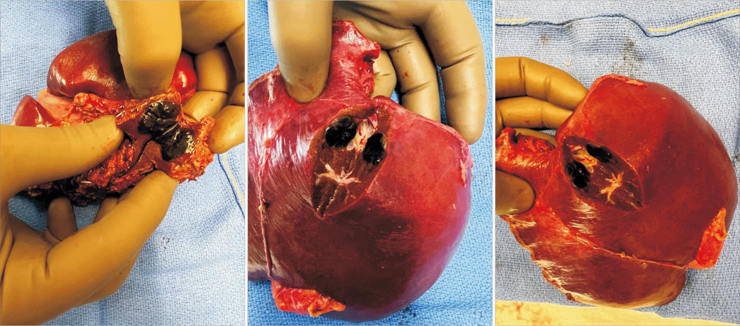
We started the procedure by fully mobilizing the liver and taping the infra- and supra-hepatic IVC. The right superior hepatic vein and the common trunk of the middle and left hepatic veins were also successfully isolated. Subsequent steps included dissection of the hepatic pedicle, division of the left hepatic artery branch, and further isolation and division of the left portal branch. This clearly demarcated the left liver. Despite intermittent clamping of the hepatic pedicle, liver transection resulted in significant hemorrhage. Therefore, completion of the left hepatectomy extended to segment 8 was achieved under vascular exclusion of the liver (Fig. 5). Posteriorly, a completely free plane along the IVC was observed without exposure of the second metastasis, eliminating the need for IVC resection. Estimated intraoperative blood loss was approximately 500 mL, with no transfusion required. The hepatic pedicle was clamped for 30 minutes, and the liver vascular exclusion lasted 20 minutes. On sectioning, both lesions appeared characteristic of melanoma, displaying intense pigmentation (Fig. 6). The postoperative course was uneventful. Histopathological analysis confirmed the presence of melanoma metastases and verified a complete R0 resection. Imaging at 6 months follow-up demonstrated no evidence of disease recurrence.
Case 3
The third case involved a 40-year-old woman who was referred to our center 31-months following enucleation of UM. During follow-up, imaging detected a 2 cm hepatic metastasis in segment 1 that gradually enlarged and was situated adjacent to the IVC. After multidisciplinary evaluation, the lesion was assessed as resectable, and a minimally invasive robotic approach was recommended.
Surgical technique
The patient was positioned supine, and robotic surgery was conducted using the da Vinci Xi platform (Intuitive Surgical Inc.) with a five-trocar approach.
No peritoneal effusion or carcinomatosis was present. The liver displayed normal morphology, size, and coloration. After fully mobilizing the left lobe, which was retracted toward the right, intraoperative ultrasound confirmed a lesion in the Spigelian lobe, extensively contacting the IVC and internally abutting the middle hepatic vein.
The dissection plane between the Spiegelian lobe and the IVC was entirely free. This maneuver exposed the anterior surface of the vena cava to its right margin. Two Spiegelian veins were divided, allowing completion of segmentectomy 1 with achievement of a “vascular R1” resection, without intraoperative blood loss or vascular clamping. Histopathology confirmed a liver metastasis derived from UM.
DISCUSSION
Recent studies have shown the benefit of surgical resection for liver metastases originating from various digestive tumors, even for non-colorectal, non-neuroendocrine types [9].
The optimal treatment of liver metastases from UM remains a topic of debate. This clinical scenario continues to pose significant multidisciplinary challenges as efforts aim to identify prognostic factors that improve survival. A single-institution study describing a 16-year experience demonstrated that an aggressive surgical strategy for liver metastases from UM led to promising outcomes [10]. The findings indicated that OS was enhanced following resection. Specifically, aggressive resection was associated with 2-year survival rates of 54.4% after R0, 30.7% for R1, and 14.4% for R2 resections. Moreover, a disease-free interval exceeding 24 months between primary UM and the appearance of metastases was found to be a favorable prognostic indicator. Nevertheless, patients with more than 4 metastases and those with bilobar involvement exhibited poorer survival.
A thorough evaluation of liver metastases following treatment of the primary tumor is essential. A notable investigation into the preoperative staging of UMLM using MRI and fluorodeoxyglucose-positron emission tomography (FDG-PET) demonstrated that MRI is the most effective imaging technique for detecting liver metastases from UM and, in some cases, is the only modality capable of indicating the presence of miliary metastatic disease [11]. Nevertheless, MRI has limited sensitivity for lesions smaller than 5 mm. In our first case (case 1), as observed, no miliary lesions were detected during the preoperative staging. During surgical exploration, seven pigmented subcapsular lesions measuring 1 to 3 mm were unexpectedly found in the right liver and were subsequently excised using electrocauterization. At this point, we intentionally opted for a vascular R1 type resection without reconstruction of the right hepatic vein, as the lesion was easily separated from the vein and considering the potential existence of multiple additional millimeter-sized melanocytic lesions within the right liver. This underscores how meticulous preoperative staging can impact the extent of surgical resection or, as in our case, prompt modifications to the plan. Favorable survival outcomes for established liver metastases have been achieved with the greatest possible tumor reduction when technically feasible.
The approach of “debulking” surgery has been suggested for specific scenarios involving liver metastases from colorectal cancer [8], but current literature does not support the application of this concept to metastases originating from UM.
It has been shown that vascular R1 resection, defined as tumor detachment from intrahepatic vessels, provides local disease control comparable to R0 resection and offers satisfactory survival in patients with liver metastases from colorectal cancer [12] and, more recently, in intrahepatic cholangiocarcinoma [13]. In our UMLM cases, recurrence of disease appeared 8 years (case 1) and 11 years (case 2) following enucleation of the primary tumor. The disease-free interval between diagnosis of primary UM and diagnosis of liver metastases is prolonged, which we believe supports the rationale for a more aggressive surgical approach (Table 1). At present, there is a lack of data in the literature regarding the use of debulking surgery for melanoma metastases, particularly those originating from UM, which metastasize exclusively via hematogenous dissemination. Nevertheless, given the positive outcomes reported in the context of colorectal liver metastases (CRLMs), exploring the feasibility of this surgical approach in patients with liver metastases from UM appears warranted, particularly in those with an extended disease-free interval following primary UM. The broadening of indications for hepatic surgery in cases of advanced liver metastases, facilitated by improved chemotherapy, may justify the consideration of cytoreductive surgery in combination with systemic therapy. In our experience, the choice between performing vascular R1 resection or vascular reconstruction was finalized intraoperatively after thorough exploration and ultrasound evaluation. As detailed in Table 1, differences between planned and performed procedures demonstrate the incremental nature of intraoperative decision-making. The surgical plan was revised in response to intraoperative findings, with careful consideration of oncologic parameters (such as metastasis number and distribution, and disease-free interval) and technical aspects (including the volume of future liver remnant and overall patient status).
All patients included in this series presented with liver-limited disease and exhibited no evidence of extrahepatic metastases. We recognize that this therapeutic approach may not apply to patients with concomitant extrahepatic involvement, for whom thorough multidisciplinary evaluation remains essential. In the context of liver-directed therapy for patients with isolated UMLM, the SCANDIUM randomized controlled trial recently demonstrated that a single administration of IHP yields statistically superior anti-tumor responses and improved progression-free survival compared to chemotherapy or immune checkpoint inhibition, while also providing a better health-related quality of life [14]. Following treatment, establishing robust follow‐up programs after resection of UM and subsequent liver metastases is crucial to achieving optimal patient outcomes. Currently, there is no established consensus regarding the most effective follow‐up protocols in terms of screening modality or time intervals [6]. Liver transplantation is increasingly being considered as an alternative in the scientific discussion on liver metastases, marking a significant development in oncologic liver surgery [15]. However, definitive data supporting other oncological indications for liver transplantation remain insufficient. In 2024, Berardi et al. [16] reported a case of liver transplantation for end-stage liver disease after multiple oncologic therapies in a patient with unresectable liver metastases from breast cancer. To date, there are no reports in the literature of liver transplantation performed for UMLM. The full potential of liver transplantation within the oncologic setting thus warrants further investigation, ideally after meticulous patient selection in specialized and tertiary care centers.
In conclusion, we have described two complex cases involving bilobar liver metastases from UV infiltrating the hepatic veins, both treated with aggressive surgical management, and one case of a single S1 metastasis addressed using minimally invasive robotic surgery. In these instances, maximal tumor reduction, when technically feasible, offers the most favorable opportunity for disease-free survival. Preoperative evaluation of liver metastases is often complicated by the presence of numerous miliary lesions. Both pre- and postoperative liver-targeted therapies are required. Currently, robust evidence to guide the management of liver metastases from UV is lacking. Based on the positive outcomes seen in CRLMs, exploring the possible application of a similar approach in UMLM may be worthwhile, particularly for patients with a prolonged disease-free interval between primary UM and the onset of metastases, with each case evaluated by a multidisciplinary team. Additional potential indications for transplant oncology may also be identified and merit further investigation in future studies.
 XML Download