



 PDF
 Citation
 Print


INTRODUCTION
Over 70% of hepatocellular carcinomas (HCCs) are not amenable to surgical resection at the time of diagnosis [1]. Ex vivo liver resection and autotransplantation (ELRAT) provides an opportunity for selected patients by facilitating total hepatectomy, ex situ tumor excision, and reimplantation of the organ using transplantation-based methods. Although surgical advances have been made, ELRAT remains technically intensive with considerable risks during the perioperative period [2]. This complexity has restricted the procedure to a limited number of specialized centers possessing both transplant and hepatobiliary surgical proficiency.
Conversion therapy has simultaneously arisen as a means to downstage borderline or initially unresectable HCC, utilizing transarterial chemoembolization (TACE) as a single modality or in conjunction with systemic agents. Nevertheless, the effectiveness of these approaches is still under assessment, and standardized treatment algorithms are yet to be established.
Although ELRAT and conversion therapy share the overarching goal of achieving curative oncologic resection, there has been only a single case report integrating these approaches; in that instance, ELRAT was conducted following TACE and radiofrequency ablation to address advanced HCC [3]. That patient, however, succumbed to respiratory failure on postoperative day (POD) 4.
In this report, we present the first documented successful case of ELRAT performed after TACE and stereotactic body radiation therapy (SBRT) to treat a large HCC with presumed vascular invasion, resulting in 24 months of recurrence-free survival postoperatively.
Written informed consent for publication of this case report, including accompanying images and clinical details, was obtained from the patient.
CASES
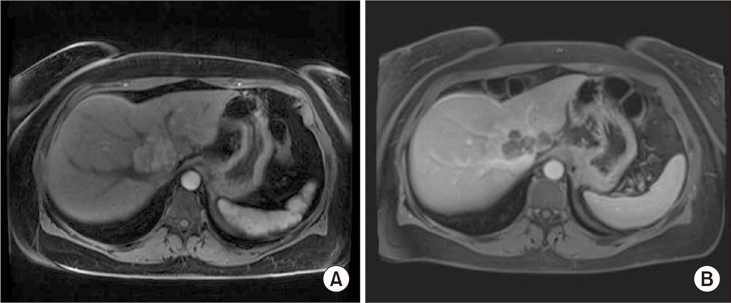
A 50-year-old female with a history of hyperlipidemia presented with upper abdominal pain. MRI demonstrated a 7.9 cm lesion involving liver segments 1, 4, and 8, with invasion of the retrohepatic inferior vena cava (IVC), and the right, middle, and left hepatic veins, without clear evidence of vascular occlusion (Fig. 1A). Biopsy identified moderately differentiated HCC, and her alpha-fetoprotein (AFP) was measured at 7,026.2 ng/mL. Due to the involvement of the IVC and all three hepatic veins, conventional resection was not considered feasible. TACE was performed with the intention of considering extended resection if response was observed. After one month, CT imaging showed central necrosis without a reduction in size, and AFP decreased to 412 ng/mL but remained elevated. Following a multidisciplinary review, SBRT (45 Gy in 5 fractions) was incorporated into the treatment plan. Two months after SBRT, AFP declined to 13.8 ng/mL, and MRI demonstrated a reduction in tumor size to 6.0 cm (Fig. 1B). PET-CT showed no evidence of extrahepatic disease. ELRAT was therefore considered.
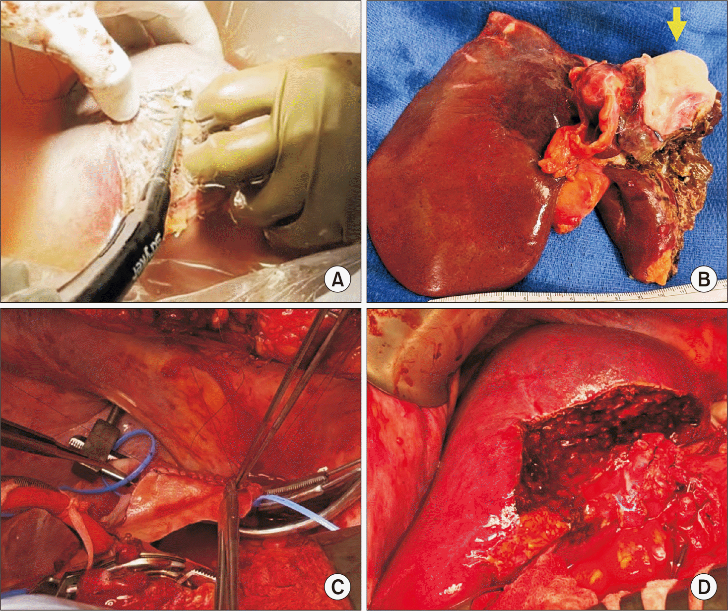
To ensure hemodynamic stability during the anticipated anhepatic phase, a veno-venous bypass was first established between the right femoral vein and the right internal jugular vein. Next, a Mercedes Benz incision was made. Intraoperative ultrasound confirmed tumor extension into the IVC and the left and middle hepatic veins, in close proximity to the right hepatic vein. Portal dissection and liver mobilization were performed, and in-situ transection was initiated but discontinued when nearing the tumor. The entire liver, together with a cuff of the anterior IVC wall, was resected and perfused using cold University of Wisconsin solution. The portal vein was then cannulated and incorporated into the bypass circuit to decompress the splanchnic system. On the back table, excision of the left lobe and tumor, along with a portion of segment 8, was accomplished (Fig. 2A, 2B). The IVC defect was repaired with bovine pericardium (Fig. 2C), and piggyback autograft implantation of the right lobe was completed by anastomosing the right hepatic vein of the graft to the reconstructed IVC, followed by an end-to-end portal vein anastomosis (Fig. 2D). After reperfusion, end-to-end arterial and bile duct anastomoses were performed. Total operation time was 534 minutes, estimated blood loss was 2,500 mL, cold ischemia time was 1 hour 57 minutes, and warm ischemia was 23 minutes. Histopathology confirmed residual HCC within a necrotic nodule, with clear resection margins and no lymph node involvement.
The patient was extubated on POD 1 and transferred out of the ICU on POD 2. She was discharged on POD 10 after an uneventful postoperative course. Three months later, she underwent mesh repair for an incisional hernia. At 19 months, she developed lower extremity edema secondary to IVC stenosis, which was managed with stent placement. At 24 months postoperatively, she remains recurrence-free, with normal AFP values.
DISCUSSION
Although technically feasible from the outset, upfront ELRAT was considered inappropriate in this instance because of elevated AFP levels and the presence of vascular invasion. Due to its substantial complication rate (24.5%) and a 30-day mortality of 9.5%, ELRAT necessitates meticulous patient selection [2]. Disease recurrence represents a significant contributor to mortality following ELRAT for malignant tumors, emphasizing the need for thorough preoperative evaluation of tumor biology and disease control. Locoregional therapy—possibly in combination with systemic therapy—may be valuable for determining therapeutic response and surveilling for metastases prior to ELRAT, thereby improving patient selection and outcomes.
Selection of ideal candidates for ELRAT after conversion therapy must be rigorous and based on multiple criteria. These include: 1) exclusion of extrahepatic disease confirmed by comprehensive imaging such as PET-CT, 2) evidence of favorable tumor biology demonstrated by robust serological response (notable reduction of AFP) and radiological findings (tumor necrosis and decreased size) after preoperative treatment, 3) assurance of sufficient future liver remnant volume and hepatic function, and 4) verification of patient performance status adequate to withstand this extensive surgical intervention. The use of a multidisciplinary team is critical in evaluating these factors for optimal candidate identification.
TACE remains the principal conversion therapy for HCC, although its effectiveness is limited, with only about a 10% conversion rate. When TACE is combined with SBRT, local tumor control and overall survival are improved; however, the conversion rate increases only modestly to 13%. Another emerging strategy is the combination of TACE with tyrosine kinase inhibitors and immune checkpoint inhibitors, which has demonstrated a conversion rate of 42%, indicating a potential advantage for ELRAT outcomes [4]. However, these preoperative interventions may result in adverse effects, including post-embolization syndrome, hepatic dysfunction, and complications related to radiation. Therefore, a collaborative multidisciplinary team is necessary to balance the benefits of therapy and determine optimal surgical timing.
In summary, we document the first successful ELRAT performed after TACE and SBRT for large HCC presenting with high AFP and vascular invasion. In advanced HCC cases where primary surgical resection is not initially possible, neoadjuvant treatment and stringent candidate selection may make ELRAT feasible with satisfactory outcomes.
 XML Download