



 PDF
 Citation
 Print


INTRODUCTION
Alveolar echinococcosis (AE) is a rare and potentially life-threatening zoonotic disease caused by the parasite Echinococcus multilocularis (EM). While EM is most commonly found in Central Asia, Russia, and Western-Central Europe, its distribution extends across the Northern Hemisphere, and it is increasingly recognized as an emerging infectious disease in North America [1,2]. During 2000–2020, approximately 30–35 confirmed cases of AE due to EM were reported in Canada [3]. Humans serve as intermediate hosts and acquire infection through contact with definitive hosts (foxes, wolves, coyotes, domestic dogs) or by ingesting contaminated food and water [4]. EM metacestodes develop initially in the liver, creating foci that may vary from several millimeters up to lesions exceeding 20 cm in size, often with central necrosis [2]. After establishing in the liver, larvae may spread to other organs either by direct extension or, less commonly, by hematogenous metastasis. After an asymptomatic latency period of 10–15 years, patients typically present with advanced disease. Management generally includes prolonged anthelminthic therapy as well as surgical resection. Although surgery is often not pursued in patients with distant metastases, the varied nature of AE presentations necessitates individualized management strategies for each patient [5]. The authors report a case of metastatic AE affecting the gallbladder, biliary tract, and spleen, originally considered for transplantation but ultimately managed with simultaneous left hepatic lobectomy and cholecystectomy. This case examines the role of liver transplantation in AE patients and underscores the challenges inherent in treating advanced infections.
CASE
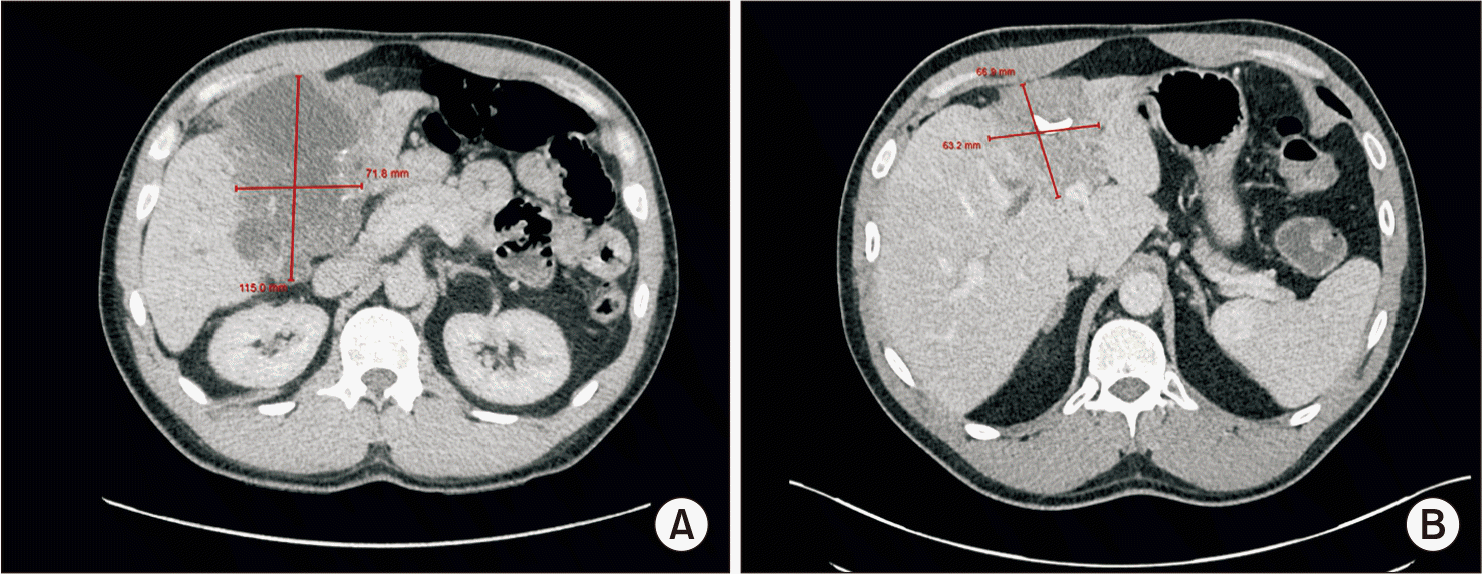
A previously healthy 48-year-old gentleman presented to the hospital in July 2022 with a 2-week history of abdominal pain, jaundice, and an unintentional weight loss of 14 kg over two months. He worked as a cattle rancher and had horses, dogs, sheep, and coyotes on his property. Notably, the patient mentioned that his previous dog had died from an unspecified intra-abdominal tumour. He was admitted to the hospital, where initial laboratory results revealed: alkaline phosphatase 607 U/L, total bilirubin 80 µmol/L, alanine transaminase 404 U/L, aspartate aminotransferase 102 U/L, international normalized ratio 1.0, and albumin 34 g/L. Computed tomography of the abdomen and pelvis (CT AP) demonstrated a large cystic and necrotic mass with internal calcifications (Fig. 1A) located in the gallbladder fossa, exhibiting extensive hepatic involvement. The gallbladder could not be distinguished separately from the mass, which was adjacent to the first portion of the duodenum and the hepatic flexure. The CT AP also identified intrahepatic biliary duct dilatation, splenic lesions, as well as peritoneal thickening and nodules.
A hepatic drain was inserted for biliary decompression, and an ultrasound-guided liver biopsy was performed. The patient was discharged with a preliminary diagnosis of metastatic cholangiocarcinoma. However, biopsy findings subsequently revealed pronounced inflammation, scattered eosinophils, and cystic material with positive Gomori methenamine silver (GMS) and Ziehl-Neelsen staining, findings suggestive of AE. He was referred to an Infectious Diseases specialist and underwent further evaluation, including two additional liver biopsies, nucleic acid testing, and serology. These investigations confirmed AE, and the patient was diagnosed with advanced hepatic AE metastasizing to the gallbladder, biliary tree, spleen, and peritoneum. No metastatic lesions were detected on CT of the chest and brain. His disease burden was too extensive for surgical resection as determined by General Surgery review. In September 2022, the patient was readmitted for treatment of biliary obstruction, necessitating placement of a common biliary duct stent via endoscopic retrograde cholangiopancreatography, and initiation of anthelminthic chemotherapy with albendazole. Following stent insertion and commencement of antiparasitic chemotherapy, his bilirubin levels decreased to 21 µmol/L from 75 µmol/L. Other than developing a persistently draining biliary fistula, his clinical condition remained stable during subsequent months. Repeat CT AP in August 2023 demonstrated modest interval regression of the hepatic mass (Fig. 1B).
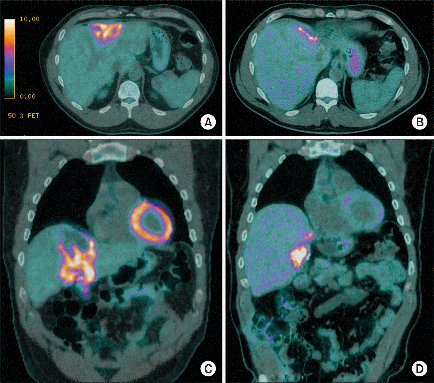
The patient underwent a liver transplant assessment in August 2023 after his follow up CT was completed. His case was evaluated by a multidisciplinary team, which included specialists from Hepatology, Hepatobiliary Surgery, Transplant Infectious Diseases, and an Infectious Disease physician with expertise in managing AE. They concluded that the patient would require a liver transplantation (LT), splenectomy, and complete excision of the entire fistulous tract from the liver to the skin to achieve full infection removal. Considering the estimated 1-year mortality risk of 10%–15% for a combined LT and splenectomy, along with the absence of additional indications for urgent transplantation, both the patient and the care team decided to continue albendazole therapy while monitoring disease activity with annual positron emission tomography–computed tomography (PET-CT) imaging. The PET-CT performed in September 2024 showed no new hypermetabolic lesions in the liver, and the previously detected splenic lesions had resolved (Fig. 2A, 2C).
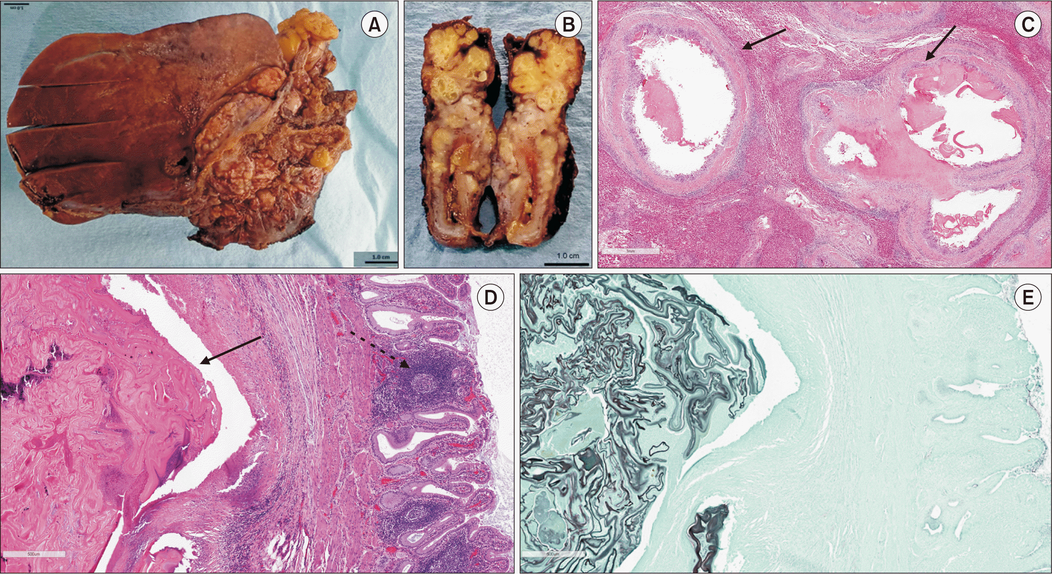
Given significant interval regression, surgical interventions were reconsidered with the patient, leading to the choice to resect sites of active disease. In December 2024, the patient was taken to the operating room for a planned open left hepatectomy and biliary fistula excision. Intraoperatively, marked inflammation was observed surrounding the gallbladder, with telangiectatic venous vessels visible in the biliary tree and small bowel. Due to concerns about potential intraperitoneal spillage of biliary contents, the surgical team proceeded with left hepatectomy and extended the resection margins, along with an en-bloc cholecystectomy. An intraoperative cholangiogram verified there was no biliary leakage from either the cystic duct stump or the right resection plane. Gross pathology revealed a cystic lesion 0.5 cm from the right hepatic parenchymal margin and a uniformly indurated gallbladder located 1.8 cm from the cystic duct margin. Histopathology confirmed the presence of multiple cystic lesions and hooklets in both the liver and gallbladder parenchyma, findings further visualized with GMS staining (Fig. 3).
The procedure was well tolerated, and the patient was discharged two weeks after surgery with normal liver enzyme and function test results. The patient’s latest PET-CT in May 2025 at six months post-surgery demonstrated hypermetabolic activity in the liver resection bed, indicating possible residual infection (Fig. 2B, 2D). The patient remains asymptomatic and will persist with indefinite albendazole therapy with therapeutic drug level monitoring, accompanied by regular clinical follow up and annual PET scans to assess disease activity and monitor long term albendazole therapy tolerability.
DISCUSSION
Despite its rising incidence, AE continues to be an underrecognized infectious disease in North America [4,6]. Patients with symptomatic AE frequently present similarly to the individual described in this case, typically exhibiting advanced disease with signs of hepatic dysfunction, such as jaundice, abdominal distention, right upper quadrant pain, fevers, and manifestations of portal hypertension. At first presentation, AE is commonly misidentified as a malignant tumor due to radiographic characteristics including necrosis, local invasion, and metastatic dissemination [2]. A high disease burden accompanied by frequent misdiagnosis during initial evaluation renders AE a difficult condition to manage. The World Health Organization’s Informal Working Group on Echinococcus (WHO-IWGE) defines a case of AE as confirmed when at least two of the following diagnostic criteria are met: I) characteristic lesions observed on imaging; II) histopathological confirmation of EM metacestodes; III) detection of EM-specific serum antibodies; or IV) presence of EM nucleic acid in a clinical specimen [7]. Once the diagnosis is established, management of AE should involve a multidisciplinary team and consists of chemotherapy with albendazole or mebendazole, benzimidazole anthelminthic therapy, surgical excision, and endoscopic or percutaneous interventions with prolonged follow-up using cross-sectional imaging [5]. Therapeutic strategy is guided by the PNM classification (Table 1), which was established by the WHO-IWGE [5]. This system relies on imaging findings to determine the extent of the parasitic lesion within the liver (P), involvement of adjacent organs (N), and the presence of metastasis (M) [2,7].
The patient in this case was classified as P3N1M1. He commenced therapy with albendazole. Benzimidazole anthelminthics, including mebendazole and albendazole, are the only antimicrobial agents presently recommended for AE; albendazole is generally favored over mebendazole due to reduced pill burden, greater cost-effectiveness, and broader availability. Although emerging evidence has demonstrated potential cures with extended therapy [8], albendazole is most often regarded as a suppressive rather than curative treatment [9,10]. Recent studies indicate a 10–15-year survival rate exceeding 80–90% with albendazole monotherapy, a notable improvement compared to survival rates below 10% observed in untreated or inadequately managed patients [5,11,12]. Approximately 10% of individuals prescribed albendazole cannot tolerate the therapy, with drug-induced hepatitis representing the most frequent adverse effect necessitating discontinuation [12]. In patients unable to tolerate prolonged albendazole monotherapy, case reports have described the use of salvage agents such as pembrolizumab, nitazoxanide, amphotericin B, and mefloquine, though none are currently recommended due to insufficient evidence of efficacy and the potential for significant medication-related adverse effects [13].
Radical surgical resection provides a potentially curative approach for AE and remains the preferred strategy in patients with localized hepatic involvement. However, because of the prolonged latent clinical phase, the majority of cases are unfortunately identified at an advanced stage, at which point curative resection is rarely achievable. While earlier studies advised against palliative resection [5,14], more recent evidence indicates a possible survival advantage associated with surgical resection [15] .
The management of advanced hepatic AE remains uncertain, and liver transplantation (LT) serves as a last-resort therapy when the lesion surpasses the limits for surgical resection. Approximately 150 cases of liver transplantation for AE have been reported globally, with 25 patients identified as having metastatic disease prior to transplantation [5,16-19]. Most of the individuals who underwent LT had additional indications for transplantation, including end stage liver disease resulting from biliary cirrhosis, Budd-Chiari syndrome, or recurrent cholangitis [5]. When AE is associated with extrahepatic disease, LT necessitates more extensive surgical resection. Alongside the inherent hazards of this complex procedure—such as a mortality rate of 10%–15% during the first year post-liver transplantation and up to 50% at 10 years—the required immunosuppression may accelerate larval proliferation at extrahepatic sites, resulting in an increased risk of aggressive disease recurrence [5,20]. Literature describes significant variability, with post-LT AE recurrence rates estimated to be as high as 60% [16]. Due to the substantial risk of recurrence, practitioners who are contemplating transplantation must ascertain the patient’s ability to tolerate long term albendazole therapy. Ultimately, LT may be appropriate for patients with chronic liver failure and lesions that cannot be resected, provided they can tolerate long term albendazole; however, it should be proposed with caution to those who have alternative therapeutic options, given the elevated mortality and risk of aggressive disease recurrence, most notably in the setting of immunosuppression.
The patient described in this case experienced a favorable response to long term albendazole monotherapy combined with percutaneous drainage over a 2.5-year period, resulting in disease regression and creating an opportunity for surgical resection with curative intent. Given that this patient achieved resolution of liver dysfunction through biliary stenting and percutaneous hepatic drain placement, there were no further indications for LT, and transplantation might have led to less optimal outcomes. The observation of residual or recurrent PET positive disease on the most recent CT scan underscores the chronic nature of this illness and highlights the complexity of multimodal management strategies. This case report contributes to the growing body of literature on AE treatment by detailing both the advantages and disadvantages of liver transplantation in advanced disease, demonstrating the importance of multidisciplinary team involvement in treatment planning, and addressing considerations for re-evaluating surgical intervention candidacy.
 XML Download