



 PDF
 Citation
 Print


INTRODUCTION
Choledochal cysts are congenital abnormalities of the biliary tract, associated with a considerable risk for malignant transformation, even following complete surgical removal. The combined malignancy incidence among adults with choledochal cysts is reported to be approximately 10%, with 3% developing malignancy after surgical intervention. Among these, cholangiocarcinoma has been identified as the most prevalent neoplasm [1]. This oncogenic risk is attributed to persistent inflammation, pancreaticobiliary maljunction, and residual dysplastic changes in the biliary epithelium.
While open pancreatoduodenectomy remains the standard treatment for periampullary malignancies, minimally invasive techniques—including laparoscopic and robotic pancreatoduodenectomy—have shown equivalent oncologic outcomes and may offer benefits in postoperative recovery [2,3]. However, performing minimally invasive pancreatoduodenectomy in patients with a history of minimally invasive choledochal cyst excision introduces notable technical difficulties due to postoperative adhesions and anatomical alterations within the hepatoduodenal ligament region. The current literature specifically addressing robotic or laparoscopic pancreatoduodenectomy in this subset of patients is limited, with only sporadic case reports describing its feasibility [4,5]. In this report, we present a case of robot-assisted pancreatoduodenectomy performed for periampullary cancer in an individual with a prior robotic choledochal cyst excision. Neoadjuvant chemotherapy was initially administered due to advanced disease at diagnosis. Ultimately, pathological complete remission was achieved. This case adds to the limited literature by providing evidence of the feasibility and success of minimally invasive surgery for periampullary cancer following choledochal cyst excision.
CASES
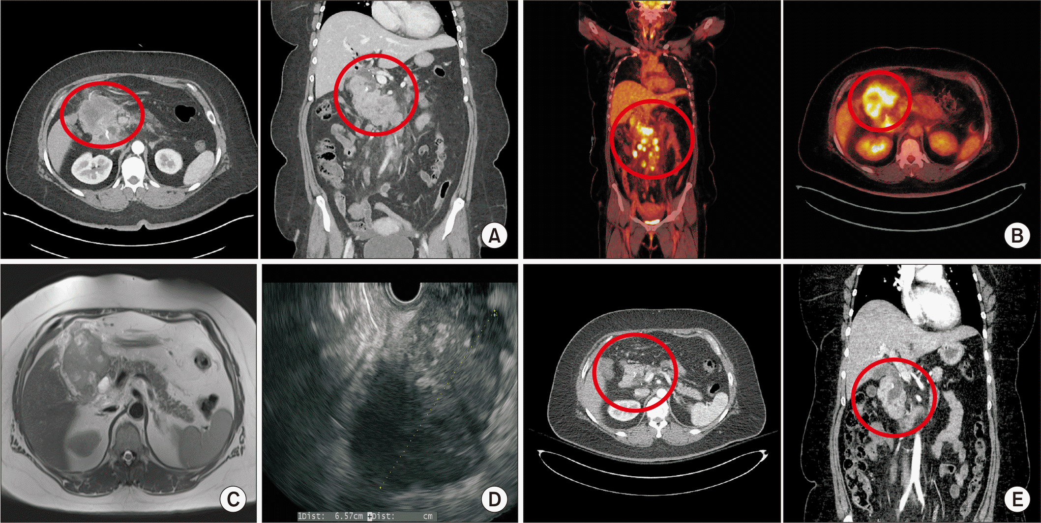
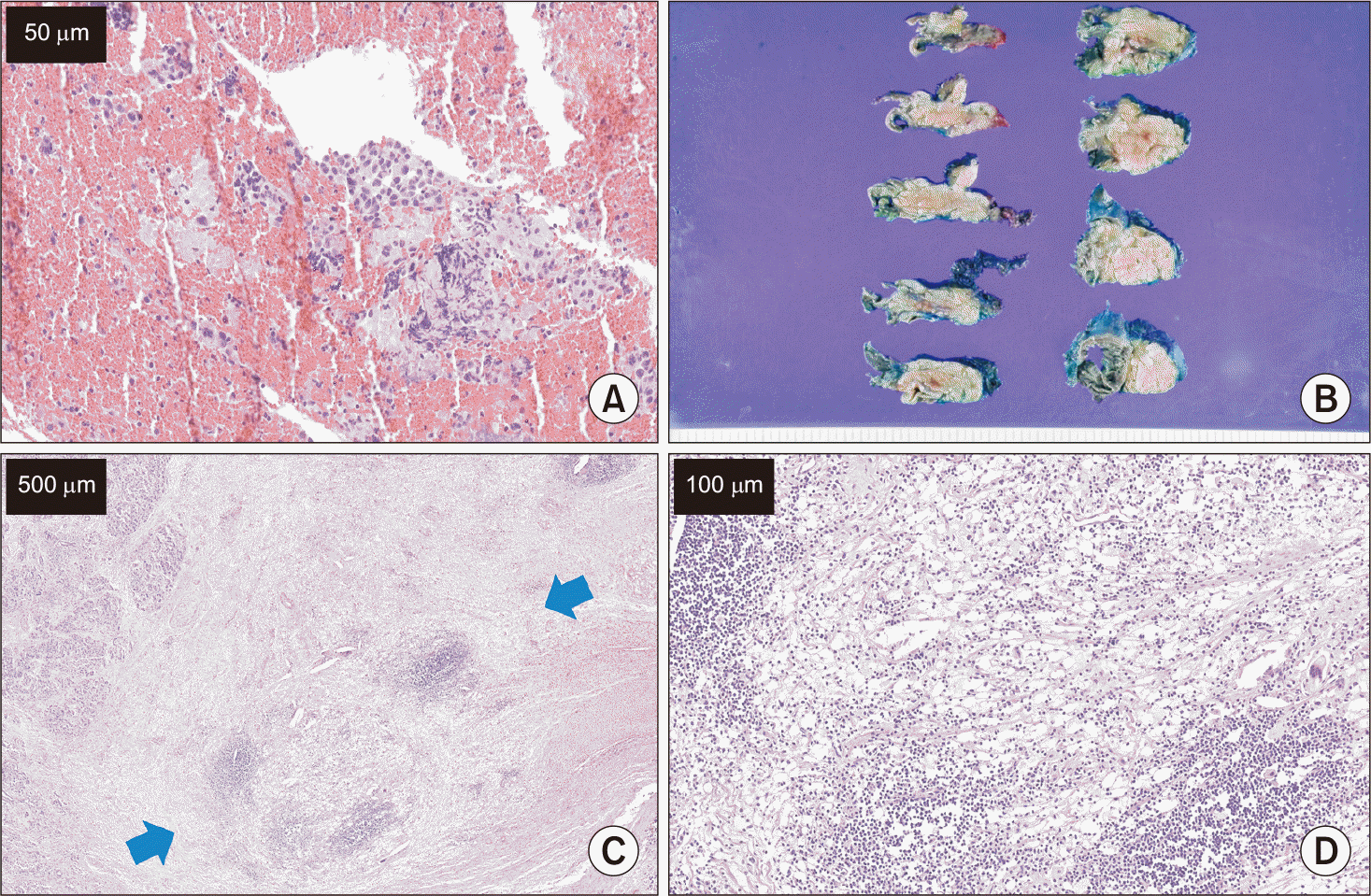
A 44 years old female with a history of choledochal cyst presented to our outpatient clinic. The patient had previously undergone robotic excision of the bile duct for a choledochal cyst in November 2022. She reported experiencing abdominal discomfort for 1 month prior to her visit. An initial computed tomography (CT) scan and magnetic resonance image (MRI) demonstrated an 8 cm mass-like lesion in the pancreatic head that raised suspicion for either exophytic pancreatic head cancer or conglomerated metastatic lymph nodes (Fig. 1A, 1C). Additionally, multiple metastatic lymph nodes were identified in the epigastric area, mesentery, retroperitoneal space, and both common iliac chains. Positron emission tomography (PET)-CT scan revealed significant uptake in the primary mass and lymph nodes, with additional suspicion for peritoneal metastasis (Fig. 1B). The initial carbohydrate antigen 19-9 (CA19-9) level was 3.3 U/mL (normal range 0–35). An endoscopic ultrasonographic biopsy of the pancreatic mass was performed (Fig. 1D), and the biopsy demonstrated atypical cells with necrosis, favoring poorly differentiated carcinoma (Fig. 2A). Due to the past history of choledochal cyst excision and inconclusive immunohistochemistry for pancreatic origin, the case was discussed in a multidisciplinary meeting involving surgeons, medical oncologists, radiologists, pathologists, and nuclear medicine physicians, who concluded that the patient likely had bile duct cancer originating from the remnant distal bile duct. Neoadjuvant therapy with durvalumab, gemcitabine, and cisplatin was planned before surgery. In total, 11 cycles of neoadjuvant chemotherapy were administered, which the patient tolerated well throughout the clinical course. Upon follow up CT scan revealing resolution of metastatic lymph nodes and a decrease in size of the main mass, surgical intervention was planned (Fig. 1E). CA19-9 level prior to surgery was 8.4 U/mL.
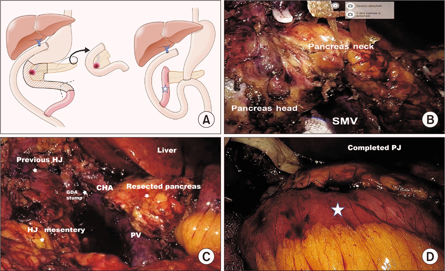
Robot assisted pylorus-preserving pancreaticoduodenectomy was scheduled for April 2025. The surgical approach involved resection of the right side pancreaticoduodenal unit along with the jejunum, and anastomosis of the pancreatic duct to the remnant A-loop from the previous operation (Fig. 3A). Moderate to severe adhesions of abdominal structures were observed around the wound and previous hepaticojejunostomy site, as well as the hepatoduodenal ligament. Adhesiolysis was performed to optimize the operative field. Suspicious nodules in the peritoneum and mesenteric lymph nodes were resected for intraoperative frozen biopsy, which confirmed absence of carcinoma. The Roux-limb mesentery and pancreaticoduodenal unit were adherent due to inflammation, necessitating careful dissection while preserving the mesenteric vascular arcade. Tunneling of the pancreas was accomplished as the final step of the resection phase (Fig. 3B). Following removal of the main mass and duodenum, the surgical specimen was retrieved (Fig. 3C). The Revo-I robotic system (Meerecompany Inc.) was utilized for the reconstruction phase. Reconstruction of the pancreatic duct was performed using the remaining A-loop from the previous jejunojejunostomy. The resected end of the A-loop was positioned near the pancreatic resection site in a retrocolic manner, and end-to-side duct-to-mucosa pancreaticojejunostomy anastomosis was completed (Fig. 3D).
The patient tolerated the immediate postoperative phase well. On postoperative day 3, the patient developed a fever, and empirical piperacillin-tazobactam therapy was initiated. CT imaging was performed, revealing only expected postoperative changes without complications. The patient’s diet was advanced to a soft diet the following day, which was well tolerated. The intraabdominal drain was removed on the 6th postoperative day, and the patient was discharged the next day. At the outpatient clinic, the patient remained stable, and the final biopsy demonstrated a complete pathological response with no residual carcinoma, along with extensive fibrosis, necrosis, and granulomatous inflammation containing multinucleated giant cells (Fig. 2B–2D). A total of 8 lymph nodes were retrieved, all negative for carcinoma, resulting in a final stage of ypT0N0. The patient is currently under surveillance and continues to receive immune checkpoint inhibitor therapy without further chemotherapy.
DISCUSSION
This case report demonstrates the feasibility of minimally invasive pancreatoduodenectomy in a patient with advanced periampullary cancer after previous robotic choledochal cyst excision. Table 1 provides an overview of reported cases in which pancreatoduodenectomy was performed for periampullary cancer developing after choledochal cyst excision and subsequent hepaticojejunostomy [4-15]. To our knowledge, this represents the first reported instance of a minimally invasive reoperation for metachronous periampullary carcinoma following prior choledochal cyst excision. In anticipation of potential conversion to open surgery, the resection phase for the pancreatoduodenal unit was performed laparoscopically, followed by robotic pancreatojejunostomy reconstruction. Laparoscopic resection was accomplished successfully, despite significant adhesions in the previous abdominal wound and hepatoduodenal ligament, without causing definitive injury to intraabdominal structures.
The occurrence of periampullary malignancy years after choledochal cyst resection underscores the critical need for long-term surveillance in this patient cohort. Although prophylactic cyst excision is performed, residual biliary epithelium or pancreaticobiliary maljunction can persist as potential sites for malignant transformation. A large-scale study conducted by Watanabe et al. [16] evaluated 1,353 patients with choledochal cyst and/or pancreaticobiliary malunion in Japan. In this investigation, the post-excision cancer risk was reported as 0.7%, with a mean interval to diagnosis of 9 years. Similarly, a multicenter study by Lee et al. [17] in South Korea identified a comparable incidence of 0.6%. While the incidence of malignancy remains low, it is highly fatal upon occurrence, thus warranting lifelong surveillance in these patient groups.
Notably, this case illustrated that the periampullary cancer was detected at a relatively advanced stage with multiple lymph node metastases at diagnosis; consequently, neoadjuvant chemotherapy was administered prior to surgery to increase the probability of achieving a curative outcome. The effectiveness of neoadjuvant therapy involving chemotherapy combined with an immune checkpoint inhibitor was demonstrated in the TOPAZ-1 trial [18]. The addition of durvalumab led to a significant improvement in overall survival, progression-free survival, and the objective response rate compared to gemcitabine and cisplatin only. Application of real-world data further confirmed the feasibility, safety, and efficacy of neoadjuvant immunotherapy [19]. Ultimately, this patient achieved pathological complete remission as indicated in the final pathology report. Although multiple stigmata suggested peritoneal seeding, no viable tumor cells were identified in any of the frozen section biopsies. This young female patient benefitted from the combined advantages of minimally invasive surgery and neoadjuvant chemotherapy, resulting in optimal oncologic outcomes.
There are two important surgical technical considerations. First, the presence of fibrosis and adhesions caused by previous surgery increases the complexity of tissue dissection, especially in the area surrounding the porta hepatis. Meticulous technique is required to avoid injury to major vascular structures such as the proper hepatic artery and portal vein during the laparoscopic resection phase. Additionally, careful preservation of the vascular supply to the Roux-en-Y limb was essential to minimize the risk of inadvertent disruption of the previous choledochojejunostomy. Second, we utilized a segment of residual proximal jejunum, specifically the part of the afferent loop from the earlier jejunojejunostomy, for reconstruction of the remnant pancreas. Surgery on a soft pancreatic remnant with a small duct poses unique technical challenges and demands fine surgical control. Under these circumstances, robotic assistance proved advantageous for executing a minimally invasive approach. We successfully employed robotic techniques for the pancreaticojejunostomy anastomosis. This procedure requires advanced laparoscopic expertise and a thorough understanding of hepatobiliary anatomy to minimize potential complications such as vascular injury or bile duct damage.
There is a paucity of comparative studies evaluating open versus minimally invasive pancreatoduodenectomy in patients with a history of choledochal cyst cancer, which limits the ability to establish clear conclusions about long-term oncologic outcomes. However, short-term data from this and similar cases indicate that minimally invasive procedures can yield equivalent surgical margins and lymph node retrieval when performed by skilled surgeons [20]. Future investigations should aim to develop standardized surveillance guidelines for patients with prior choledochal cysts to facilitate early cancer detection, while further evaluation of the safety of minimally invasive techniques in this patient population could enhance our understanding of long-term survival and recurrence.
In summary, minimally invasive pancreatoduodenectomy is a feasible alternative for periampullary cancers developing after choledochal cyst excision when undertaken at specialized centers with hepatobiliary expertise. This case contributes further evidence that robotic and laparoscopic approaches can be selectively and effectively applied in complex reoperative scenarios.
 XML Download