Abstract
Pancreaticoduodenectomy remains the only curative intervention for periampullary and pancreatic head cancers, with R0 resection being essential for long-term survival. Nonetheless, the predictive value of preoperative imaging, particularly following neoadjuvant therapy, often remains inadequate. Committing to irreversible surgical steps too early can lead to futile procedures associated with significant morbidity. Here, we introduce the innovative “Total Non-Division Technique,” which strategically combines the superior mesenteric artery (SMA)-first approach and total mesopancreas excision (TMpE) to ensure resectability prior to performing any irreversible maneuvers. The procedure initiates with the Cattell Braasch Valdoni manoeuvre, Kocherisation and division of the ligament of Treitz which facilitates a 270-degree duodenal-jejunal derotation, clarifying the SMA and SMV anatomical relationship. Employing a combined posterior and right medial SMA-first approach allows for early vascular control while safeguarding aberrant hepatic arteries. TMpE (type 2 or 3) is achieved en bloc, providing radical clearance within pl-Ph-II between the SMA and celiac axis. Irreversible surgical actions are intentionally deferred until R0 resectability is established, supporting intraoperative decision-making and allowing for procedure abandonment should oncological criteria not be fulfilled. This surgical method enhances rates of R0 resection and reduces morbidity linked to non-curative operations. By avoiding early bile duct transection, contamination risk is minimized, and delayed pancreatic division optimizes margin status and haemostasis. This innovative approach is safe and follows established principles of oncologic surgery, while introducing an intraoperative “path of escape,” analogous to Sun Tzu’s doctrine of strategic flexibility, ensuring that irreversible commitments are undertaken only upon clear oncological justification.
Pancreaticoduodenectomy (PD), also commonly known as the ‘Whipple’s procedure’, has undergone significant evolution over several decades since its original description by pioneers such as Kausch and Whipple [1]. As this remains the only established procedure offering curative potential for patients with periampullary and pancreatic head malignancies, substantial efforts have been made to refine and standardize this technically demanding surgery to reduce its historically high morbidity. Achieving an R0 resection is widely recognized as a critical prognostic determinant for survival in pancreatic head and periampullary cancer cases [2,3]. For preoperative evaluation and assessment of resectability, the National Comprehensive Cancer Network (NCCN) guidelines recommend use of a pancreatic protocol contrast-enhanced computed tomography (CECT) [4]. However, the positive predictive value of CECT for determining resectability is limited to 81%, and this figure declines further to 60% in the context of borderline resectable pancreatic cancers [5,6]. Additionally, the diagnostic accuracy of CECT for determining resectability is further compromised after administration of neo-adjuvant therapy [7,8]. Because only direct intraoperative assessment can definitively determine resectability and facilitate avoidance of an R1 resection, it is essential to refrain from committing to any irreversible steps prior to clear establishment of resectability. Initiating an irreversible step before confirmation of resectability increases the likelihood of futile surgery and necessitates potentially harmful palliative interventions, which may worsen morbidity without improving prognosis. To address these challenges, we present our technique, which facilitates early assessment of resectability via the ‘SMA (superior mesenteric artery) first approach,’ ensures oncological adequacy through ‘total mesopancreas excision (TMpE),’ and simultaneously prevents any irreversible steps until resectability is fully established by the end of the procedure.
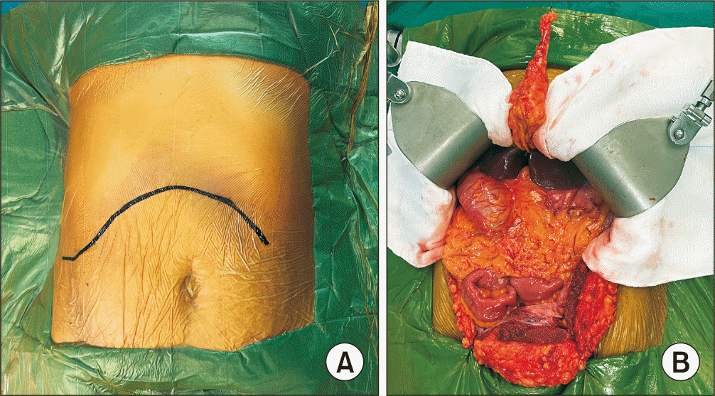
A rooftop/chevron incision is utilized to perform the laparotomy. Thorough intra-abdominal exploration is undertaken to rule out the presence of metastatic disease. The falciform ligament is transected, ensuring that a long proximal segment is preserved and tied securely to facilitate cranial retraction of the liver. The lower abdominal flap is anchored to the left iliac fossa using sutures to provide improved operative exposure. A Thompson retractor is then placed as shown in (Fig. 1).
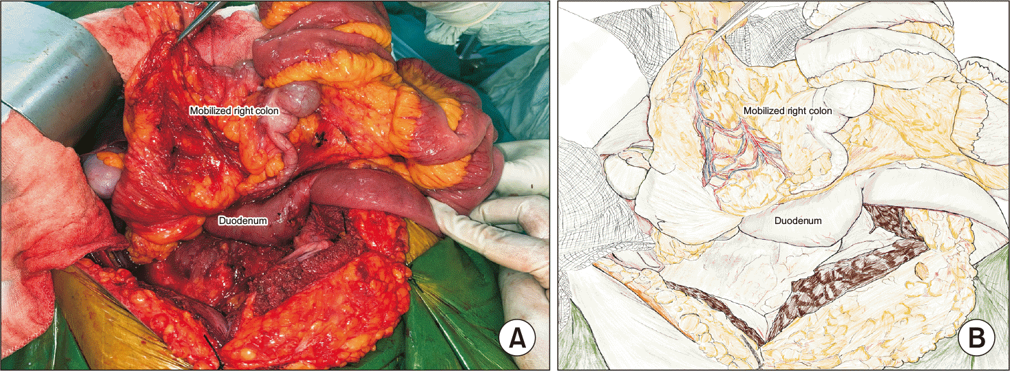
The right Toldt’s line is incised from the hepatic flexure to the cecum in a reverse J-shaped manner, followed by lateral-to-medial mobilization of the ascending colon and cecum. Dissection then proceeds medially along the root of the mesentery toward the ligament of Treitz, carefully preserving the plane within Toldt’s fascia [11]. The infracolic greater omentum is dissected from the transverse mesocolon, allowing entry into the right lesser sac through the fourth layer of the greater omentum. The dissection is carried out anterior to the supramesocolic pancreas and the second part of the duodenum. Next, the hepatocolic and phrenocolic ligaments are divided, and the inframesocolic duodenum and pancreas are separated from the colon by dividing Fredet’s fascia [11]. Special care must be taken to avoid injury to the venous branches of the loop of Henle. Ligation of the loop of Henle and/or right gastroepiploic vein and anterosuperior pancreaticoduodenal veins may be required in select situations. This sequence leads to complete cranial and lateral mobilization of the right colon and small bowel (Fig. 2).
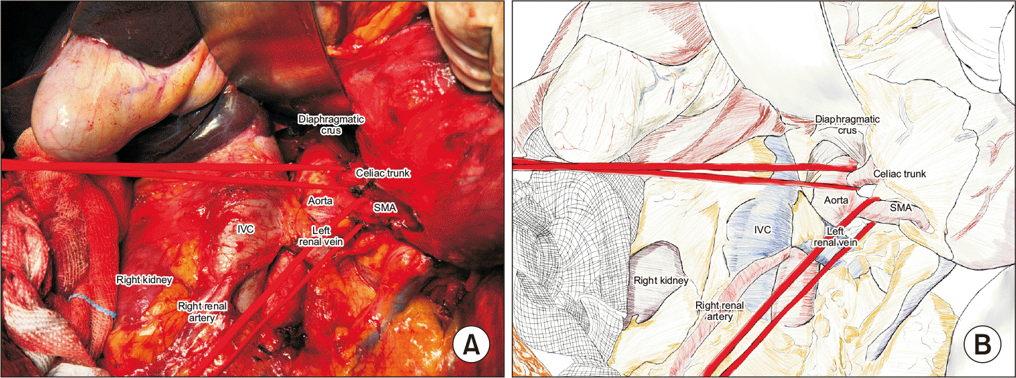
Kocherization is accomplished by incising lateral to the second part of the duodenum and dissecting within the Treitz fascia, thus mobilizing the duodenum and pancreatic head away from the inferior vena cava and aorta. The dissection is extended medially past the left border of the aorta. Para-aortic lymph node sampling (stations 16a2, 16b1) is performed and sent for frozen section analysis. If metastases are identified, resection may be halted based on tumor biology [13,14]. If no metastases are present, the procedure continues (Fig. 3).
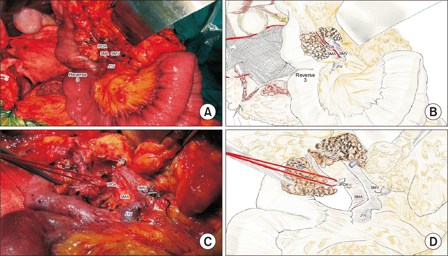
The ligament of Treitz is divided to mobilize the duodenojejunal flexure. Subsequently, the small bowel is rotated cranially and rightward, resulting in a 270-degree counter-clockwise derotation along the SMA/superior mesenteric vein (SMV) axis. This configuration, known as “reverse 3,” repositions the duodenojejunal flexure to the right of the SMA and moves the right colon to its left. As a result, the SMA-SMV relationship is altered, facilitating anatomic orientation and enhancing exposure of the mesopancreas for complete resection (Fig. 4).
A hybrid technique combining the posterior approach (as described by Pessaux et al. [15]) and the medial uncinate approach (as detailed by Hackert et al. [16]) is utilized. Dissection is initiated at the SMA root, progressing caudally along its right border. If a replaced right hepatic artery is present, it is carefully protected. The inferior pancreaticoduodenal artery (IPDA) is encountered and encircled during the dissection. The celiac trunk is exposed cranially up to the right diaphragmatic crus, clearly identifying both the SMA and celiac origins in a “V” configuration (Fig. 3).
A mesenteric window is developed between the first and second jejunal arteries. The first jejunal vein is followed to its convergence with the SMV to facilitate identification of the dorsally positioned SMA [17]. The inferior pancreaticoduodenal vein and IPDA are meticulously isolated but remain unligated at this stage. In the presence of root involvement, an inferior infracolic technique as described by Weitz et al. [18] may be employed.
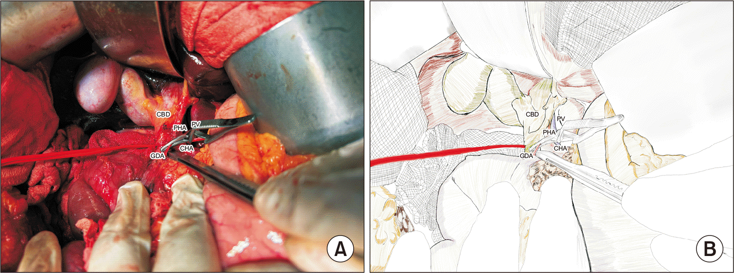
Dissection is initiated from the left to the right over the common hepatic artery (CHA). Station 8a lymph nodes are removed, and the right gastric artery may be ligated to facilitate station 5 lymphadenectomy. The gastroduodenal artery (GDA) is isolated and encircled with a loop (Fig. 5). Within the anatomical triangle defined by the CHA medially, the GDA laterally, and the superior border of the pancreas inferiorly, the portal vein is located posteriorly [19]. Nodes at stations 8p, 12b, 12a, and 12p are then dissected. The bile duct is precisely looped, which is especially important in the presence of an aberrant right hepatic artery. For technical convenience, the gallbladder is separated from its fossa. Subpyloric station 6 nodes are accessed by dissecting and ligating the right gastroepiploic vessels.
A bulldog clamp is positioned on the GDA, and hepatic artery flow is confirmed (Fig. 5). With the GDA clamped and the IPDA looped, an en bloc excision of the mesopancreas commences between the SMA and the celiac trunk. Dissection moves toward the pancreatic head and uncinate process. Based on the tumor characteristics, either a type 2 or 3 mesopancreas excision with plPh-II nerve plexus dissection is conducted [20,21]. Irreversible maneuvers are deferred until resectability is definitively established.
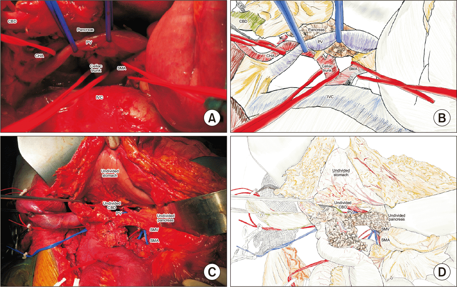
Circumferential clearance of the right aspect of the SMA is achieved, including removal of station 14t nodes. The GDA and IPDA are ligated if this has not been carried out earlier. Following neoadjuvant therapy, suspected SMA involvement is further assessed by periadventitial dissection and intraoperative frozen section [22]. If malignancy is identified, the surgical approach is tailored accordingly. The mesopancreas, uncinate process, and pancreatic head are then mobilized en bloc from the portal vein (Fig. 6). Venous invasion is managed with prior vascular control of the portal vein, SMV, and splenic vein.
The jejunum is divided using a linear stapler, while the stomach is resected proximal to the pylorus. Before transecting the pancreas at the neck or body, stay sutures are applied to the superior and inferior pancreatic borders. The order of transection—stomach → pancreas → bile duct (if stented), or stomach → bile duct → pancreas—depends on the presence of biliary stenting. For cases requiring complex venous resections, all relevant structures are transected before venous resection is attempted.
Following haemostasis and copious lavage, the small bowel and right colon are restored to their anatomical positions. The distal jejunal limb is delivered into the supracolic compartment via a mesenteric window to the right of the middle colic artery. Pancreaticojejunostomy is performed using either the Heidelberg or modified Blumgart technique, guided by the pancreatic duct diameter and gland consistency [23,24]. Hepaticojejunostomy is performed with interrupted 4-0 or 5-0 polydioxanone sutures. An antecolic gastrojejunostomy is constructed, and a naso jejunal tube is placed distally. A Jackson-Pratt drain is inserted posterior to the pancreaticojejunostomy, with a second drain positioned in the pelvis.
The mean operative time for this procedure is 450 ± 40 minutes. Detailed key steps and critical aspects of the procedure are presented in Table 1. Furthermore, Table 2 and 3 provide a review and comparison of various types of pancreaticoduodenectomies described in the literature with our technique. Supplementary Video 1 includes a demonstration of the principal operative steps.
PD remains the only definitive and potentially curative intervention for patients with periampullary and pancreatic head carcinomas. Although only 20% of these malignancies are initially resectable, nearly 30% to 40% of borderline resectable and locally advanced tumors can become amenable to surgical resection following neoadjuvant therapy [25-27]. As surgical techniques have become increasingly advanced, accurate assessment of both resectability and the radical nature of the procedure has become essential for predicting prognostic outcomes. While a pancreatic protocol CECT is highly valuable for predicting resectability and planning for potential vascular resections, we contend that its predictive value may diminish after neoadjuvant treatment, making definitive assessment of resectability dependent upon intraoperative findings.
Our approach begins with the Cattell Braasch Veldoni maneuver [9,10]. This technique, combined with the Kocher’s maneuver, substantially clarifies the anatomy of the principal vessels and their relationships with the mesopancreas and uncinate process [28,29]. To achieve early vascular control, we utilize both the ‘posterior SMA-first approach’ initially described by Pessaux et al. [15] and the ‘right medial uncinate approach’ introduced by Hackert et al. [16]. This strategy facilitates preservation of the aberrant right hepatic artery—encountered in 10% to 21% of patients—and enables early control of the IPDA, thereby reducing venous congestion and minimizing intraoperative blood loss from the pancreatic head and uncinate process [30,31]. Additional benefits of the SMA-first approach, which include improved R0 resection rates, reduced postoperative complications, and enhanced overall survival, as recently reported in meta-analyses, are also achieved by our method [32,33].
Another crucial element of ‘radicality’ in our approach involves the complete excision of the mesopancreas. The mesopancreas was initially described by Gockel et al. [34] and is composed of fibrofatty tissue containing neurovascular bundles (perineural plexus of pancreatic head; plPh-I and plPh-II), lymph nodes, IPDA, and the first jejunal artery and vein [20,35]. Undertaking TMpE leads to a marked improvement in R0 resection rates and lymph node retrieval while reducing locoregional recurrence, all with acceptable complication rates compared to the standard PD [36]. Within our method, the ‘reverse 3’ arrangement of D3 and DJ flexure enables clearer anatomical orientation and facilitates an en bloc type 2 or type 3 mesopancreas excision (type 2: mesopancreas divided along the right side of the SMA without nerve plexus; type 3: mesopancreas division includes periadventitial dissection to the right of SMA incorporating the nerve plexus) depending on the underlying pathology, and achieves complete clearance of tissue between the SMA and CHA over the right 180 degrees [20,21]. The dissection level, either periadventitial or sub adventitial, is determined by the specific disease context [22].
Additional benefits of our approach include minimized bile contamination and reduced intraoperative blood loss. Bile duct division is performed at the end of the operation, which significantly reduces the risk of contamination, particularly important for stented patients who are more prone to surgical site infections [37]. This also shortens the duration of bulldog clamp application on the common bile duct, potentially lowering rates of ischemic injury and subsequent strictures. Delayed pancreatic transection permits the attainment of broader surgical margins beyond the pancreatic neck and further decreases blood loss by enabling greater control over the vasculature.
The combination of the SMA-first approach with TMpE has now been adopted at several institutions and has demonstrated improved oncological results [38,39]. We implement this technique alongside the principle of maintaining the intactness of all critical structures until the end of the operation. As per our review, this precise combination is not previously reported in the literature (Table 2, 3). Our approach aligns with the strategic principle of the ‘path of escape’ as articulated by Sun Tzu, the Chinese military strategist and philosopher: “Always leave an escape route and make it obvious, hence the ‘golden bridge’ to retreat across.” This philosophy highlights the value of maintaining an option for tactical withdrawal to prevent disastrous confrontations.
This innovative PD technique is both safe and oncologically robust, achieving adequate radicality while preserving the opportunity for retreat when R0 resection cannot be attained. We plan to report our short-term and long-term outcomes using this technique in subsequent publications. Additional comparative and multicenter research is needed to confirm the efficacy and broader relevance of this PD method.
Supplementary data related to this article can be found at https://doi.org/10.14701/ahbps.25-097.
References
1. Are C, Dhir M, Ravipati L. History of pancreaticoduodenectomy: early misconceptions, initial milestones and the pioneers. HPB (Oxford) 2011;13:377-384.
10.1111/j.1477-2574.2011.00305.x
21609369
PMC3103093
2. Tummala P, Howard T, Agarwal B. Dramatic survival benefit related to R0 resection of pancreatic adenocarcinoma in patients with tumor ≤25 mm in size and ≤1 involved lymph nodes. Clin Transl Gastroenterol 2013;4:e33.
10.1038/ctg.2013.4
23515131
PMC3615697
3. Menon KV, Gomez D, Smith AM, Anthoney A, Verbeke CS. Impact of margin status on survival following pancreatoduodenectomy for cancer: the Leeds Pathology Protocol (LEEPP). HPB (Oxford) 2009;11:18-24.
10.1111/j.1477-2574.2008.00013.x
19590619
PMC2697870
4. Kulkarni NM, Soloff EV, Tolat PP, Sangster GP, Fleming JB, Brook OR, et al. White paper on pancreatic ductal adenocarcinoma from society of abdominal radiology's disease-focused panel for pancreatic ductal adenocarcinoma: Part I, AJCC staging system, NCCN guidelines, and borderline resectable disease. Abdom Radiol (NY) 2020;45:716-728.
10.1007/s00261-019-02289-5
31748823
5. Somers I, Bipat S. Contrast-enhanced CT in determining resectability in patients with pancreatic carcinoma: a meta-analysis of the positive predictive values of CT. Eur Radiol 2017;27:3408-3435.
10.1007/s00330-016-4708-5
28093626
PMC5491588
6. Allen VB, Gurusamy KS, Takwoingi Y, Kalia A, Davidson BR. Diagnostic accuracy of laparoscopy following computed tomography (CT) scanning for assessing the resectability with curative intent in pancreatic and periampullary cancer. Cochrane Database Syst Rev 2016;7(7):CD009323.
10.1002/14651858.CD009323.pub3
27383694
7. Park S, Jang JK, Byun JH, Kim JH, Lee SS, Kim HJ, et al. CT in the prediction of margin-negative resection in pancreatic cancer following neoadjuvant treatment: a systematic review and meta-analysis. Eur Radiol 2021;31:3383-3393.
10.1007/s00330-020-07433-0
33123793
8. Cassinotto C, Cortade J, Belleannée G, Lapuyade B, Terrebonne E, Vendrely V, et al. An evaluation of the accuracy of CT when determining resectability of pancreatic head adenocarcinoma after neoadjuvant treatment. Eur J Radiol 2013;82:589-593.
10.1016/j.ejrad.2012.12.002
23287712
9. Cattell RB, Braasch JW. A technique for the exposure of the third and fourth portions of the duodenum. Surg Gynecol Obstet 1960;111:378-379.
10. Valdoni P. Derotazione intestinale. In: Valdoni P, ed. Chirurgia addominale: tecniche operatorie. Vallardi; 1974;143-151. Italian.
11. Brachini G, Cirillo B, Matteucci M, Cirocchi R, Tebala GD, Cavaliere D, et al. A systematic review of varying definitions and the clinical significance of fredet's fascia in the era of complete mesocolic excision. J Clin Med 2023;12:6233.
10.3390/jcm12196233
37834876
PMC10573991
12. Livani A, Angelis S, Skandalakis PN, Filippou D. The story retold: the Kocher manoeuvre. Cureus 2022;14:e29409.
10.7759/cureus.29409
36304342
PMC9586190
13. Schwarz L, Lupinacci RM, Svrcek M, Lesurtel M, Bubenheim M, Vuarnesson H, et al. Para-aortic lymph node sampling in pancreatic head adenocarcinoma. Br J Surg 2014;101:530-538.
10.1002/bjs.9444
24633831
14. Sillesen M, Hansen CP, Burgdorf SK, Dencker EE, Krohn PS, Gisela Kollbeck SL, et al. Impact of para aortic lymph node removal on survival following resection for pancreatic adenocarcinoma. BMC Surg 2023;23:214.
10.1186/s12893-023-02123-2
37528360
PMC10394933
15. Pessaux P, Varma D, Arnaud J-P. Pancreaticoduodenectomy: superior mesenteric artery first approach. J Gastrointest Surg 2006;10:607-611.
10.1016/j.gassur.2005.05.001
16627229
16. Hackert T, Werner J, Weitz J, Schmidt J, Büchler MW. Uncinate process first: a novel approach for pancreatic head resection. Langenbecks Arch Surg 2010;395:1161-1164.
10.1007/s00423-010-0663-9
20582600
17. Nagakawa Y, Hosokawa Y, Sahara Y, Takishita C, Hijikata Y, Osakabe H, et al. Approaching the superior mesenteric artery from the right side using the proximal-dorsal jejunal vein preisolation method during laparoscopic pancreaticoduodenectomy. Surg Endosc 2018;32:4044-4051.
10.1007/s00464-018-6118-z
29484553
18. Weitz J, Rahbari N, Koch M, Büchler MW. The "artery first" approach for resection of pancreatic head cancer. J Am Coll Surg 2010;210:e1-e4.
10.1016/j.jamcollsurg.2009.10.019
20113929
19. Kalayarasan R, Gautham MS, Gnanasekaran S, Pottakkat B. The technique of precise and systematic vascular control during robotic pancreaticoduodenectomy for periampullary and pancreatic tumours. J Minim Access Surg 2021;17:399-404.
10.4103/jmas.JMAS_239_20
33885010
PMC8270053
20. Inoue Y, Saiura A, Oba A, Kawakatsu S, Ono Y, Sato T, et al. Optimal extent of superior mesenteric artery dissection during pancreaticoduodenectomy for pancreatic cancer: Balancing surgical and oncological safety. J Gastrointest Surg 2019;23:1373-1383.
10.1007/s11605-018-3995-3
30306451
21. Shyr BS, Wang SE, Chen SC, Shyr YM, Shyr BU. Mesopancreas dissection level 3 for pancreatic head cancer in combined robotic/open pancreatoduodenectomy: a propensity score-matched study. Surg Endosc 2025;39:1191-1199.
10.1007/s00464-024-11475-6
39739104
PMC11794409
22. Miao Y, Cai B, Lu Z. Technical options in surgery for artery-involving pancreatic cancer: Invasion depth matters. Surg Open Sci 2023;12:55-61.
10.1016/j.sopen.2023.03.001
36936450
PMC10020102
23. Büchler MW, Friess H, Wagner M, Kulli C, Wagener V, Z'Graggen K. Pancreatic fistula after pancreatic head resection. Br J Surg 2000;87:883-889.
10.1046/j.1365-2168.2000.01465.x
10931023
24. Fujii T, Sugimoto H, Yamada S, Kanda M, Suenaga M, Takami H, et al. Modified Blumgart anastomosis for pancreaticojejunostomy: technical improvement in matched historical control study. J Gastrointest Surg 2014;18:1108-1115.
10.1007/s11605-014-2523-3
24733259
25. Rompen IF, Habib JR, Wolfgang CL, Javed AA. Anatomical and biological considerations to determine resectability in pancreatic cancer. Cancers (Basel) 2024;16:489.
10.3390/cancers16030489
38339242
PMC10854859
26. Roselló S, Pizzo C, Huerta M, Muñoz E, Aliaga R, Vera A, et al. Neoadjuvant treatment for locally advanced unresectable and borderline resectable pancreatic cancer: oncological outcomes at a single academic centre. ESMO Open 2020;5:e000929.
10.1136/esmoopen-2020-000929
33229503
PMC7684818
27. Aljubran AH, Alata MK, Badran AA, Alshamsan B, Alnemary Y, Bazarbashi S, et al. Resectability rate of pancreatic cancer after neoadjuvant therapy. J Clin Oncol 2024;42(16_suppl):e16348-e16348.
10.1200/JCO.2024.42.16_suppl.e16348
28. Del Chiaro M, Segersvärd R, Rangelova E, Coppola A, Scandavini CM, Ansorge C, et al. Cattell-braasch maneuver combined with artery-first approach for superior mesenteric-portal vein resection during pancreatectomy. J Gastrointest Surg 2015;19:2264-2268.
10.1007/s11605-015-2958-1
26423804
29. Virgilio E, Amodio PM, Scorsi A, Goglia A, Macarone Palmieri R. Advantages of the maneuver of intestinal derotation for pancreaticoduodenectomy. J Invest Surg 2016;29:359-365.
10.3109/08941939.2016.1160166
27096254
30. Yang SH, Yin YH, Jang JY, Lee SE, Chung JW, Suh KS, et al. Assessment of hepatic arterial anatomy in keeping with preservation of the vasculature while performing pancreatoduodenectomy: an opinion. World J Surg 2007;31:2384-2391.
10.1007/s00268-007-9246-5
17922256
31. Sanjay P, Takaori K, Govil S, Shrikhande SV, Windsor JA. 'Artery-first' approaches to pancreatoduodenectomy. Br J Surg 2012;99:1027-1035.
10.1002/bjs.8763
22569924
32. Ironside N, Barreto SG, Loveday B, Shrikhande SV, Windsor JA, Pandanaboyana S. Meta-analysis of an artery-first approach versus standard pancreatoduodenectomy on perioperative outcomes and survival. Br J Surg 2018;105:628-636.
10.1002/bjs.10832
29652079
33. Jiang X, Yu Z, Ma Z, Deng H, Ren W, Shi W, et al. Superior mesenteric artery first approach can improve the clinical outcomes of pancreaticoduodenectomy: a meta-analysis. Int J Surg 2020;73:14-24.
10.1016/j.ijsu.2019.11.007
31751791
34. Gockel I, Domeyer M, Wolloscheck T, Konerding MA, Junginger T. Resection of the mesopancreas (RMP): a new surgical classification of a known anatomical space. World J Surg Oncol 2007;5:44.
10.1186/1477-7819-5-44
17459163
PMC1865381
35. Adham M, Singhirunnusorn J. Surgical technique and results of total mesopancreas excision (TMpE) in pancreatic tumors. Eur J Surg Oncol 2012;38:340-345.
10.1016/j.ejso.2011.12.015
22264964
36. da Silva LFL, Belotto M, de Almeida LFC, Samuel J, Pereira LH, Albagli RO, et al. Radicality and safety of total mesopancreatic excision in pancreatoduodenectomy: a systematic review and meta-analysis. World J Surg Oncol 2024;22:217.
10.1186/s12957-024-03495-2
39180093
PMC11342630
37. Sasmal PK, Das Poddar KK, Mishra TS, Kumar P. Surgical site infections in patients of periampullary carcinoma undergoing delayed bile duct division (COMBILAST) in Whipple's procedure: a prospective cohort study. Pathogens 2023;12:448.
10.3390/pathogens12030448
36986370
PMC10058893
38. Nguyen TK, Nguyen HH, Luong TH, Dang KK, Le VD, Tran DD, et al. Pancreaticoduodenectomy with superior mesenteric artery first-approach combined total meso-pancreas excision for periampullary malignancies: a high-volume single-center experience with short-term outcomes. Ann Hepatobiliary Pancreat Surg 2024;28:59-69.
10.14701/ahbps.23-068
38049111
PMC10896681
39. Vasiliadis KD. "Mesopancreas-first" radical resection of pancreatic head cancer following the Cattell-Braasch-Valdoni maneuver: appreciating the legacy of pioneers in visceral surgery. Ann Hepatobiliary Pancreat Surg 2021;25:376-385.
10.14701/ahbps.2021.25.3.376
34402439
PMC8382854
Fig. 1
Incision and exposure for the procedure. (A) Chevron incision. (B) Application of Thompson retractor and placement of full thickness lower abdominal wall sutures to ensure optimal exposure of the abdominal cavity.
Fig. 2
Complete Cattell Braasch Valdoni manoeuvre; the right colon, caecum, and terminal ileum are fully mobilized and rotated from the right lower to the left upper quadrant. The incision line extends from the right side of the hepatic flexure to below the caecum and continues medially and cranially towards the ligament of Treitz. (A) Intraoperative photograph. (B) Schematic diagram corresponding to the photograph.
Fig. 3
Kocher manoeuvre; after dissection of the Treitz fascia, the duodenum and the pancreatic head are elevated. Lymph nodes 16a2 and 16b1 are excised. The celiac trunk and SMA are identified at their origins and encircled. (A) Intraoperative photograph. (B) Schematic representation of the corresponding photograph. IVC, inferior vena cava; SMA, superior mesenteric artery.
Fig. 4
Division of the ligament of Treitz and full 270-degree derotation of the duodenum and small intestine along the superior mesenteric vessels. The ‘Reverse 3’ configuration is achieved, with the duodenojejunal flexure and small bowel repositioned to the right of the SMA and the right hemicolon to the left of the SMA. (A) Actual photograph showing ‘Reverse 3’ configuration. (B) Schematic representation of the ‘Reverse 3’ configuration. (C) IPDA looped and preserved, employing the medial uncinate approach. (D) Schematic diagram of (C). IPDA, inferior pancreaticoduodenal artery; SMA, superior mesenteric artery; SMV, superior mesenteric vein; J1V, first jejunal vein.
Fig. 5
Dissection of the hepatoduodenal ligament to address station 8 and 12 lymph nodes. The GDA is encircled and preserved. A Bulldog clamp is temporarily applied to assess aberrant hepatic arterial supply and to minimize blood loss during excision of the mesopancreas. (A) Intraoperative photograph. (B) Schematic diagram of (B). GDA, gastroduodenal artery; PHA, proper hepatic artery; CHA, common hepatic artery; PV, portal vein; CBD, common bile duct.
Fig. 6
(A, B) Intraoperative photograph and corresponding schematic illustration, respectively, showing medial mobilization of the head of pancreas and mesopancreas from the PV, achieving clearance of the mesopancreas triangle. (C, D) Intraoperative photograph and schematic illustration, respectively, demonstrating the complete non-division technique, preserving the pancreatic and duodenal blood supply, pancreas, stomach, CBD, and jejunum. PV, portal vein; IVC, inferior vena cava; SMA, superior mesenteric artery; CHA, common hepatic artery; CBD, common bile duct.
Table 1
Detailed surgical steps and clinical implications of the “total non-division technique” in pancreaticoduodenectomy
SMA, superior mesenteric artery; SMV, superior mesenteric vein; IVC, inferior vena cava; DJ, duodenojejunal; IPDA, inferior pancreaticoduodenal artery; RHA, right hepatic artery; CHA, common hepatic artery; GDA, gastroduodenal artery; CA, celiac artery; TMpE, total mesopancreas excision; PJ, pancreaticojejunostomy; HJ, hepaticojejunostomy; GJ, gastrojejunostomy; NJ, nasojejunal.
Table 2
Comparative analysis of surgical approaches for pancreaticoduodenectomy (Whipple’s Procedure)
| Study/technique | SMA approach | Mesopancreas handling | Bile duct/pancreas transection | Extent of dissection | Unique contribution | Difference from present study |
|---|---|---|---|---|---|---|
| Pessaux et al. [15], 2006 (posterior SMA-first) | Posterior | Partial mesopancreas clearance | Performed after SMA assessment but before uncinate dissection | Focused on SMA root | Early arterial control; decreased margin positivity | Lacked mesopancreas en bloc strategy; division of structures occurred earlier |
| Hackert et al. [16], 2010 (right medial uncinate first) | Medial-right approach to SMA | Partial; done during uncinate dissection | Pancreas and bile duct divided before full clearance | Emphasis on uncinate dissection safety | Protection of replaced RHA | No structured non-division policy; SMA looped late |
| Weitz et al. [18], 2010 (infracolic SMA-first) | Infracolic + upward | SMA exposed early | Pancreas divided after artery dissection | Systematic early arterial dissection | Avoids unnecessary dissection if tumor is unresectable | No reverse 3 configuration; mesopancreas not excised in totality |
| Gockel et al. [34], 2007 (mesopancreas concept introduction) | Not specified | Anatomical definition of mesopancreas | No guidance on sequence | Descriptive anatomical framework | Defined “mesopancreas” as key oncological zone | Did not integrate dissection strategy with SMA-first approach |
| Adham et al. [35], 2012 (TMpE) | Variable | TMpE formalized | Division done after TMpE | Radical resection strategy for oncologic margin | Introduced type 1–3 TMpE levels | Did not incorporate non-division or strategic retreat option |
| Shyr et al. [21], 2025 (level 3 TMpE robotic/open) | Dual: robotic SMA-first + mesopancreas | TMpE level 3 (extensive) | Transection done after perivascular clearance | Maximal plPh-II and SMA clearance | High-volume robotic experience | Our technique formalizes non-division + SMA-first + TMpE in open approach |
| Nguyen et al. [38], 2024 (SMA-first + TMpE) | SMA-first | TMpE included | Division occurs after vascular control | Combined open SMA-first + radical excision | Improved short-term outcomes | Similar principles but lacks codified non-division timing |
| Current study: “total non-division technique” | Posterior + right medial ± infracolic SMA-first | Formalized TMpE (type 2/3) with plPh-II clearance | Explicitly delayed until resectability is confirmed (non-division strategy) | Clearance between SMA and CA; venous preparation | Codified avoidance of any irreversible steps until oncologic viability is proven; “path of escape” | Synthesizes and sequences known strategies into a structured, fail-safe approach not previously defined |
Table 3
Major distinctions between the total non-division technique pancreaticoduodenectomy (TNT-PD) and conventional pancreaticoduodenectomy


PDF
Citation
Print


XML Download