Journal List > Ann Hepatobiliary Pancreat Surg > v.29(4) > 1516093404

Barros, Murillo, Vozmediano, Vásquez, and Marchán: Transcystic management of choledocholithiasis: Outcomes, factors associated with bile duct injury and implications for surgical practice

Abstract

Backgrounds/Aims

Choledocholithiasis with gallbladder in situ presents a complex surgical challenge. The transcystic approach offers a minimally invasive alternative to choledochotomy; however, its adoption remains limited. This study assessed the safety, efficacy, and reproducibility of this method.

Methods

This retrospective study involved 71 patients diagnosed with choledocholithiasis and gallbladder in situ, all of whom underwent transcystic bile duct exploration at a primary care hospital. Demographic, clinical, and intraoperative variables were analyzed to determine predictors of bile duct injury.

Results

Transcystic exploration achieved successful completion in 92.9% of cases and a duct clearance rate of 94%. The most common complication was bile duct injury, showing a significant association with previous endoscopic or percutaneous procedures (p = 0.031), increased preoperative leukocyte count (p = 0.050), and advanced age (median 72.7 vs. 60.4 years; p = 0.031). Conversion to choledochotomy elevated the risk of injury, and incomplete duct clearance correlated with higher complication rates. No specific intraoperative techniques or devices exhibited a significant impact on outcomes. Imaging at six months demonstrated no persistent strictures, supporting the likelihood of transient inflammatory changes.

Conclusions

The transcystic approach is a safe, effective, and reproducible first-line intervention for choledocholithiasis with gallbladder in situ. Patient-specific and disease-related factors primarily determine bile duct injury risk, rather than the surgical technique itself. Further prospective randomized studies are needed to confirm these findings.

INTRODUCTION

Cholelithiasis is a prevalent digestive disorder, accounting for up to one-third of general surgery procedures [1]. Approximately 5%–10% of these patients develop choledocholithiasis, although the true incidence may be underestimated. The disease is dynamic—gallstones may spontaneously migrate into the intestine—so imaging provides only a snapshot. It is not uncommon to observe disappearance of previously identified stones or the appearance of new stones during surgery [1].
Improvements in minimally invasive surgery and diagnostic modalities have contributed to earlier and often incidental detection. In conjunction with contemporary guidelines advocating treatment of all confirmed cases, this has notably shortened the disease’s natural history [1].
Endoscopic retrograde cholangiopancreatography (ERCP) has conventionally been the treatment of choice, either prior to or following cholecystectomy (two-stage approach). Laparoscopic bile duct exploration (LBDE), however, provides a one-stage alternative and has potential to alter standard practice [2,3]. Evidence indicates that stone clearance, morbidity, and mortality are comparable for ERCP and LBDE. ERCP is more frequently associated with pancreatitis, whereas bile leakage is the principal risk with LBDE [4-12].
There are two types of LBDE: choledochotomy and transcystic. The transcystic approach, employing 2.8–3.5 mm choledochoscopes and utilized in approximately 30% of reported cases, is associated with improved outcomes—including reduced incidence of bile leaks, shorter operative times, decreased length of hospital stay, and lower overall costs [12-18]. Therefore, it is essential that studies distinguish between these techniques; failure to do so may mask the specific benefits offered by minimally invasive approaches [17].

Study objectives

• To establish transcystic LBDE as the first-line treatment for choledocholithiasis in patients with an in situ gallbladder.
• To define criteria for appropriate patient selection for this technique.
• To determine risk factors associated with bile duct injury in individuals undergoing this procedure.

MATERIALS AND METHODS

Study design and sample

This prospective observational cohort study included 71 patients who underwent elective laparoscopic cholecystectomy with transcystic LBDE at our institution between June 2020 and June 2024. Diagnosis of choledocholithiasis was established through ultrasound, computerized tomography, magnetic resonance cholangiopancreatography (MRCP), or prior ERCP. All patients were followed for a minimum of 6 months, with clinical and laboratory assessments performed at 1 month and imaging (predominantly MRCP) at 6 months.
Two groups were compared in the analysis:
• Group A: Patients who developed bile duct injury
• Group B: Patients who did not experience bile duct injury

Definition of bile duct injury

Bile duct injury was defined as the occurrence of either postoperative bile leak or biliary stricture. Biliary stricture was diagnosed based on follow-up imaging, commonly using MRCP [19]. The diagnosis of bile leak was established if drain bilirubin measured at least 3x the serum level, or when additional intervention, such as drainage, reoperation, or procedures for biliary peritonitis, was required [20]. The severity of bile leaks was graded according to the International Study Group of Liver Surgery (ISGLS) criteria [21]:
• Type A: No change in management and no associated infection.
• Type B: Requires active therapeutic measures not involving laparotomy or laparoscopy, and is associated with mild to moderate infection.
• Type C: Necessitates surgical intervention and is accompanied by severe infection.

Exclusion criteria

The exclusion criteria included patients with neoplasms of the biliary tract and those initially managed with non-transcystic approaches (such as choledochotomy or transinfundibular techniques).

Data and ethics

The study adhered to STROBE guidelines and received approval from our hospital’s Ethics Committee (EO260-23_HIE), in accordance with the principles outlined in the Declaration of Helsinki.

Surgical technique

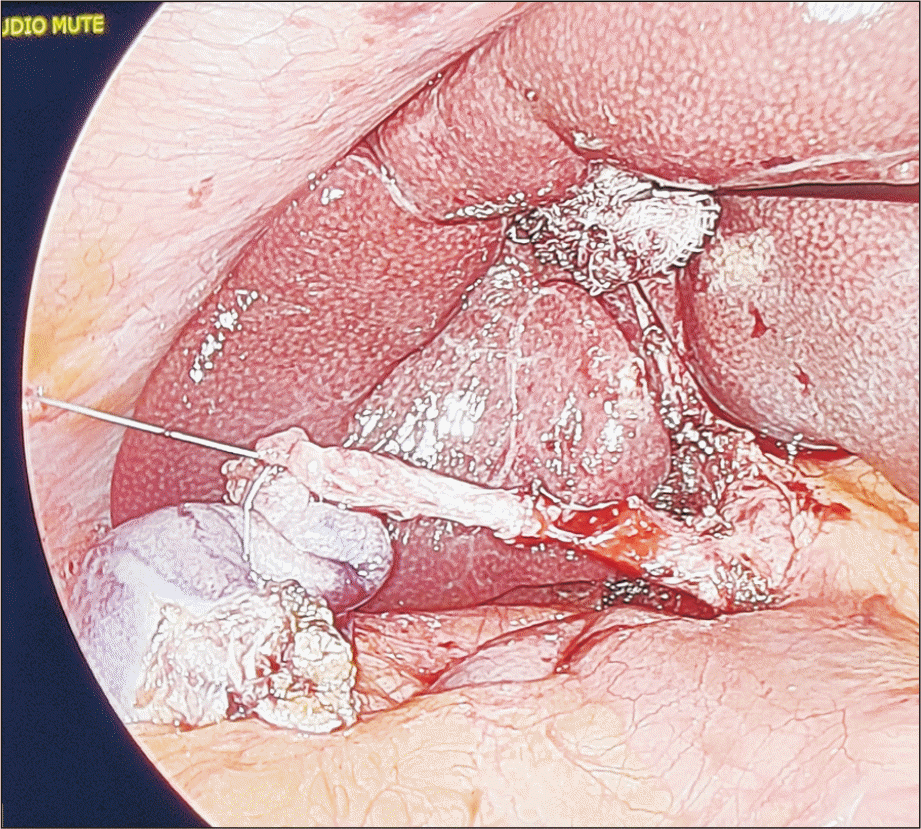
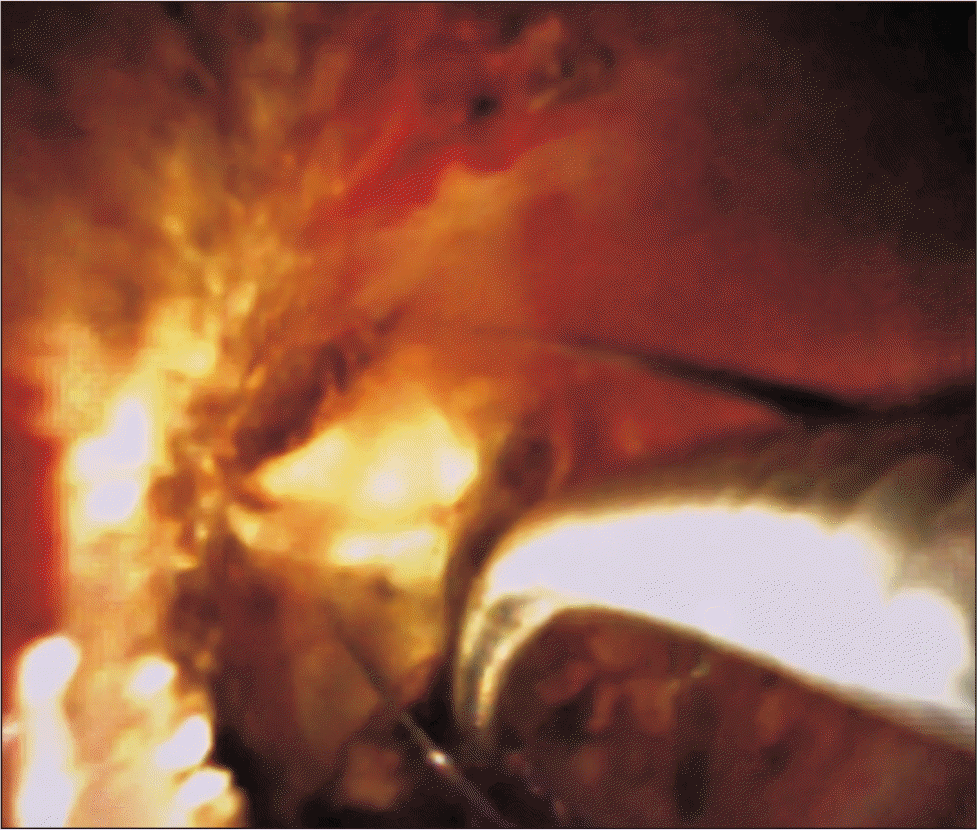
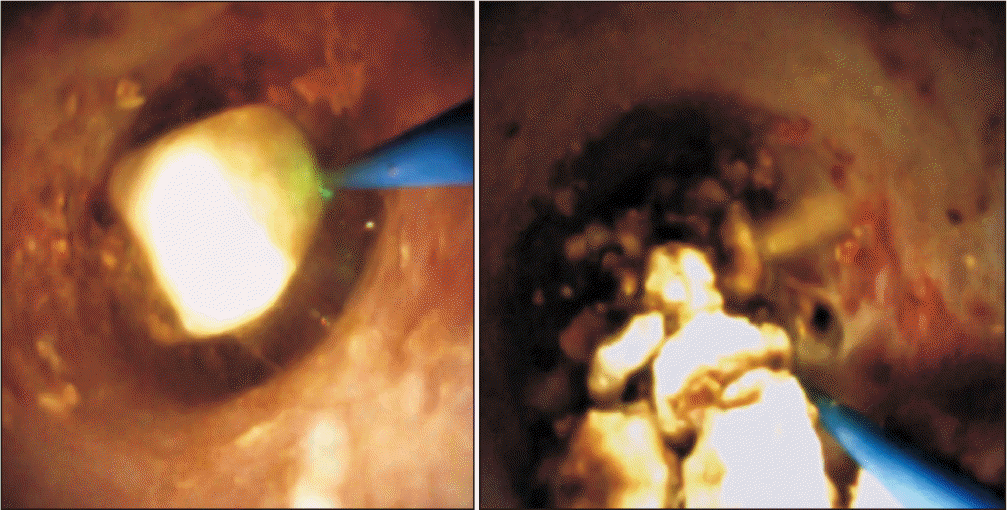
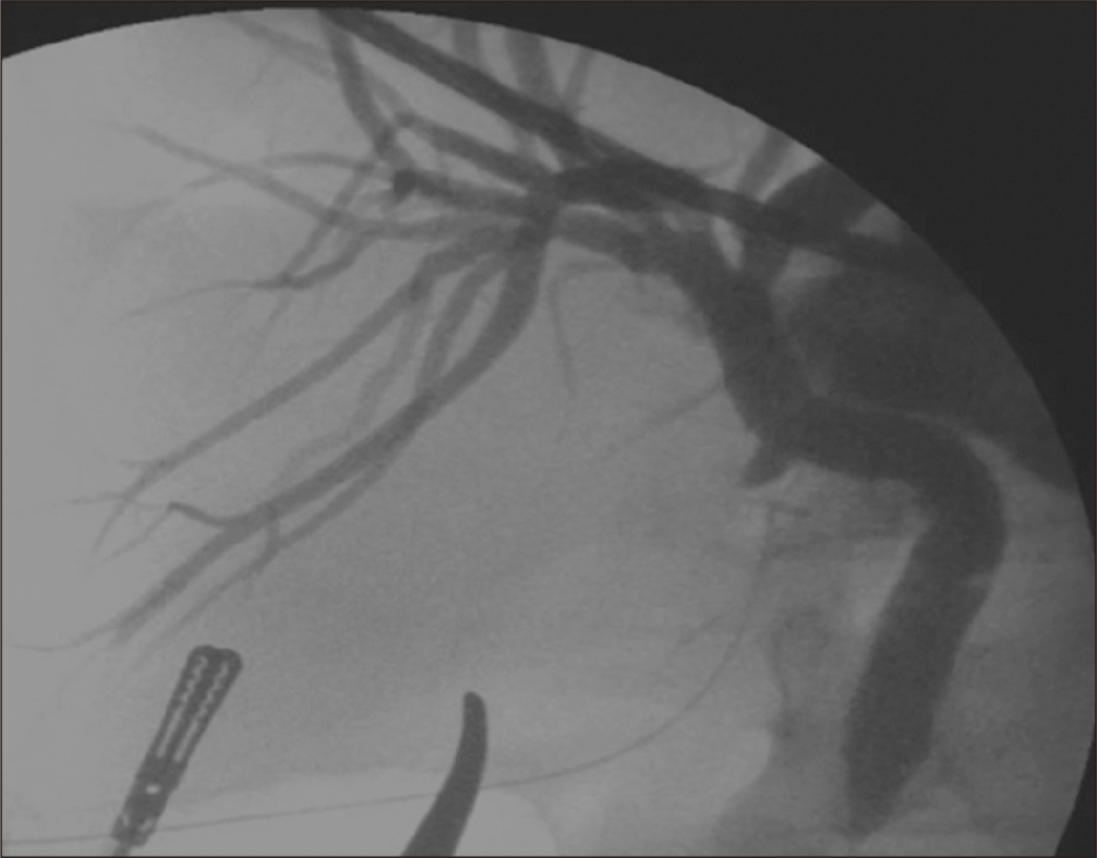
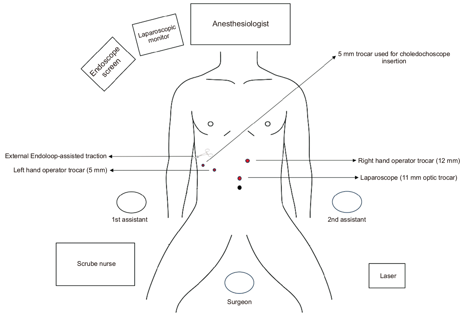
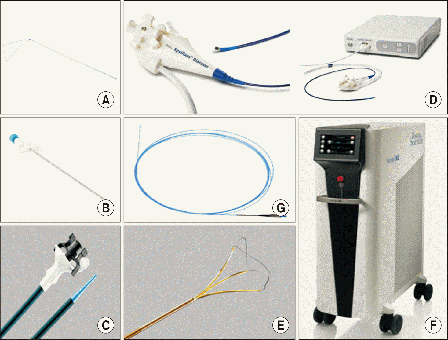
Fig. 1 and 2 depict the arrangement of the patient, surgical team, and instruments during the procedure. The operation utilized a laparoscopic approach with four trocars. Initially, an anterograde cholecystectomy was performed, after which the cystic–common bile duct angle was carefully dissected, aiming to limit the use to three trocars during this phase. Following this, an Endoloop (Ethicon, Johnson & Johnson) (Fig. 2A) was attached at the junction between the infundibulum and cystic duct, then brought out through the right hypochondrium using the assistance of an EndoClose (Covidien, Medtronic) (Fig. 2B). This enabled application of traction on the gallbladder to achieve a 90° angle between the cystic duct and common bile duct, as illustrated in Fig. 3. Next, a partial transverse incision was made on the anterior wall of the cystic duct 1–2 centimetres from the hepatic duct. At this stage, the fourth trocar was inserted, intended for introducing the choledochoscope, which should be advanced along the same traction line created by the Endoloop on the cystic duct. In selected cases, a Flexor Parallel access system (Cook Medical) (Fig. 2C) functioned as the introducer. The choledochoscopy was generally carried out using the SpyGlass Discover system (Boston Scientific Corporation) (Fig. 2D). If stones were visualized, extraction was attempted with the NGage device (Cook Medical) (Fig. 2E, 4). For stones that were large or impacted, lithotripsy was performed with the Auriga XL laser system (Boston Scientific Corporation) and a 270-micron Holmium:YAG fiber (Cook Medical) (Fig. 2F, 2G, 5). When appropriate, crushing maneuvers were employed. Management of microlithiasis or sludge involved flushing with pressurized saline. Intraoperative cholangiography (Fig. 6) was selectively performed when bile duct clearance remained uncertain, especially if any portion of the exploration—either distal or proximal to the cystic duct—could not be fully accomplished. Correlation between imaging findings and number of stones removed was not emphasized, as the pathological condition is dynamic. The cystic duct stump was closed with double non-absorbable clips. In cases where transcystic access was unsuccessful, an infundibular approach was considered. Choledochotomy was reserved for situations in which distal exploration or stone extraction via the cystic duct could not be achieved.

Study variables

A comprehensive range of variables was examined to determine possible risk factors for bile duct injury.

Epidemiological and preoperative variables

We collected data on sex, age, American Society of Anestheologist (ASA) classification [21], and Charlson Comorbidity Index, which estimates 1-year mortality risk from comorbidities and age [22]. Nutritional status was assessed using serum albumin, in addition to evaluating diabetes, international normalized ratio (INR), and imaging characteristics such as bile duct diameter and number/size of stones. Preoperative interventions—including ERCP, percutaneous transhepatic cholangiography (THPC), or radiological drainage (e.g., cholecystostomy)—were documented. Laboratory results from within 48 hours before surgery included amylase, bilirubin, aspartate aminotransferase (AST), alamine aminotransferase (ALT), gammaglutamyl transferase (GGT), alkaline phosphatase (ALP), white blood cell (WBC) count, and hemoglobin.

Intraoperative variables

We examined potential effects of cholecystitis, cystic/common bile duct diameter, and characteristics of stones, as well as the surgical approach utilized. Additional intraoperative aspects included the type of introducer, instruments, cranial exploration, flushing or crushing techniques, details on lithotripsy use (including frequency and energy settings), duodenoscopy, duration of surgery, and intra-abdominal drainage placement.

Postoperative variables

Complications were classified according to the Clavien-Dindo system [23], including bleeding, pancreatitis, and cholangitis. We documented admissions to the intensive care unit (ICU), need for ERCP or THPC, and detected residual stones or sludge. Duration of hospitalization and early postoperative laboratory values (amylase, bilirubin, AST, ALT, GGT, ALP, WBC, hemoglobin) were also analyzed.

Post-discharge follow-up

We recorded data on hospital readmissions, additional procedures, mortality, and follow-up laboratory values (at 1 and 6 months). Associations with bile duct injury were analyzed. MRCP findings at 6 months were included when obtained.

Statistical analysis

Categorical variables, including sex and readmission, were summarized using frequencies and percentages, while quantitative variables, such as age, stone size, and days until reintervention, were reported as median and interquartile range. The assumption of normality for numeric variables was evaluated using the Anderson-Darling test. Associations between biliary injury and clinical or sociodemographic variables were examined with the chi-squared test and Fisher’s exact test when appropriate. Comparisons of quantitative variables between groups with and without biliary injury were conducted using the Mann-Whitney U test. Cramer’s V was calculated to assess strength of association (classified as small if <0.2, moderate if 0.2–0.6, and large if >0.6, per Cramer, 1946), and Cohen’s d was used for effect size (small if <0.5, moderate if 0.5–0.8, and large if >0.8, in accordance with Cohen, 1988). For variables demonstrating statistical significance in the preceding analyses, a univariate logistic regression model was applied to estimate the risk associated with each in relation to biliary injury. A type I error of 0.05 was used for all tests. Statistical analyses were performed using R software (version 4.4.1).

RESULTS

Bile sludge (n = 5) or choledocholithiasis (n = 52) was identified in 57 patients (80.2%), while 12 patients (16.9%) had a clear bile duct. Malignant biliary dilation was observed in 2 patients (2.8%), including one distal cholangiocarcinoma and one gallbladder carcinoma. The mean operative time was 209 ± 92.5 minutes, and the transcystic approach was successful in 92.9% of cases. In cases where this approach failed (7.1%), incomplete stone fragmentation necessitated conversion either to a transinfundibular approach (1.4%) or to choledochotomy (5.7%). The transinfundibular route was chosen in a single case due to significant cystic duct distortion from scleroatrophic cholecystitis. The mean bile duct diameter was 10.1 ± 2.9 mm, with laser lithotripsy utilized in 51.4% of patients. Complete bile duct clearance was obtained in 94.4% of cases. The mean postoperative hospital stay was 5.3 ± 4.2 days.
Among the 71 patients, 15.4% (n = 11: 9 leaks, 2 strictures) experienced biliary injuries. Reintervention was necessary in 3 patients (4.3%). Malignancy accounted for 2 of these cases: one patient with high-grade dysplasia required pancreaticoduodenectomy and another, with pT2 gallbladder carcinoma and a positive cystic margin, was treated with margin re-excision with laparoscopic resection of segments IVb and V and hilar lymphadenectomy.
Bile leakage was recorded in 12.7% (n = 9) and represented the most common complication, with a range of severities observed: 1 type A (managed conservatively), 7 type B cases (treated using percutaneous/ERCP/THPC drainage), and 1 type C that required surgery and resulted in death from biliary sepsis and biliohemorrhage. In this case, choledochotomy with T-tube drainage was performed. Biliary stricture was observed in 4.2% (3 patients).

Sample and factors analysis

Descriptive data and preoperative factors are presented in Table 1. Table 2 provides a comparison between patients with (Group A) and without (Group B) bile duct injury.

Intraoperative factors

A group-wise comparison of intraoperative variables is provided in Table 3.

Postoperative factors

Comparisons of postoperative outcomes between the groups are outlined in Table 4.

Post-discharge follow-up

A comparative assessment of post-discharge findings between groups is shown in Table 5.

DISCUSSION

To our knowledge, this represents the largest cohort study of transcystic LBDE in our country. Our data support the practical feasibility of a one-stage transcystic approach for the majority of patients, irrespective of stone burden or bile duct diameter—even in cases involving nondilated ducts [24]. The strategy described here, which avoids both papillary manipulation and choledochotomy—and therefore reduces the associated complications [14]—was successfully completed in 92.3% of cases, with a bile duct clearance rate of 94.4%, closely matching results reported in larger cohorts [25]. We recommend the transcystic approach as the first-line option for patients with choledocholithiasis and cholecystolithiasis and suggest that strict selection based on cystic duct diameter is unnecessary. Noteworthy, the conversion rate to choledochotomy remained low, despite our experience stemming from an earlier series, and these conversion rates are comparable to those reported elsewhere [24].
The incidence of postoperative pancreatitis was slightly higher than previously described in other series [17,25], but this did not result in adverse clinical consequences. This finding may be attributable to our protocol of systematically measuring amylase levels during the first 24 hours postoperatively, which allowed for the identification of hyperamylasemia and for distinguishing true pancreatitis from postoperative abdominal pain.
The incidence of biliary stricture following laparoscopic common bile duct exploration remains low, consistently falling within the 0%–2% range in recent literature, regardless of prior abdominal operations [26]. Reported bile leak rates after transcystic LBDE are generally 1%–3% in studies using the ISGLS definition [27]. This value corresponds to those from large cohorts of LBDE, for which bile leak constitutes the most frequent complication; most episodes are low-grade and resolve with conservative or endoscopic measures [27]. In our series, bile leak was also the most frequent complication. The marginally increased rates of leaks and strictures may be related to a higher proportion of individuals with a history of biliary interventions (primarily ERCP), which could contribute to heightened local inflammation and technical difficulty. Additionally, our sample included a greater number of patients with higher ASA physical status scores, reflecting an increased burden of systemic comorbidities, previously identified as a risk factor for biliary complications after surgery [25,27].
Our study demonstrates that bile duct injury is associated with increased complications, prolonged hospitalization, higher rates of ICU admission and readmission, and a greater reliance on postoperative THPC and ERCP. These findings highlight the substantial clinical and economic consequences of bile duct injury. One of the primary aims was to assess, specifically through evaluation of preoperative and intraoperative factors, the potential for optimizing patient selection and identifying predictors of bile duct injury risk.
In our cohort, the transcystic approach, irrespective of cystic duct or common bile duct diameter, was linked to a reduced risk of bile duct injury. Consistent with prior research [28,29], the identification of residual choledocholithiasis or biliary sludge—diagnosed postoperatively using ERCP, THPC, or MRCP—correlated with a higher occurrence of bile duct injury. This relationship may clarify the significant differences seen in preoperative liver enzyme profiles. Normal transaminase results may cause clinically significant choledocholithiasis to be overlooked, increasing the likelihood of incomplete bile duct clearance and subsequent bile duct injury. Notably, lower preoperative levels of AST, ALT, and GGT were significantly correlated with bile duct injury, with affected patients displaying lower median values than those without injury. Conversely, this correlation was not observed for other markers of cholestasis, such as bilirubin or ALP, which did not show significant variation between groups.
We did not detect significant differences with respect to intraoperative cholecystitis. Nonetheless, higher preoperative leukocyte counts might be linked to the occurrence of bile duct injury. An unexpected observation, given that none of the patients underwent surgery with coagulopathy, was the association between elevated INR values and a heightened risk of bile duct injury.
Once choledocholithiasis was identified, 67.6% of patients underwent immediate surgery. For the remaining patients, factors such as healthcare logistics, staffing constraints, resource limitations, or initial management by other departments necessitated preoperative interventions, as outlined in Table 1. All forms of prior interventions were independently associated with an increased risk of bile duct injury. The notable frequency of these interventions in our cohort likely reflects the initial absence of clear, standardized protocols. ERCP was most frequently performed, but was unsuccessful in cases involving residual stones (n = 6), papillary ectropion (n = 3), procedural difficulties or intolerance (n = 2), and a history of subtotal gastrectomy (n = 1). THPC was the next most common and was mainly used for emergent biliary drainage in patients with cholangitis. In four patients, THPC was conducted following unsuccessful ERCP. Two patients at high surgical risk received percutaneous cholecystostomy before undergoing definitive surgery. We propose that both endoscopic and percutaneous interventions contribute to local inflammation and increase the likelihood of stricture formation, complicating subsequent bile duct exploration and elevating the risk of injury. As clinical experience increased and adherence to protocols improved, the necessity for preoperative procedures decreased. LBDE—particularly via the transcystic route—offers the advantage of definitive management in a single procedure, reducing the need for additional interventions and their associated risks. Implementation of this approach has been associated with superior outcomes, shorter hospitalization, and reduced healthcare expenditures. Our results support routine transcystic exploration in patients with suspected choledocholithiasis and gallbladder in situ, including in acute situations.
Examination of demographic variables showed no significant associations between sex, ASA status, Charlson score, nutritional assessment, or presence of diabetes and the occurrence of bile duct injury. Notably, patient age was significantly correlated with bile leakage, with those sustaining duct injuries exhibiting a higher median age than their counterparts. While aging is frequently associated with bile duct dilation, duct diameter was not significantly related to injury rates in our cohort. This age effect may indicate that older patients experience delays in diagnosis and intervention, possibly due to their initial management by specialized departments such as geriatrics. Additionally, atypical clinical presentations in these patients may contribute to the challenges of early diagnosis.
Other than the choice of surgical technique, analysis revealed no significant differences between patient groups concerning the particular intraoperative methods employed. The risk for bile duct injury was unaffected by maneuvers such as cystic duct stenting, the crush and flush method, proximal stone retrieval, laparoscopic irrigation, or duodenoscopy. Similarly, neither the auxiliary devices utilized, type or frequency of lithotripsy, drain use, nor operative duration showed any measurable influence on the risk of bile duct injury.
In the analysis of postoperative laboratory values obtained 24–48 hours after surgery, no significant differences were identified between groups. Therefore, early postoperative indicators—such as amylase, hemoglobin, bilirubin, transaminases, ALP, and leukocyte counts—do not seem to predict bile duct injury. Nonetheless, significant differences in bilirubin at one month and GGT at six months were observed postoperatively. The finding of lower bilirubin values at one month in the bile injury group is probably a methodological artifact, as our laboratory does not provide precise bilirubin measures in outpatient settings when values fall within the normal range. Consequently, we assumed these as 1 mg/dL, which may have led to an overestimation of levels in the non-injury group. The observation of lower GGT levels at six months in the injury group lacks a credible clinical basis and most likely represents a spurious association.
Despite substantial loss to follow-up, no radiological differences were identified between groups on MRCP performed at six months. Importantly, patients experiencing early postoperative biliary strictures did not exhibit lasting abnormalities on follow-up imaging, supporting the interpretation that these were likely transient inflammatory events rather than established lesions.
Our study is subject to several limitations. Being a retrospective observational investigation, it carries inherent selection bias and reflects the clinical practice of a single primary-level institution. As such, the management strategy for choledocholithiasis with gallbladder in situ described herein may not be applicable to other clinical environments. A notable limitation is that the intervention could not always be conducted within 72 hours after diagnosis, which may permit progression of inflammation and complicate surgical dissection. Additionally, our series included a high proportion of patients who had undergone prior interventional procedures, as previously discussed. Moreover, the study comprises the first 71 cases performed, representing the complete learning curve. This factor may explain certain outcomes, such as increased operative times, higher bile leak rates, or longer hospitalizations, compared to other published series [12,25].
In summary, the transcystic approach represents a valid, reproducible, and safe first-line technique for patients presenting with choledocholithiasis and gallbladder in situ—regardless of stone number or size, cystic duct or bile duct diameter, clinical or biochemical presentation, or individual patient characteristics. Based on these results, we propose that routine preselection of patients is unnecessary; only factors such as patient age and preoperative transaminase and leukocyte levels require closer monitoring. The intraoperative techniques employed and devices used do not seem to impact the incidence of bile duct injury. Nevertheless, there is a need for prospective randomized studies to evaluate long-term outcomes in comparison with alternative techniques.
ACKNOWLEDGMENTS

The authors would like to express their gratitude to the entire Department of General Surgery at Hospital Universitario Infanta Elena for their valuable collaboration, clinical support, and commitment throughout the development of this study. We would like to thank Dr. Daniel Cuenca for his assistance in the design of the figures.

Notes

CONFLICT OF INTEREST

No potential conflict of interest relevant to this article was reported.

AUTHOR CONTRIBUTIONS

Conceptualization: SJGM. Data curation: RCV. Methodology: VNB, LAM. Visualization: VNB, CGV. Writing - original draft: VNB, LAM. Writing - review & editing: VNB.

References

1. Ramia JM, Parra-Membrives P. Cirugía de la litiasis biliar. In: Paricio PP, Ximénez EG, Pérez EM, Conde SM, Soto SN, Soler EMT, ed. Cirugía AEC. 3rd ed. Editorial Médica Panamericana, 2022: 819-827. Spainish.
2. Collins C, Maguire D, Ireland A, Fitzgerald E, O'Sullivan GC. A prospective study of common bile duct calculi in patients undergoing laparoscopic cholecystectomy: natural history of choledocholithiasis revisited. Ann Surg 2004;239:28-33. 10.1097/01.sla.0000103069.00170.9c 14685097 PMC1356189
3. Parra-Membrives P, Díaz-Gómez D, Vilegas-Portero R, Molina-Linde M, Gómez-Bujedo L, Lacalle-Remigio JR. Appropriate management of common bile duct stones: a RAND Corporation/UCLA Appropriateness Method statistical analysis. Surg Endosc 2010;24:1187-1194. 10.1007/s00464-009-0748-0 19915905
4. Bekheit M, Smith R, Ramsay G, Soggiu F, Ghazanfar M, Ahmed I. Meta-analysis of laparoscopic transcystic versus transcholedochal common bile duct exploration for choledocholithiasis. BJS Open 2019;3:242-251. 10.1002/bjs5.50132 31183439 PMC6551404
5. Liu JG, Wang YJ, Shu GM, Lou C, Zhang J, Du Z. Laparoscopic versus endoscopic management of choledocholithiasis in patients undergoing laparoscopic cholecystectomy: a meta-analysis. J Laparoendosc Adv Surg Tech A 2014;24:287-294. 10.1089/lap.2013.0546 24809784
6. Lu J, Cheng Y, Xiong XZ, Lin YX, Wu SJ, Cheng NS. Two-stage vs single-stage management for concomitant gallstones and common bile duct stones. World J Gastroenterol 2012;18:3156-3166. 10.3748/wjg.v18.i24.3156 22791952 PMC3386330
7. Martinez-Isla A, Navaratne L. Laparoscopic common bile duct exploration. Springer, 2022. 10.1007/978-3-030-95504-5
8. Pan L, Chen M, Ji L, Zheng L, Yan P, Fang J, et al. The safety and efficacy of laparoscopic common bile duct exploration combined with cholecystectomy for the management of cholecysto-choledocholithiasis: an up-to-date meta-analysis. Ann Surg 2018;268:247-253. 10.1097/SLA.0000000000002731 29533266
9. Prasson P, Bai X, Zhang Q, Liang T. One-stage laproendoscopic procedure versus two-stage procedure in the management for gallstone disease and biliary duct calculi: a systemic review and meta-analysis. Surg Endosc 2016;30:3582-3590. 10.1007/s00464-015-4657-0 26718360
10. Ricci C, Pagano N, Taffurelli G, Pacilio CA, Migliori M, Bazzoli F, et al. Comparison of efficacy and safety of 4 combinations of laparoscopic and intraoperative techniques for management of gallstone disease with biliary duct calculi: a systematic review and network meta-analysis. JAMA Surg 2018;153:e181167. 10.1001/jamasurg.2018.1167 29847616 PMC6137518
11. Singh AN, Kilambi R. Single-stage laparoscopic common bile duct exploration and cholecystectomy versus two-stage endoscopic stone extraction followed by laparoscopic cholecystectomy for patients with gallbladder stones with common bile duct stones: systematic review and meta-analysis of randomized trials with trial sequential analysis. Surg Endosc 2018;32:3763-3776. 10.1007/s00464-018-6170-8 29603004
12. Wahi JE, Warmack T, Barghout R, Kashif K, Rosario LE, Davies J, et al. Five-year experience with transcystic laparoscopic common bile duct exploration. J Laparoendosc Adv Surg Tech A 2023;33:276-280. 10.1089/lap.2022.0408 36459625
13. Zhu HY, Xu M, Shen HJ, Yang C, Li F, Li KW, et al. A meta-analysis of single-stage versus two-stage management for concomitant gallstones and common bile duct stones. Clin Res Hepatol Gastroenterol 2015;39:584-593. 10.1016/j.clinre.2015.02.002 25936687
14. Feng Q, Huang Y, Wang K, Yuan R, Xiong X, Wu L. Laparoscopic transcystic common bile duct exploration: advantages over laparoscopic choledochotomy. PLoS One 2016;11:e0162885. 10.1371/journal.pone.0162885 27668730 PMC5036868
15. Hajibandeh S, Hajibandeh S, Sarma DR, Balakrishnan S, Eltair M, Mankotia R, et al. Laparoscopic transcystic versus transductal common bile duct exploration: a systematic review and meta-analysis. World J Surg 2019;43:1935-1948. 10.1007/s00268-019-05005-y 30993390
16. Pang L, Zhang Y, Wang Y, Kong J. Transcystic versus traditional laparoscopic common bile duct exploration: its advantages and a meta-analysis. Surg Endosc 2018;32:4363-4376. 10.1007/s00464-018-6286-x 29943056
17. Marks B, Al Samaraee A. Laparoscopic exploration of the common bile duct: a systematic review of the published evidence over the last 10 years. Am Surg 2021;87:404-418. 10.1177/0003134820949527 33022185
18. Navaratne L, Martinez Isla A. Transductal versus transcystic laparoscopic common bile duct exploration: an institutional review of over four hundred cases. Surg Endosc 2021;35:437-448. 10.1007/s00464-020-07522-7 32246237
19. Cantù P, Mauro A, Cassinotti E, Boni L, Vecchi M, Penagini R. Post-operative biliary strictures. Dig Liver Dis 2020;52:1421-1427. 10.1016/j.dld.2020.07.026 32868211
20. Koch M, Garden OJ, Padbury R, Rahbari NN, Adam R, Capussotti L, et al. Bile leakage after hepatobiliary and pancreatic surgery: a definition and grading of severity by the International Study Group of Liver Surgery. Surgery 2011;149:680-688. 10.1016/j.surg.2010.12.002 21316725
21. Hackett NJ, De Oliveira GS, Jain UK, Kim JY. ASA class is a reliable independent predictor of medical complications and mortality following surgery. Int J Surg 2015;18:184-190. 10.1016/j.ijsu.2015.04.079 25937154
22. Charlson ME, Pompei P, Ales KL, MacKenzie CR. A new method of classifying prognostic comorbidity in longitudinal studies: development and validation. J Chronic Dis 1987;40:373-383. 10.1016/0021-9681(87)90171-8 3558716
23. Clavien PA, Barkun J, de Oliveira ML, Vauthey JN, Dindo D, Schulick RD, et al. The Clavien-Dindo classification of surgical complications: five-year experience. Ann Surg 2009;250:187-196. 10.1097/SLA.0b013e3181b13ca2 19638912
24. Fang L, Huang J, Zheng H, Duan ZQ, Liu TD, Xiong H, et al. Laparoscopic transcystic common bile duct exploration in patients with a nondilated common bile duct. Ann Palliat Med 2021;10:12845-12856. 10.21037/apm-21-3609 35016436
25. Huang J, Hu W, Liu J, Tang X, Fan Y, Xu L, et al. Laparoscopic transcystic common bile duct exploration: 8-year experience at a single institution. J Gastrointest Surg 2023;27:555-564. 10.1007/s11605-023-05594-z 36652180
26. Huang Y, Feng Q, Wang K, Xiong X, Zou S. The safety and feasibility of laparoscopic common bile duct exploration for treatment patients with previous abdominal surgery. Sci Rep 2017;7:15372. 10.1038/s41598-017-15782-y 29133895 PMC5684132
27. Hua J, Meng H, Yao L, Gong J, Xu B, Yang T, et al. Five hundred consecutive laparoscopic common bile duct explorations: 5-year experience at a single institution. Surg Endosc 2017;31:3581-3589. 10.1007/s00464-016-5388-6 28039642
28. Zhang JF, Du ZQ, Lu Q, Liu XM, Lv Y, Zhang XF. Risk factors associated with residual stones in common bile duct via T tube cholangiography after common bile duct exploration. Medicine (Baltimore) 2015;94:e1043. 10.1097/MD.0000000000001043 26131813 PMC4504534
29. Noble H, Whitley E, Norton S, Thompson M. A study of preoperative factors associated with a poor outcome following laparoscopic bile duct exploration. Surg Endosc 2011;25:130-139. 10.1007/s00464-010-1146-3 20567852

Fig. 1
Arrangement of the patient, operating team, and instruments utilized during the procedure. Endoloop-assisted traction (Ethicon, Johnson & Johnson), which is externally maintained using a hemostatic clamp, is demonstrated.
ahbps-29-4-449-f1.tif
Fig. 2
Various devices employed during the procedure. (A) Endoloop (Ethicon, Johnson & Johnson). (B) EndoClose (Covidien, Medtronic). (C) Flexor Parallel access system (Cook Medical). (D) SpyGlass Discover system (Boston Scientific Corporation). (E) NGage device (Cook Medical). (F) Auriga XL laser system (Boston Scientific Corporation). (G) 270-micron Holmium:YAG fiber (Cook Medical).
ahbps-29-4-449-f2.tif
Fig. 3
Final exposure of the cystic duct, appropriately oriented to facilitate access to the working channel with multiple devices. Although infrequent, an introducer may sometimes be utilized to enhance access to the lumen.
ahbps-29-4-449-f3.tif
Fig. 4
Application of a basket to grasp and extract the lithiasis.
ahbps-29-4-449-f4.tif
Fig. 5
Process of lithotripsy for stone fragmentation, enabling its subsequent removal using a basket or by expulsion into the duodenum through irrigation.
ahbps-29-4-449-f5.tif
Fig. 6
Cholangiography obtained at the end of the exploration reveals no filling defects, consistent with complete duct clearance.
ahbps-29-4-449-f6.tif
Table 1
Demographic and preoperative characteristics
Mean ± SD Median [Q1–Q3] Min, Max n %
Age (yr) 61.4 ± 17.3 62.1 [50.9–76.0] 21.2, 89.9 - -
Sex Female - - - 41 58.6%
Male - - - 29 41.4%
ASA physical status scale 1 - - - 13 22%
2 - - - 28 47.5%
3 - - - 15 25.4%
4 - - - 3 5.1%
Age-adjusted Charlson Comorbidity Index 0 - - - 15 21.4%
1 - - - 6 8.6%
2 - - - 12 17.1%
3 - - - 6 8.6%
4 - - - 13 18.6%
5 - - - 8 11.4%
6 - - - 6 8.6%
7 - - - 1 1.4%
8 - - - 2 2.9%
9 - - - 1 1.4%
Diabetes mellitus Yes - - - 15 22.4%
No - - - 52 77.6%
Unknown - - - 4 -
Albumin (g/dL) 3.9 ± 0.5 3.9 [3.5–4.3] 2.8, 5.0 - -
INR 1.1 ± 0.1 1.1 [1.0–1.2] 0.9, 1.4 - -
CBD diameter (mm) 9.0 ± 3.7 8.0 [7.0–11.0] 3.0, 19.0 - -
Choledocolitiasis size (mm) 6.3 ± 4.8 6.0 [3.0–9.0] 0.0, 20.0 - -
Choledocolitiasis number 1 - - - 19 27.5%
2 - - - 9 13%
3 - - - 15 21.7%
4 or more - - - 7 10.1%
Debris - - - 5 7.2%
Interventional preoperative procedures No - - - 48 67.6%
ERCP - - - 12 16.9%
PTC - - - 5 7%
IR - - - 1 1.4%
ERCP + - - - 4 5.6%
PTC
ERCP + - - - 1 1.4%
IR - - - - -
Amylasea) (U/L) 170.4 ± 761.5 56.5 [37.0–82.0] 9.0, 5,856.0 - -
Bilirrubinea) (mg/dL) 2.6 ± 2.7 1.6 [0.6–3.6] 0.2, 14.1 - -
GGTa) (U/L) 463.1 ± 611.4 348.0 [99.0–593.0] 11.0, 4,418.0 - -
ALPa) (U/L) 256.1 ± 280.8 163.5 [105.5–342.5] 10.0, 2,022.0 - -
ASTa) (U/L) 137.1 ± 222.2 61.0 [23.0–140.0] 12.0, 1,482.0 - -
ALTa) (U/L) 198.2 ± 235.1 115.0 [27.0–310.0] 5.0, 1,233.0 - -
Leukocytosisa) (×103 μL) 7,928 ± 3,368 7,510 [6,050–9,030] 2,650, 25,140 - -
Hemoglobinea) (g/dL) 13.1 ± 1.5 13.2 [12.1–14.3] 9.6, 16.6 - -

SD, standard deviation; ASA, American Society of Anesthesiologist; INR, international normalized ratio; CBD, common bile duct; ERCP, endoscopic retrograde cholangiopancreatography; PTC, percutaneous transhepatic cholangiography; IR, interventional radiology; GGT, gammaglutamil transferase; ALP, alkaline phosphatase; AST, aspartate aminotransferase; ALT, alanine aminotransferase; -, not available.

a)Laboratory values taken from a blood test performed 24–48 hours before surgery.

Table 2
Demographic and preoperative characteristics (comparative analysis)
Bile injury p-value Effect size

Yes (n = 11) No (n = 60)
Age (yr) Mean ± SD 71.7 ± 9.0 59.5 ± 17.8 0.031* 0.256
Median [Q1–Q3] 72.7 [67.1–78.6] 60.4 [45.8–75.1]
Min, Max 53.7, 82.1 21.2, 89.9
Sex Female 5 (45.5%) 37 (61.7%) 0.338 0.080
Male 6 (54.5%) 23 (38.3%)
ASA physical status scale 2 6 (60.0%) 22 (44.9%) 0.768 0.180
3 3 (30.0%) 12 (24.5%)
1 1 (10.0%) 12 (24.5%)
4 0 (0.0%) 3 (6.1%)
Unknown 1 11
Age-adjusted Charlson Comorbidity Index 0 0 (0.0%) 15 (25.4%) 0.451 0.321
4 2 (18.2%) 11 (18.6%)
2 3 (27.3%) 9 (15.3%)
5 3 (27.3%) 5 (8.5%)
1 1 (9.1%) 5 (8.5%)
3 1 (9.1%) 5 (8.5%)
6 1 (9.1%) 5 (8.5%)
8 0 (0.0%) 2 (3.4%)
7 0 (0.0%) 1 (1.7%)
9 0 (0.0%) 1 (1.7%)
Diabetes mellitus Yes 8 (72.7%) 44 (78.6%) 0.699 0.004
No 3 (27.3%) 12 (21.4%)
Unknown 0 4
Albumin (g/dL) Mean ± SD 4.0 ± 0.7 3.8 ± 0.5 0.537 0.081
Median [Q1–Q3] 4.0 [3.6–4.5] 3.8 [3.5–4.3]
Min, Max 2.8, 5.0 3.0, 4.6
INR Mean ± SD 1.2 ± 0.1 1.1 ± 0.1 0.023* 0.280
Median [Q1–Q3] 1.2 [1.1–1.2] 1.1 [1.0–1.1]
Min, Max 1.0, 1.4 0.9, 1.4
CBD diameter (mm) Mean ± SD 9.5 ± 3.6 8.9 ± 3.8 0.507 0.082
Median [Q1–Q3] 9.0 [7.0–11.0] 8.0 [6.5–11.0]
Min, Max 4.0, 17.0 3.0, 19.0
Choledocolitiasis size (mm) Mean ± SD 8.3 ± 3.3 6.4 ± 4.2 0.225 0.170
Median [Q1–Q3] 7.0 [6.0–10.0] 7.0 [3.0–8.0]
Min, Max 4.0, 14.0 0.0, 20.0
Choledocolitiasis number 1 5 (50.0%) 31 (52.5%) 0.292 0.351
2 1 (10.0%) 8 (13.6%)
3 3 (30.0%) 3 (5.1%)
4 or more 0 (0.0%) 6 (10.2%)
Clear CBD 0 (0.0%) 5 (8.5%)
Debris 1 (10.0%) 4 (6.8%)
Dilatated CBD without sludge/lithiasis 0 (0.0%) 2 (3.4%)
Interventional preoperative procedures No 4 (36.4%) 44 (73.3%) 0.031* 0.244
Yes 7 (63.6%) 16 (26.7%)
Amylasea) (U/L) Mean ± SD 86.1 ± 76.8 188.0 ± 836.8 0.365 0.120
Median [Q1–Q3] 66.0 [40.0–104.0] 55.0 [37.0–81.0]
Min, Max 9.0, 287.0 14.0, 5,856.0
Bilirrubinea) (mg/dL) Mean ± SD 2.1 ± 2.5 2.7 ± 2.8 0.479 0.085
Median [Q1–Q3] 1.0 [0.5–2.2] 1.7 [0.6–3.7]
Min, Max 0.2, 8.2 0.2, 14.1
GGTa) (U/L) Mean ± SD 205.2 ± 235.2 512.0 ± 648.9 0.014* 0.296
Median [Q1–Q3] 106.0 [14.0–328.0] 404.5 [130.0–609.0]
Min, Max 11.0, 765.0 12.0, 4,418.0
ALPa) (U/L) Mean ± SD 199.5 ± 221.8 267.1 ± 291.2 0.152 0.175
Median [Q1–Q3] 109.0 [81.0–241.0] 177.0 [110.0–351.0]
Min, Max 10.0, 743.0 53.0, 2,022.0
ASTa) (U/L) Mean ± SD 42.9 ± 43.7 153.1 ± 236.2 0.021* 0.279
Median [Q1–Q3] 21.5 [14.0–67.0] 74.0 [24.0–156.0]
Min, Max 12.0, 140.0 12.0, 1,482.0
ALTa) (U/L) Mean ± SD 74.9 ± 94.9 220.8 ± 246.4 0.018* 0.283
Median [Q1–Q3] 28.0 [14.0–143.0] 143.0 [33.0–376.0]
Min, Max 7.0, 314.0 5.0, 1,233.0
Leukocytosisa) (×103 μL) Mean ± SD 10,232 ± 5,538 7,506 ± 2,663 0.050 0.234
Median [Q1–Q3] 9,390 [6,870–12,150] 7,135 [5,895–8,470]
Min, Max 4,550, 25,140 2,650, 18,853
Hemoglobinea) (g/dL) Mean ± SD 13.0 ± 1.3 13.1 ± 1.6 0.892 0.017
Median [Q1–Q3] 13.3 [12.3–13.9] 13.2 [12.1–14.3]
Min, Max 10.5, 14.8 9.6, 16.6

SD, standard deviation; ASA, American Society of Anesthesiologist; INR, international normalized ratio; CBD, common bile duct; ERCP, endoscopic retrograde cholangiopancreatography; PTC, percutaneous transhepatic cholangiography; IR, interventional radiology; GGT, gammaglutamil transferase; ALP, alkaline phosphatase; AST, aspartate aminotransferase; ALT, alanine aminotransferase.

a)Laboratory values taken from a blood test performed 24–48 hours before surgery.

*Statistically significant difference (p < 0.05).

Table 3
Intra-operative characteristics (comparative analysis)
Bile injury p-value Effect size

Yes (n = 11) No (n = 60)
Intraoperative cholecistitis Yes 8 (72.7%) 31 (55.4%) 0.337 0.090
No 3 (27.3%) 25 (44.6%)
Unknown 0 4
Cystic duct length (mm) Mean ± SD 28.3 ± 20.2 23.1 ± 11.3 0.732 0.068
Median [Q1–Q3] 25.0 [10.0–50.0] 20.0 [20.0–30.0]
Min, Max 10.0, 50.0 4.0, 50.0
Cystic duct diameter (mm) Mean ± SD 3.8 ± 2.5 4.4 ± 3.4 0.373 0.144
Median [Q1–Q3] 3.0 [2.0–4.0] 3.0 [3.0–4.0]
Min, Max 2.0, 8.0 2.0, 20.0
CBD diameter (mm) Mean ± SD 9.7 ± 3.1 10.1 ± 2.9 0.580 0.074
Median [Q1–Q3] 9.0 [7.0–12.0] 10.0 [8.0–12.0]
Min, Max 6.0, 15.0 5.0, 20.0
Choledocolitiasis (number) Clear CBD 1 (9.1%) 11 (19.0%) 0.293 0.359
Debris 0 (0.0%) 5 (8.6%)
1 2 (18.2%) 17 (29.3%)
2 1 (9.1%) 8 (13.8%)
3 6 (54.5%) 9 (15.5%)
4 or more 1 (9.1%) 6 (10.3%)
Tumoral dilatation 0 (0.0%) 2 (3.4%)
Choledocolitiasis size Clear CBD 1 (9.1%) 11 (19.6%) 0.611 0.198
Non clear CBD 10 (90.9%) 38 (67.9%)
Debris 0 (0.0%) 5 (8.9%)
Tumoral dilatation 0 (0.0%) 2 (3.6%)
Intraoperative image Choledocoscopy 4 (36.4%) 30 (50.8%) 0.261 -
IOC 1 (9.1%) 2 (3.4%)
Choledocoscopy + IOC 5 (45.5%) 26 (44.1%)
Choledocoscopy + PTC 1 (9.1%) 1 (1.7%)
Unknown 0 1
Surgical approach Transcystic 8 (72.7%) 57 (96.6%) 0.025* 0.368
Coledochotomy 2 (18.2%) 2 (3.4%)
Trans-infundibular 1 (9.1%) 0 (0.0%)
Cystic duct introducer No introducer 4 (36.4%) 39 (66.1%) 0.092 0.182
Introducer 7 (63.6%) 20 (33.9%)
Unknown 0 1
Working instruments employed NGage Nitinol Stone Extractor 4 (44.4%) 23 (59.0%) 0.308 0.368
Dormia NStone 1 (11.1%) 3 (7.7%)
Dormia + Fogarty Catheter 1 (11.1%) 3 (7.7%)
SpyBite Biopsy Forceps 0 (0.0%) 4 (10.3%)
Dormia + NGage 1 (11.1%) 3 (7.7%)
Fogarty 2 (22.2%) 1 (2.6%)
Pediatrics NG tube/Elefant 0 (0.0%) 2 (5.1%)
Laparoscopic flush No 7 (63.9%) 34 (60.7%) > 0.999 0.000
Yes 4 (36.4%) 22 (39.3%)
Unknown 0 1
Craneal to the cystic duct CBD exploration Yes 5 (45.5%) 33 (66.0%) 0.507 0.069
No 6 (54.55) 22 (40.0%)
Unknown 0 4
Crush and flush No 10 (90.9%) 51 (91.1%) > 0.999 0.000
Yes 1 (91.1%) 5 (8.9%)
Unknown 1 4
Lithotripsy Yes 8 (72.7%) 28 (47.5%) 0.124 0.145
No 3 (27.3%) 31 (52.5%)
Lithotrispsy: energy (joules) Mean ± SD 0.8 ± 0.4 1.0 ± 0.5 0.217 0.212
Median [Q1–Q3] 0.5 [0.5–1.2] 1.2 [0.5–1.2]
Min, Max 0.5, 1.2 0.5, 1.8
Lithotripsy: frequency (hertzs) Mean ± SD 5.5 ± 1.1 6.3 ± 1.5 0.195 0.223
Median [Q1–Q3] 5.0 [5.0–5.5] 5.0 [5.0–8.0]
Min, Max 5.0, 8.0 5.0, 8.0
Duodenoscopy No 9 (81.8%) 31 (56.4%) 0.178 0.153
Yes 2 (18.2%) 24 (43.6%)
Operative time (min) Mean ± SD 208.6 ± 83.1 209.5 ± 94.9
Median [Q1–Q3] 185.0 [170.0–252.0] 195.0 [140.0–295.0] 0.967 0.006
Min, Max 95.0, 365.0 35.0, 385.0
Use of intra-abdominal drainage Yes 9 (81.8%) 42 (73.7%) 0.718 0.023
No 2 (18.2%) 15 (26.3%)

CBD, common bile duct; IOC, intraoperative cholangiography; PTC, percutaneous transhepatic cholangiography; -, not available.

*Statistically significant difference (p < 0.05).

Table 4
Post-operative characteristics (comparative analysis)
Bile injury p-value Effect size

Yes (n = 11) No (n = 60)
Bile leak No 2 (18.2%) 60 (100.0%) < 0.001* 0.831
Yes 9 (81.8%) 0 (0.0%)
CBD stricture No 8 (72.7%) 60 (100.0%) 0.003* 0.491
Yes (ERCP) 1 (9.1%) 0 (0.0%)
Yes (MRI) 1 (9.1%) 0 (0.0%)
Yes (PTC) 1 (9.1%) 0 (0.0%)
Morbidity (Clavien-Dindo) 0 1 (9.1%) 41 (68.3% < 0.001* -
1 0 (0.0%) 5 (8.3%)
2 1 (9.1%) 13 (21.7%)
3a 6 (54.5%) 1 (1.7%)
3b 2 (18.2%) 0 (0.0%)
4a 0 (0.0%) 0 (0.0%)
4b 0 (0.0%) 0 (0.0%)
Bleeding No 10 (90.9%) 60 (100.0%) 0.155 0.114
Yes 1 (9.1%) 0 (0.0%)
Pancreatitis No 9 (81.8%) 59 (98.3%) 0.061 0.200
Yes 2 (18.2%) 1 (1.7%)
Cholangitis No 9 (81.8%) 54 (90.0%) 0.601 0.032
Yes 2 (18.2%) 6 (10.0%)
ICU admission No 9 (81.8%) 60 (100.0%) 0.022* 0.280
Yes 2 (18.2%) 0 (0.0%)
CBD clearance Yes 8 (72.7%) 59 (98.3%) 0.011* 0.317
No 3 (27.3%) 1 (1.7%)
Postoperative ERCP No 8 (72.7%) 58 (96.7%) 0.024* 0.262
Yes 3 (27.3%) 2 (3.3%)
Postoperative PTC No 7 (63.6%) 58 (96.7%) 0.004* 0.360
Yes 4 (36.4%) 2 (3.3%)
Length of staya) (day) Mean ± SD 10.3 ± 5.6 4.4 ± 3.2 0.001* 0.390
Median [Q1–Q3] 11.0 [7.0–14.0] 4.0 [3.0–5.0]
Min, Max 2.0, 21.0 1.0, 18.0
Total stayb) (day) Mean ± SD 12.6 ± 8.0 7.1 ± 5.9 0.017* 0.285
Median [Q1–Q3] 12.0 [7.0–15.0] 4.5 [3.0–9.5]
Min, Max 3.0, 28.0 1.0, 30.0
Amylasec) (U/L) Mean ± SD 120.5 ± 208.8 97.4 ± 181.0 0.211 0.156
Median [Q1–Q3] 32.0 [24.0–92.0] 56.5 [33.0–80.0]
Min, Max 14.0, 703.0 14.0, 1,221.0
GGTc) (U/L) Mean ± SD 184.5 ± 103.5 355.7 ± 488.0 0.260 0.14
Median [Q1–Q3] 180.0 [122.0–219.0] 236.0 [121.0–431.0]
Min, Max 7.0, 374.0 18.0, 3,254.0
ASTc) (U/L) Mean ± SD 129.8 ± 123.3 122.0 ± 132.7 0.949 0.009
Median [Q1–Q3] 65.0 [30.0–190.0] 70.0 [44.0–153.0]
Min, Max 14.0, 374.0 15.0, 650.0
ALTc) (U/L) Mean ± SD 152.7 ± 110.5 161.4 ± 138.4 0.965 0.007
Median [Q1–Q3] 144.0 [51.0–260.0] 120.5 [66.0–211.0]
Min, Max 21.0, 322.0 13.0, 597.0
ALPc) (U/L) Mean ± SD 147.2 ± 64.5 211.0 ± 276.2 0.594 0.067
Median [Q1–Q3] 138.0 [97.0–175.0] 147.5 [94.0–221.0]
Min, Max 59.0, 291.0 51.0, 2,042.0
Leukocytosisc) (×103 μL) Mean ± SD 11,707.3 ± 5,161.1 11,779.6 ± 3,453.1 0.404 0.104
Median [Q1–Q3] 10,450.0 [8,670.0–12,690.0] 11,710.0 [9,670.0–14,470.0]
Min, Max 5,770.0, 23,950.0 4,420.0, 20,500.0
Hemoglobinec) (g/dL) Mean ± SD 11.7 ± 1.2 12.5 ± 1.6 0.194 0.161
Median [Q1–Q3] 11.5 [10.8–11.9] 12.7 [11.2–13.9]
Min, Max 10.5, 14.1 9.4, 17.0

CBD, common bile duct; ERCP, endoscopic retrograde cholangiopancreatography; MRI, magnetic resonance imaging; PTC, percutaneous transhepatic cholangiography; ICU, intensive care unit; SD, standard deviation; GGT, gammaglutamil transferase; ALP, alkaline phosphatase; AST, aspartate aminotransferase; ALT, alanine aminotransferase; -, not available.

a)Length of stay defined as the number of post-operative days the patient stayed in hospital.

b)Length of stay defined as the number of days the patient stayed in hospital.

c)Laboratory values taken from a blood test performed 24–48 hours after surgery.

*Statistically significant difference (p < 0.05).

Table 5
Follow-up characteristics (comparative analysis)
Bile injury p-value Effect size

Yes (n = 11) No (n = 60)
Re-admission No 6 (54.5%) 55 (94.8%) 0.002* 0.399
Yes 5 (45.5%) 3 (5.2%)
Re-operation No 10 (90.9%) 57 (96.6%) 0.406 0.006
Yes 1 (9.1%) 2 (3.4%)
Mortality (90 days) No 7 (87.5%) 49 (100.0%) 0.140 0.138
Yes 1 (12.5%) 0 (0.0%)
Amylasea) (U/L) Mean ± SD 53.2 ± 25.9 74.7 ± 37.0 0.209 0.226
Median [Q1–Q3] 38.5 [36.0–79.0] 73.5 [44.0–91.0]
Min, Max 34.0, 93.0 6.0, 155.0
Bilirrubinea) (mg/dL) Mean ± SD 0.4 ± 0.2 0.9 ± 0.8 < 0.001* 0.483
Median [Q1–Q3] 0.4 [0.2–0.5] 1.0 [0.5–1.0]
Min, Max 0.2, 0.6 0.2, 5.4
ASTa) (U/L) Mean ± SD 19.0 ± 6.5 23.3 ± 13.1 0.159 0.195
Median [Q1–Q3] 17.5 [15.5–19.5] 22.0 [17.0–25.0]
Min, Max 13.0, 34.0 9.0, 93.0
ALTa) (U/L) Mean ± SD 23.4 ± 12.3 33.4 ± 42.5 0.546 0.083
Median [Q1–Q3] 22.0 [16.0–28.0] 22.5 [16.0–34.0]
Min, Max 11.0, 50.0 5.0, 286.0
GGTa) (U/L) Mean ± SD 86.1 ± 78.3 91.5 ± 90.6 0.811 0.034
Median [Q1–Q3] 51.0 [27.0–140.0] 75.5 [30.0–108.0]
Min, Max 12.0, 230.0 8.0, 468.0
ALPa) (U/L) Mean ± SD 115.6 ± 42.6 112.7 ± 61.9 0.559 0.082
Median [Q1–Q3] 111.0 [82.0–140.0] 99.0 [76.0–138.0]
Min, Max 65.0, 194.0 40.0, 374.0
Leukocytosisa) (×103 μL) Mean ± SD 8,662.0 ± 3,969.1 7,554.4 ± 2,792.2 0.455 0.106
Median [Q1–Q3] 8,000.0 [5,320.0–10,750.0] 7,200.0 [5,590.0–8,780.0]
Min, Max 3,330.0, 16,380.0 3,500.0, 15,180.0
Hemoglobinea) (g/dL) Mean ± SD 12.5 ± 1.4 13.5 ± 1.5 0.114 0.223
Median [Q1–Q3] 12.9 [11.8–13.5] 13.5 [12.2–14.5]
Min, Max 10.2, 14.4 10.9, 16.5
MRIb) (findings) No 5 (83.3%) 22 (86.4%) 0.673 0.405
abnormality
Stricture 0 (0%) 0 (0%)
Retained 0 (0%) 1 (3.8%)
stones
Others 1 (16.7%) 3 (11.4%)
Unknown 5 34
Amylasec) (U/L) Mean ± SD 60.6 ± 23.1 60.5 ± 25.8 0.874 0.039
Median [Q1–Q3] 57.0 [52.0–86.0] 61.0 [41.0–66.0]
Min, Max 21.0, 89.0 23.0, 117.0
Bilirrubinec) (mg/dL) Mean ± SD 0.7 ± 0.3 0.8 ± 0.4 0.821 0.039
Median [Q1–Q3] 0.6 [0.4–1.0] 1.0 [0.4–1.0]
Min, Max 0.4, 1.0 0.2, 2.1
ASTc) (U/L) Mean ± SD 19.7 ± 7.2 19.9 ± 6.5 0.930 0.015
Median [Q1–Q3] 19.0 [15.0–21.0] 17.0 [15.0–26.0]
Min, Max 13.0, 37.0 11.0, 32.0
ALTc) (U/L) Mean ± SD 21.9 ± 16.0 20.9 ± 11.1 0.859 0.027
Median [Q1–Q3] 16.5 [14.0–21.0] 17.0 [14.0–26.0]
Min, Max 8.0, 64.0 7.0, 60.0
GGTc) (U/L) Mean ± SD 30.9 ± 39.4 39.2 ± 34.5 0.040* 0.294
Median [Q1–Q3] 12.5 [10.0–20.0] 28.0 [18.0–45.0]
Min, Max 8.0, 116.0 9.0, 163.0
ALPc) (U/L) Mean ± SD 92.8 ± 27.2 91.5 ± 35.4 0.681 0.063
Median [Q1–Q3] 87.0 [74.0–120.0] 84.5 [63.5–117.5]
Min, Max 58.0, 129.0 42.0, 188.0
Leukocytosisc) (×103 μL) Mean ± SD 7,596.0 ± 2,252.2 7,433.8 ± 1,925.2 0.865 0.026
Median [Q1–Q3] 7,075.0 [6,240.0–9,750.0] 7,075.0 [6,020.0–8,485.0]
Min, Max 4,530.0, 11,410.0 4,830.0, 13,150.0
Hemoglobinec) (g/dL) Mean ± SD 13.5 ± 2.4 14.1 ± 1.5 0.780 0.041
Median [Q1–Q3] 14.3 [12.3–14.8] 14.3 [13.7–14.9]
Min, Max 8.1, 16.3 9.0, 16.8

SD, standard deviation; GGT, gammaglutamil transferase; ALP, alkaline phosphatase; AST, aspartate aminotransferase; ALT, alanine aminotransferase; MRI, magnetic resonance imaging.

a)Laboratory values taken from a blood test performed one month after surgery.

b)MRI realizad 6 months alter surgery.

c)Laboratory values taken from a blood test performed six month after surgery.

*Statistically significant difference (p < 0.05).

TOOLS
Similar articles