



 PDF
 Citation
 Print


INTRODUCTION
Living donor liver transplantation (LDLT) is an essential therapeutic approach for patients with end-stage liver disease, especially in areas where deceased donor organs are scarce. Reconstruction of hepatic venous outflow is essential to prevent graft congestion, which may cause graft dysfunction or failure. Hepatic venous outflow obstruction (HVOO), identified in 2% to 7% of LDLT recipients, is linked with considerable morbidity and mortality rates (50%–70%) [1,2]. Multiple techniques have been described for reconstructing the middle hepatic vein (MHV) and addressing accessory right inferior hepatic veins (RIHVs), and debates continue regarding the benefits of autologous, allogeneic, or synthetic materials like expanded polytetrafluoroethylene (ePTFE) [3,4].
Recent studies underscore the necessity of ensuring effective venous outflow to achieve optimal graft performance. The literature discusses the advantages of single orifice reconstruction methods in simplifying venous anastomoses, and highlights that synthetic grafts offer both reliable supply and structural durability, although concerns about chronic patency and infection risk persist [5,6]. At 108 Military Central Hospital, Vietnam, we adopted a single orifice hepatic venous outflow reconstruction method in right lobe graft LDLT, joining the MHV and right hepatic vein (RHV), with ePTFE for MHV reconstruction and a direct anastomosis between the RIHV and the inferior vena cava (IVC). This study examines the procedural details, long-term patency rates, complications, and five-year survival statistics, drawing comparisons from existing literature.
PATIENTS AND METHODS
Study design and participants
This prospective analysis enrolled 52 consecutive adult patients who underwent LDLT with right lobe grafts at 108 Military Central Hospital, Vietnam, between January 2019 and December 2020, with patient follow-up extending through December 2024. Eligibility criteria included adults over 18 years diagnosed with end-stage liver disease, such as hepatocellular carcinoma (HCC), acute-on-chronic liver failure (ACLF), or decompensated cirrhosis, necessitating LDLT. Exclusion criteria comprised procedures involving left lobe grafts, repeat transplantation, or incomplete follow-up information. All donors were in good health, aged 18 to 60 years, with compatible blood groups and a graft-recipient weight ratio of at least 0.8.
Institutional Review Board approval for this study was obtained from 108 Military Central Hospital (IRB No. 2018-042), and all aspects complied with the Helsinki Declaration (2024 revision). Written informed consent was secured from all participants. There were no illegal organ procurement activities or unethical conduct.
Surgical technique
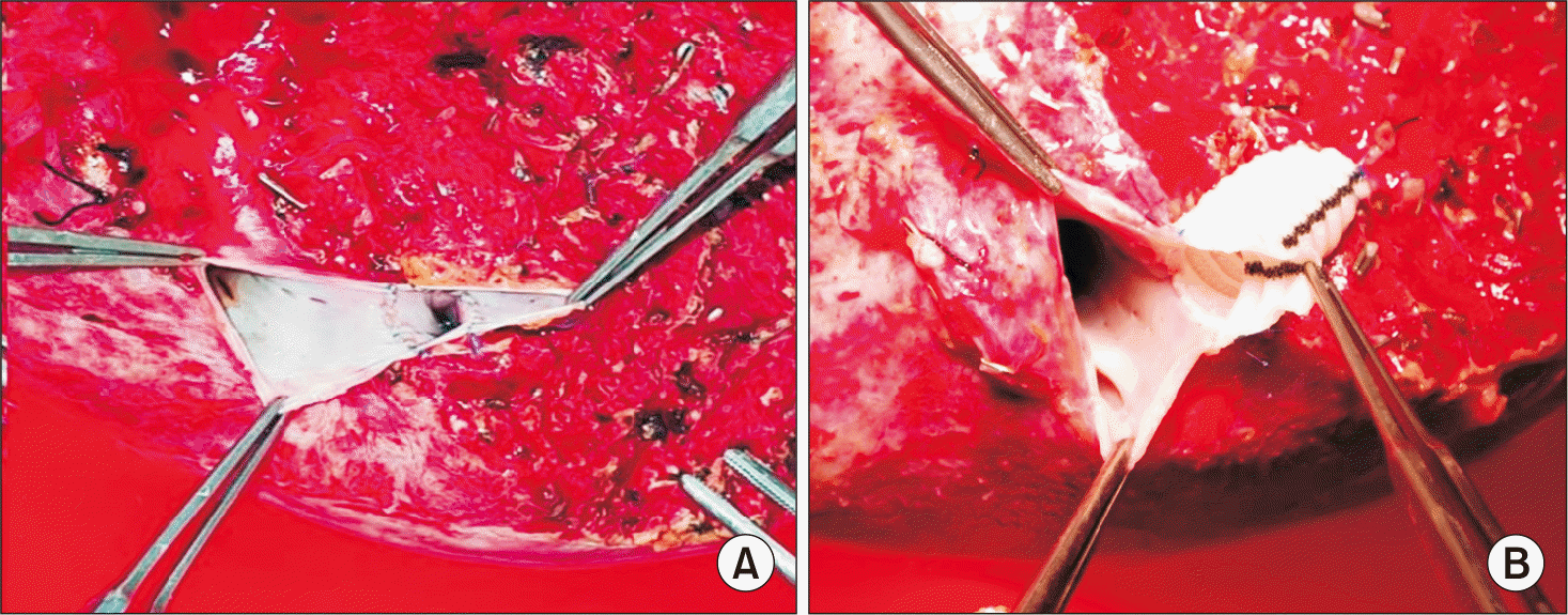
- Extended right lobe graft: the donor’s MHV was procured with the graft and conjoined with the RHV to create a single triangular orifice, which was anastomosed to the recipient’s IVC using continuous 5-0 polypropylene sutures (Fig. 1A). When the distance between the MHV and RHV surpassed 2 cm, a synthetic graft was used to lengthen the MHV trunk prior to its anastomosis with the RHV (Fig. 1B).
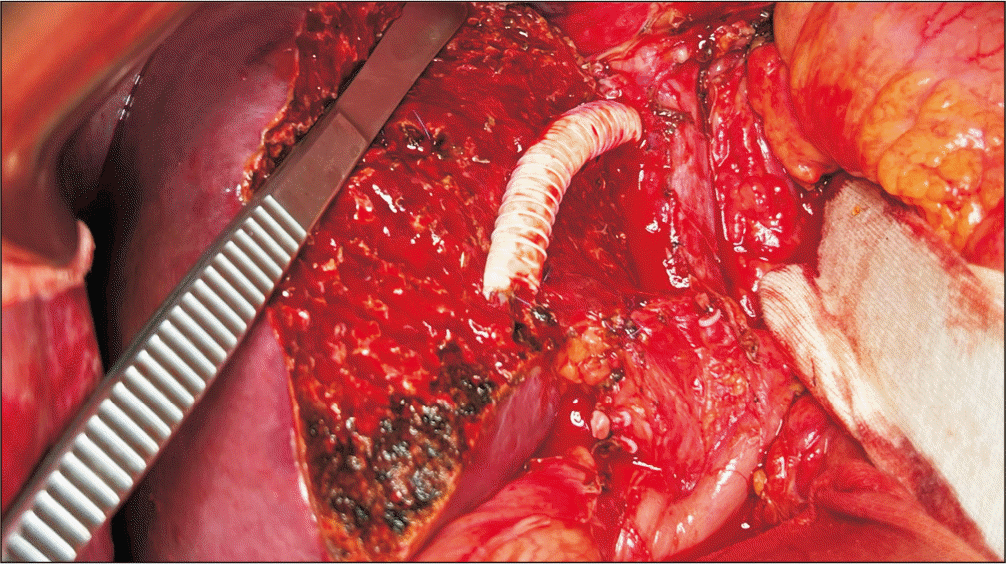
- Modified right lobe graft: MHV branches (V5, V8) > 5 mm were reconstructed using non-ringed ePTFE grafts (Fig. 2). V5 was anastomosed end-to-end and V8 end-to-side to the ePTFE graft, which was subsequently connected to the RHV via a longitudinal incision, forming a single outflow orifice. Smaller branches (< 5 mm) were ligated.
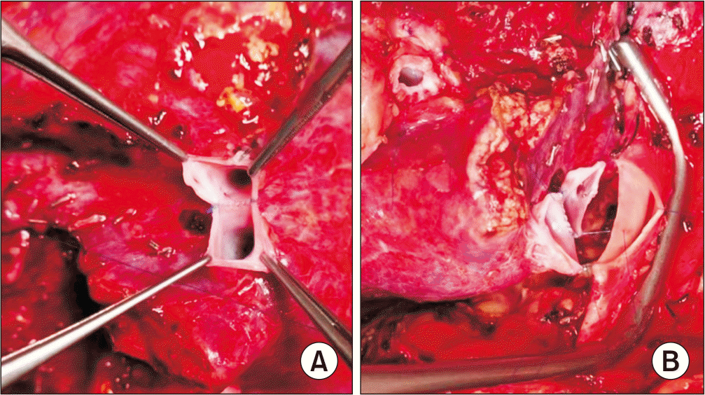
- Accessory RIHVs: veins > 5 mm were anastomosed end-to-side to the IVC using 5-0 polypropylene sutures (Fig. 3) [7].
- Intraoperative assessment: patency of all anastomoses was verified both on the back table and after reperfusion using Doppler ultrasound. The recipient’s IVC was enlarged through caudal and transverse incisions to provide an anastomotic circumference greater than 30 mm.
Follow-up and outcomes
Postoperative venous patency was monitored using Doppler ultrasound every 6 months and contrast-enhanced CT annually, or if complications such as stenosis, occlusion, or thrombosis were suspected. Long-term patency was defined as sustained hepatic vein flow without evidence of stenosis or obstruction. The primary outcomes included five-year hepatic vein patency and complication rates, while secondary outcomes assessed five-year survival stratified by indication (HCC, ACLF, cirrhosis).
Statistical analysis
Data analysis was performed using IBM SPSS 25.0 (IBM Corp.). Continuous variables are presented as means ± standard deviations, and categorical variables as frequencies and percentages. Logistic regression was utilized to evaluate risk factors for hepatic vein complications. Survival was analyzed using the Kaplan-Meier method with 95% confidence intervals (95% CIs), adopting a p-value < 0.05 as the threshold for significance.
RESULTS
Patients and hepatic vein reconstruction features
Between January 2019 and December 2020, 52 consecutive patients underwent LDLT with right lobe graft at 108 Military Central Hospital. Most patients were male (90.4%), and the mean age was 50.79 ± 11.38 years. The principal indication for LDLT was HCC (42.3%), followed by ACLF (32.7%) and decompensated cirrhosis (25.0%).
Among 52 donors, we harvested extended right lobe grafts containing the MHV trunk in 42 donors (80.7%). All hepatic veins in the grafts were categorized as type 4 based on Varotti’s classification, with 37 cases (71.2%) classified as type 4A and 15 cases (28.8%) as type 4B [8]. In four cases, the distance between the MHV and RHV exceeded 2cm, necessitating extension of the MHV trunk with a synthetic graft. Of the remaining 10 cases utilizing modified right lobe graft, we reconstructed V5 in 3 cases (5.8%), V8 in 4 cases (7.7%), and both V5 and V8 in 3 cases (5.8%). All 14/14 (100%) grafts employed ePTFE grafts. All anastomoses were completed using continuous polypropylene 5-0 suture. A single orifice hepatic venous outflow between the MHV and RHV was established in every graft (52/52).For all modified right lobe grafts, we routinely enlarged the RHV opening with a slit incision on the anterior wall, achieving a mean incision length of 9.9 ± 1.2 mm (8–12 mm). The recipient's RHV was consistently widened with a caudally vertical incision and a transverse incision, with mean lengths of 9.7 ± 1.0 mm (8–12 mm) and 14.0 ± 1.1 mm (12–16 mm), respectively. Following reperfusion, the mean circumference of the hepatic vein anastomosis was 36.5 ± 5.1 mm (28–51 mm).
Accessory RIHVs exceeding 5 mm in diameter were observed in 15 cases (28.8%); one graft presented with two large accessory RHVs requiring preservation. All accessory RHVs were reconstructed to the IVC via end-to-side anastomosis using continuous polypropylene 5-0 suture.
Intraoperative outcomes
The mean time required for hepatic vein reconstruction was 17.4 minutes, with portal vein and hepatic artery reconstructions averaging 13.8 minutes and 13.5 minutes, respectively (see Table 1 for details). The average cold ischemic time was 40.9 ± 6.3 minutes.
Following release of the IVC and portal vein clamps, complete liver parenchymal perfusion was achieved in 50 cases (96.2%), while partial perfusion, identified by a small purple patch in the anterior section, was observed in two cases (3.8%). During surgery, 51 grafts demonstrated bile secretion (98.1%). Intraoperative ultrasound confirmed that all 52/52 (100%) hepatic vein anastomoses remained patent, with no signs of congestion detected.
Long-term patency and complications
Long-term patency was maintained in 94.2% of patients. MHV-related complications developed in three patients (5.8%): two patients experienced MHV stenosis (3.8%), and one experienced MHV occlusion (1.9%). All of these cases were managed conservatively. One patient with hepatic vein occlusion died, while the other two made a complete recovery. To further assess potential risk factors, we conducted a univariate analysis on additional related variables, as summarized in Table 2. A hepatic vein anastomosis diameter less than 30mm emerged as the sole risk factor for hepatic vein complications following transplantation (odds ratio [OR], 14.286, 95% CI: 1.121–183.823, p = 0.011).
Long-term survival
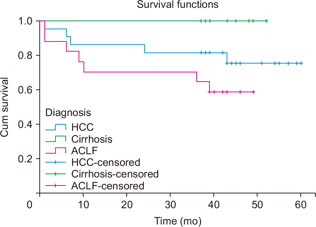
Regarding long-term outcomes after LDLT, patients with cirrhosis achieved the most favorable prognosis, displaying a 5-year survival rate of up to 100%. Conversely, patients with ACLF demonstrated the least favorable prognosis, with a 5-year survival rate of 58.8% (Fig. 4).
DISCUSSION
The single orifice hepatic venous outflow reconstruction technique showed excellent long-term patency (94.2%) and a low complication rate (5.8%) during a five-year follow-up, consistent with recent reports indicating HVOO rates of 2% to 7% in LDLT. Right lobe grafts were associated with a lower incidence (1.5%–4.2%) compared to left lobe grafts (up to 8.1%) [2,6,9]. The procedural simplicity, with a mean hepatic vein reconstruction time of 17.4 minutes and cold ischemic time of 40.9 minutes, aids in reducing graft injury, aligning with Lee et al. (2018) [10], who highlighted the importance of short ischemic intervals for favorable outcomes. Combining the MHV and RHV into a single triangular orifice, with a mean anastomotic circumference of 36.5 mm, may decrease the likelihood of stenosis compared to separate anastomoses, which are susceptible to displacement during graft hypertrophy, corroborating observations by Kim et al. (2014) [11].
The use of non-ringed ePTFE grafts in modified right lobe grafts (19.3%) demonstrated sustained durability, with no occurrences of thrombotic or infectious complications, thereby supporting the findings of Singhal et al. (2021) [6], who reported a 94% patency rate in 760 right lobe LDLTs utilizing ePTFE without an increased infection risk. In comparison, Jung et al. (2020) [5] found a lower patency rate (36%) associated with ringed PTFE grafts, indicating that non-ringed ePTFE grafts and single orifice designs may reduce flow turbulence. Additionally, Goja et al. (2018) [3] reinforced the equivalence of ePTFE to venous grafts, emphasizing its practicality and adaptability in settings with limited resources, such as Vietnam. The lack of reliance on cadaveric grafts further enhances procedural feasibility, directly addressing the challenges related to allogeneic graft availability reported by Sugawara et al. (2004) [4].
Accessory RIHV reconstruction, which was performed in 28.8% of cases, demonstrated 100% patency, supporting the findings of Thorat et al. (2016) [7], who recommended reconstructing veins with diameters greater than 5 mm to prevent posterior segment congestion. This consideration is particularly significant in LDLT, as the risk of small-for-size syndrome remains a persistent concern. Na et al. (2022) [12] highlighted this issue by reporting an 87.5% ten-year patency when wide anastomoses were performed. A notable risk factor was identified with an anastomotic diameter ≤ 30 mm (OR, 14.286; 95% CI: 1.121–183.823; p = 0.011), which is consistent with the findings of Kim et al. (2018) [13], who determined that smaller anastomoses serve as a predictor for early HVOO (4.2% occurrence in right lobe LDLTs). These results highlight the necessity for exact intraoperative sizing, which is implemented in our approach through the use of widened IVC orifices.
Survival outcomes demonstrate the efficacy of the technique, with a 100% five-year survival rate among cirrhotic patients, likely attributable to effective outflow that prevents congestion-related dysfunction, in agreement with Lee (2006) [14]. The 85.7% survival rate observed in HCC patients corresponds with favorable outcomes previously reported in selected candidates, as described by Li et al. (2017) [15]. Although the 58.8% survival rate in ACLF patients is lower, it remains consistent with prior literature that identifies systemic inflammation and coagulopathy as primary challenges [16]. The reduced survival rate in ACLF patients in this cohort primarily reflects the early phase of our transplant program, during which there was limited experience in managing ACLF cases. As illustrated in the Kaplan-Meier survival curves (Fig. 4), a majority of deaths in this group occurred within the first month after liver transplantation, indicating significant perioperative management challenges. However, in the subsequent phase, the accumulation of clinical experience and the adoption of refined management protocols led to substantial improvements, with the 3-year survival rate for ACLF patients increasing to 81.7%, as documented in our follow-up study [17]. The implementation of individualized anticoagulation and anti-inflammatory measures, in conjunction with advances in perioperative management, likely contributed to these improved results. The occurrence of mortality related to single MHV occlusion in an ACLF patient underscores the heightened prothrombotic risk in this population, underlining the importance of patient-specific anticoagulation protocols for this high-risk group [18].
While our study achieved satisfactory results, several limitations should be considered. First, the single-center design and limited sample size (n = 52) restrict the extent to which our findings may be generalized to broader populations. Second, although ePTFE grafts remained durable during the five-year follow-up, the long-term performance after this interval is not fully understood, especially due to conflicting reports regarding late occlusions [5]. Third, since there was no direct comparison with alternative reconstruction methods (e.g., separate anastomosis of V5, V8, or MHV), our assessment of the single orifice technique’s advantage remains incomplete [14,19]. Additional multicenter studies with larger, more diverse patient cohorts are needed to substantiate these results and further optimize ACLF management. The integration of advanced imaging modalities, as suggested by Kim et al. (2024) [1], may improve early detection of HVOO.
In conclusion, middle hepatic venoplasty utilizing ePTFE grafts in combination with single orifice hepatic venous outflow reconstruction represents a safe and effective strategy for right lobe LDLT, especially when the remnant liver volume is 30% to 35%. This method provides favorable long-term results, notably high patency rates, low complication incidence, and excellent survival among cirrhotic and HCC patients, while effectively addressing challenges present in resource-limited environments.
 XML Download