



 PDF
 Citation
 Print


INTRODUCTION
Obstructive jaundice is a mechanical blockage of intrahepatic or extrahepatic bile ducts, clinically manifesting as yellowing of the skin and sclera [1,2]. It interferes with bile drainage from the liver to the duodenum, damages the gut microbiota, increases bacterial translocation, and leads to hepatocellular injury. Over time, biliary obstruction may lead to progressive hepatic fibrosis and eventually cirrhosis [3-7].
Obstructive jaundice is quite common and linked to high morbidity and mortality [1,2]. The most frequent benign cause is gallstones, while the primary malignant etiologies are malignancies such as pancreatic head and gallbladder cancer [8-10]. Research studies have shown that obstructive jaundice occurs in a significant percentage of patients undergoing surgery, with incidence rates varying across different locations [8,11,12].
The illness increases the risk of life-threatening complications, including sepsis, liver failure, and multi-organ dysfunction syndrome [1,2]. The lack of bile acids in the intestines disrupts gut microbial balance, thereby enhancing gut permeability and permitting the translocation of endotoxins and bacteria. These changes exacerbate hepatocellular damage and promote fibrogenesis through inflammatory and ductular reactions that activate hepatic stellate cells to deposit collagen and extracellular matrix. As hepatocyte proliferation slows, markers such as Ki-67, a well-established nuclear antigen used to measure cell proliferation, are significantly reduced, as observed in experimental models [3-7,13-15].
Ki-67 is widely used in oncology and pathology as a proliferation marker, as it is found in the active phases of the cell cycle but not in resting cells. Since it is cell cycle-specific for dividing cells, it is an effective marker of growth fraction in tissue specimens [3,16]. Although alternative markers such as proliferating cell nuclear antigen, minichromosome maintenance, and the mitotic index are available, they face limitations due to technical issues and interpretive variability [16,17].
Indeterminate management of obstructive jaundice involves surgical repair to remove the obstructing cause [10,18]. Surgical outcomes in patients with secondary cirrhosis or liver fibrosis are typically poor. High rates of postoperative complications, such as liver failure, have occurred, mainly due to a deficiency in hepatocyte regeneration and a decline in hepatic metabolic capacity [19,20].
WHO/FAO-codified probiotics, defined as living microorganisms that confer a beneficial effect on the host, have been promoted as potential drugs for modulating functions of the gut-liver axis. They strengthen the gut barrier, suppress microbial translocation, reduce hepatic inflammation, and modify bile acid metabolism. Some strains, such as Lactobacillus rhamnosus GG and Parabacteroides distasonis, have been reported to possess antifibrotic and pro-regenerative effects by modulating cytokines IL-6 and TNF-α [1,18].
Dadih, a traditional fermented buffalo milk product from West Sumatra, is a naturally occurring source of potential probiotic lactic acid bacteria. Lactococcus lactis strain D4 (L. lactis D4), one of its microbial components, was isolated as the dominant species and has been previously shown to produce nisin with antibacterial activity. Studies have reported that L. lactis D4 can inhibit cancer-associated inflammation and growth, as well as influence apoptotic processes [21-24].
In animal models of obstructive jaundice, L. lactis D4 has demonstrated anti-inflammatory effects, prevented hepatic fibrosis, normalized liver function tests, and improved intestinal mucosal integrity. However, few studies have explored its impact on hepatocyte proliferation and histopathological changes in the liver, particularly Ki-67 expression [25-28].
Due to its availability and cost-effectiveness, L. lactis D4 from dadih has the potential to serve as a therapeutic adjunct to minimize liver injury and prevent postoperative liver failure in patients with obstructive jaundice. This study aims to investigate the effect of L. lactis D4 on Ki-67 expression and liver fibrosis in a rat model of obstructive jaundice.
MATERIALS AND METHODS
Study design
This was a purely laboratory experimental study using a post-test only randomized controlled group design involving male Wistar rats as experimental subjects. The rats were randomly divided into three groups: the sham group (laparotomy without BDL), the BDL group (laparotomy followed by BDL), and the BDL + LLD4 group (BDL plus administration of L. lactis D4 fermented milk). Each group consisted of five rats, calculated based on WHO recommendations for animal studies, with a 10% drop-out adjustment.
Animal preparation
Fifteen male Wistar rats, weighing 160 to 250 g and aged 10 to 16 weeks, were used in this study. Rats were housed individually under standard conditions, which included a 12-hour light/dark cycle, temperature maintained between 20°C and 26°C, and unlimited access to food and water. The animals were acclimated for a week before the experiment.
Preparation of the obstructive jaundice rat model
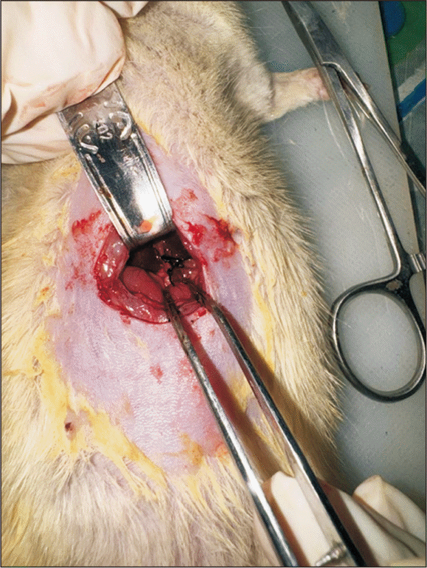
The rats were anesthetized with isoflurane, and common BDL was performed under aseptic conditions. Obstructive jaundice was experimentally induced in rats through BDL. After anesthesia was administered, the abdominal area was shaved, disinfected, and draped in a sterile manner. A midline laparotomy was performed to expose the hepatobiliary system. The common bile duct (ductus choledochus) was carefully isolated and ligated using sterile 5–0 silk sutures to obstruct bile flow from the liver to the duodenum (Fig. 1).
After successful ligation, the abdominal wall and skin were closed in layers with appropriate sutures. Postoperative recovery was closely monitored to ensure animal welfare. The bile duct obstruction led to cholestasis and elevated serum bilirubin levels, clinically manifesting as jaundice. On postoperative day 7, liver tissue specimens were harvested from the left hepatic lobe and fixed in 10% neutral-buffered formalin for subsequent histopathological examination.
Euthanasia was performed by intramuscular injection of ketamine hydrochloride (30 mg/kg) and xylazine (5 mg/kg). Following tissue collection, the animal remains were disposed of by burial at a depth of 1 meter in a plastic-lined fabric container.
Preparation and administration of L. lactis D4
L. lactis D4 was isolated from fermented Dadih produced at the Faculty of Animal Science, Universitas Andalas, with a concentration of 109 CFU/mL. The administered dose was calculated using the human-to-animal dose conversion factor (0.018), which is equivalent to 0.36 mL/200 g of body weight/day. Fermented milk was administered orally through an orogastric tube (Sonde) for seven consecutive days following BDL.
Ki-67 immunohistochemical staining
Paraffin blocks were sectioned at 4 µm and mounted on poly-L-lysine–coated slides. Sections were deparaffinized, rehydrated, and antigen retrieval was performed in citrate buffer (pH 6.0). Endogenous peroxidase was inhibited using H2O2. Non-specific binding was inhibited using 2% normal goat serum. A primary antibody, Ki-67 (1:100), was applied to the tissue sections and incubated overnight at 4°C. Detection was performed using a biotin-avidin complex and DAB as chromogen. Counterstaining was done with hematoxylin, followed by bluing, dehydration, and mounting. Ki-67 expression was examined under 200× magnification across five random fields, with the stained area quantified using ImageJ software.
Assessment of liver fibrosis
Liver tissue sections were stained with hematoxylin and eosin (H&E) and examined under a light microscope (Leica DM1000LED) at 200× magnification. The degree of hepatic fibrosis was histologically assessed by a scoring system based on the amount and pattern of fibrotic tissue accumulation within the liver lobules.
This system classifies fibrosis into five stages: F0 (normal portal area structures, most of the hepatocytes were within normal limits, and no fibrosis), F1 (mild portal fibrosis without septa), F2 (presence of a few septa), F3 (numerous septa without cirrhosis), and F4 (established cirrhosis).
Statistical analysis
After data collection, bivariate analysis was performed. Since the variables were numerical, a normality test was conducted before the bivariate analysis. Once normal data distribution was confirmed, parametric testing was carried out using One-Way Analysis of Variance (ANOVA), followed by a post-hoc test employing the least significant difference (LSD) method. A p-value of < 0.05 was considered statistically significant, with a 95% confidence interval. Data analysis was performed using SPSS software version 18.0 for Windows (IBM Corp.).
Ethical considerations
All experiments with animals were conducted in accordance with the policies of the Faculty of Medicine Research Ethics Committee, Universitas Andalas, Padang, Indonesia (Ethical Clearance Number: 26/UN.16.2/KEP-FK/2025). Animals were handled and cared for following institutional and international policies to prevent any inhumane treatment.
RESULTS
Clinical presentation of obstructive jaundice in experimental animal models
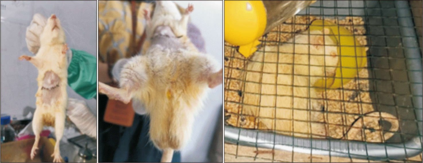
Clinical presentation of obstructive jaundice was induced in rats under BDL, characterized by icteric sclera, dark brown coloration of urine, and yellow coloration of skin (Fig. 2). These clinical presentations were confirmed by laboratory results, which showed significantly higher total bilirubin levels in the BDL group (3.17 ± 0.109 mg/dL) compared to the sham group (1.006 ± 0.14 mg/dL).
Effect of L. lactis D4 on Ki-67 expression in rats with obstructive jaundice
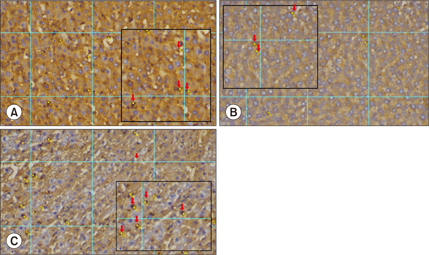
Immunohistochemistry revealed expression of Ki-67 as brown nuclear staining of hepatocytes. The strongest expression was observed in the BDL + LLD4 group, showing enhanced hepatocyte proliferation after treatment with L. lactis D4, compared to the sham and BDL groups (Fig. 3).
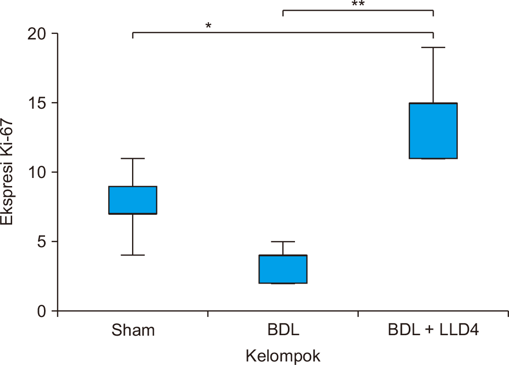
The mean expression of Ki-67 in the BDL + LLD4 group was higher than in the sham group (14.20 ± 3.347 vs. 7.60 ± 2.608), and lower in the BDL group compared to the sham group (3.40 ± 1.342). The Ki-67 expression data were homogeneous and normally distributed. One-way ANOVA analysis indicated a significant difference in Ki-67 expression among the groups (p < 0.001) (Table 1). Post-hoc LSD analysis showed significant differences between sham and BDL + LLD4 (p < 0.05) and between BDL and BDL + LLD4 groups (p < 0.01), confirming the proliferation-promoting effect of L. lactis D4 (Fig. 4).
Effect of L. lactis D4 on liver fibrosis in rats with obstructive jaundice
Fibrosis is classified into three categories based on histological staining. Predominantly progressive fibrosis is characterized by wide fibrous septa with loosely compact collagen, pale trichrome coloration, inflammatory cells, and ductular responses. Indeterminate fibrosis is a state of equilibrium between progressive and regressive alterations. Predominantly regressive fibrosis is observed in thin, compact, acellular fibrous septa with dark blue trichrome coloration, indicating recovery or healing in the liver.
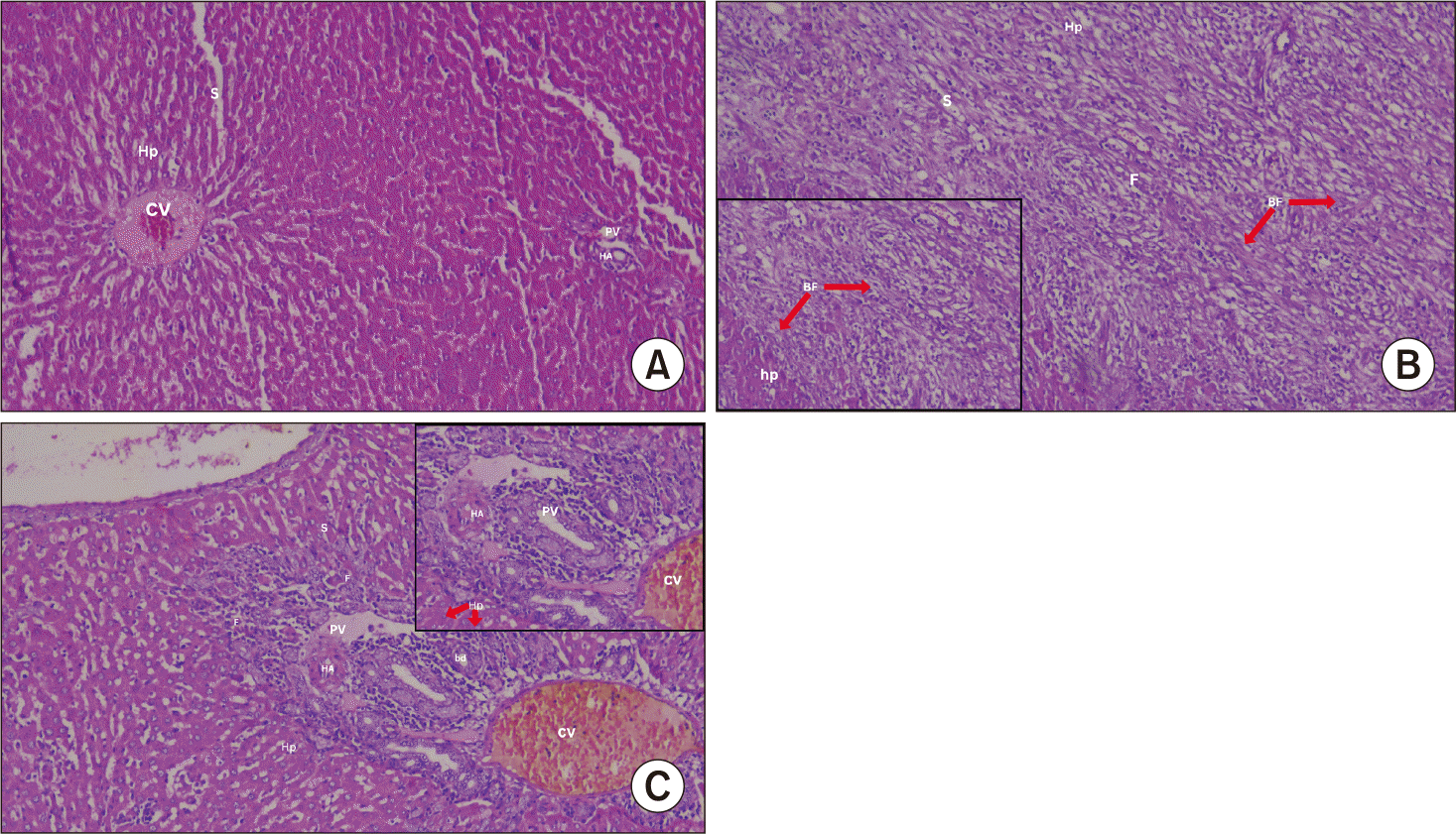
Histopathological analysis of liver fibrosis was conducted using the METAVIR scoring system, with scoring performed in five fields of view under 100× magnification (Fig. 5). The analysis revealed different levels of fibrosis among the groups. The sham group displayed normal lobular architecture with scores primarily at F0 and F1. Conversely, the BDL group exhibited extensive fibrotic changes, like portal fibrosis and bridging fibrosis, with most samples graded at F3 or F4. Treatment with L. lactis D4 in the BDL + LLD4 group maintained liver architecture with minimal fibrosis, mostly between F1 and F3, and none of the cases progressed to cirrhosis (F4). Although the p-value was not statistically significant, the fibrosis scores show a trend toward reduced fibrosis severity in the BDL + LLD4 group compared to the BDL group (Table 2).
DISCUSSION
Effect of L. lactis D4 treatment on Ki-67 expression in a rat model of obstructive jaundice
Treatment with L. lactis D4 in rats with obstructive jaundice significantly increased Ki-67 expression compared to the untreated BDL group and the sham group. Ki-67 expression, as demonstrated by brown staining in hepatocytes on immunohistochemical examination, is a marker for cell proliferation, indicating regenerative activity in liver tissue.
Previous studies have demonstrated various beneficial effects of L. lactis D4 in models of obstructive jaundice. Anami et al. (2023) [29] reported improved liver function as indicated by decreased SGOT and SGPT levels following the administration of L. lactis D4 from dadih. Suchitra et al. (2024) [30] demonstrated that L. lactis D4 reduced NF-κB and α-SMA expression, while increasing IL-6 and IL-32 expression, indicating its potential for anti-inflammatory and anti-fibrotic effects. Putri (2024) [31] also showed a significant decrease in MDA levels in rats treated with L. lactis D4, reflecting reduced oxidative stress. While these studies support the therapeutic role of L. lactis D4, our research provides novel insights by evaluating liver cell proliferation through immunohistochemical analysis of Ki-67 expression and assessing fibrosis severity using the METAVIR scoring system. To the best of our knowledge, this is the first study to explore these specific histological endpoints in the context of L. lactis D4 administration in a BDL-induced obstructive jaundice model.
The primary cells positive for Ki-67 staining were the sinusoidal liver cells, which may suggest that the initial regeneration in the liver after bile duct ligation (BDL) originates from these cells. This aligns with McConnell et al. [32], who described that liver sinusoidal endothelial cells (LSECs) play an important role in liver regeneration. They demonstrated that LSECs promote hepatocyte proliferation during the initiation phase, which subsequently declines in the angiogenic phase, as corroborated by Ki-67 staining. During the second angiogenic phase, hepatocytes experience hypoxia and produce angiogenic factors such as VEGF and angiopoietin, which stimulate the proliferation of LSEC. Hepatocyte proliferation peaks 24 to 48 hours after partial hepatectomy (PHx), while LSEC proliferation peaks on days 3 to 4. In the termination phase, LSECs secrete transforming growth factor (TGF)-β to remodel the extracellular matrix and induce hepatocyte quiescence [26].
As shown in Table 1, the BDL + LLD4 group had the highest average Ki-67 expression value (14.20 ± 3.347), which was significantly higher than that of the sham group (7.60 ± 2.608; p < 0.05) and the BDL group (3.40 ± 1.342; p < 0.01). One-way ANOVA showed a significant difference among the three groups (p < 0.001), and post-hoc LSD tests revealed a significant difference, particularly between the BDL + LLD4 group and the other two groups.
L. lactis D4, as a probiotic, also plays a vital role in regulating the gut microbiota by increasing the population of beneficial bacteria such as Lactobacillus and Bifidobacterium, which in turn, produce microbial metabolites such as short-chain fatty acids (SCFAs) like butyrate, acetate, and propionate that have hepatoprotective effects [33]. Butyrate is utilized as an energy source by hepatocytes, improves intestinal epithelial barrier function, reduces bacterial translocation and lipopolysaccharide entry into the portal circulation, thereby reducing systemic inflammation and oxidative stress, which are common in obstructive jaundice [21,22].
Furthermore, SCFAs also influence cell signaling pathways by activating G-protein-coupled receptors (GPR41/43) and inhibiting histone deacetylases, thereby leading to the upregulation of genes involved in cell proliferation such as cyclin D1 and cyclin E [27]. These pathways are likely to be the cause of the observed elevation of Ki-67 expression in the BDL + LLD4 group, indicating enhanced hepatocyte regeneration.
The accumulation of bile in obstructive jaundice leads to inflammation and oxidative damage that suppresses tissue regeneration. Probiotics, such as L. lactis D4, can attenuate this damage by promoting the production of anti-inflammatory cytokines (e.g., interleukin [IL]-10) and inhibiting the release of pro-inflammatory cytokines (e.g., tumor necrosis factor [TNF]-α, IL-6), thereby creating a microenvironment that supports hepatocyte regeneration [28]. Our previous study also found that probiotics can activate tissue regeneration signaling pathways, such as the Wnt/β-catenin pathway, implicated in promoting cell proliferation in damaged liver tissue [33].
Upregulation of Ki-67 expression in the BDL + LLD4 group supports the hypothesis that L. lactis D4 has therapeutic potential as an adjuvant in liver regeneration in obstructive jaundice with mechanisms involving attenuation of oxidative stress, modulation of inflammation, and improvement in hepatocyte and sinusoidal cell proliferation. An investigation by Kong et al. [14] on a rat model of obstructive jaundice showed that Ki-67 expression significantly decreased following liver injury, but subsequently increased again after liver regeneration induced by 76% hepatectomy and portal vein occlusion [34].
These findings highlight the potential of probiotics such as L. lactis D4 as therapeutic agents with immense potential in liver tissue repair, especially to reduce the risk of postoperative liver failure in patients with obstructive jaundice undergoing surgery.
Effect of L. lactis D4 on liver fibrosis in a rat model of obstructive jaundice
Administration of L. lactis D4 to rats with obstructive jaundice showed potential in attenuating liver fibrosis, although statistical significance was not elicited. Histopathological analysis with H&E staining revealed that the BDL + LLD4 group had larger areas of tissue regeneration compared to the BDL group, which exhibited necrosis, hemorrhage, and extensive fibrosis.
As shown in Table 2, the BDL group had a higher percentage of animals with extensive fibrosis (40% in F3 and F4), whereas the BDL + LLD4 group exhibited a uniform distribution with no animals in F4. In the sham (laparotomy control) group, the liver structure remained intact, with hepatocytes arranged in radial plates around the central vein, and there was no fibrosis or inflammation in the portal tract. There was also no bridging fibrosis. In the untreated BDL group, increased inflammatory cells, predominantly lymphocytes, were observed in the portal tract, indicating portal inflammation. Fibrotic tissue was raised, and bridging fibrosis was observed, consistent with Metavir scores of F2 to F4. Regenerative cells were only found at the margins of necrotic zones. Bridging fibrosis represents a watershed phase of liver fibrogenesis, marked by disruption of normal architecture, impaired liver function, and a risk of cirrhosis and its complications [35].
On the other hand, the BDL + LLD4 group exhibited preserved lobular architecture with minor portal fibrosis that did not penetrate the liver parenchyma and lacked bridging fibrosis, which was associated with Metavir scores F0–F2. Nevertheless, chi-square analysis revealed no significant difference between groups (p = 0.160).
A critical limitation of this study is the relatively short duration between BDL induction and evaluation, which is only 7 days. In rodent models, liver fibrosis generally progresses over a longer period, ranging from 21 to 28 days. Therefore, the histological findings in this study likely reflect early inflammatory changes and initial stages of fibrotic remodeling, rather than established or mature fibrosis. This timeframe might explain the absence of significant differences in fibrosis severity across groups, and the limited ability to detect the full antifibrotic effects of L. lactis D4.
One of the limitations was that H&E staining is insensitive and non-specific for detecting fine fibrosis and cannot distinguishably stain collagen, a specific marker of liver fibrosis. Using Masson’s Trichrome or Sirius Red would likely yield more accurate and detailed results [36]. Furthermore, the lack of significant differences according to Metavir scoring may be due to the acute phase of the obstructive injury in this study, when fibrotic changes have not yet fully developed. Kong et al. mentioned that fibrosis formation continues up to day 28 in rats with obstructive jaundice [34].
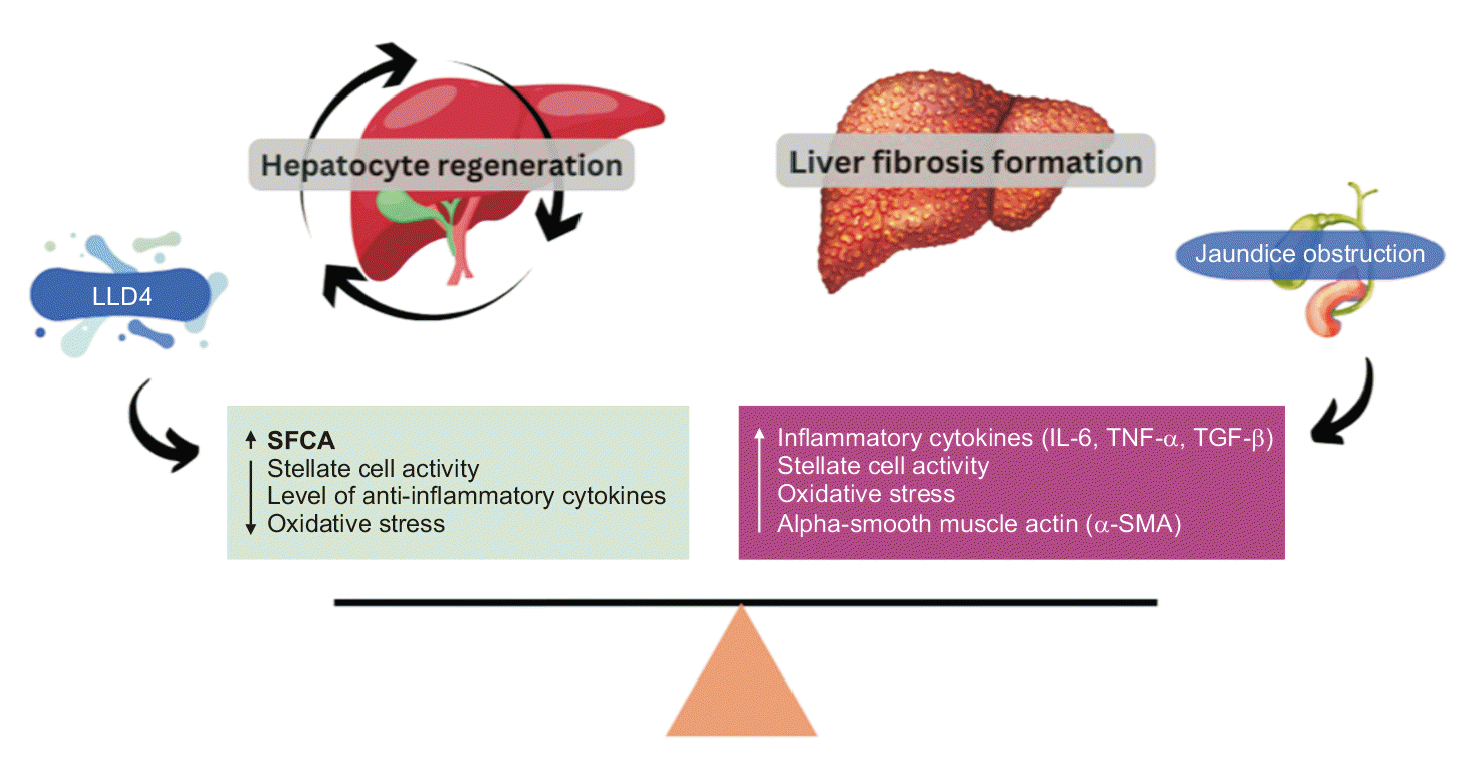
The therapeutic effect of L. lactis D4 in alleviating liver fibrosis is believed to be mediated by modulation of gut microbiota and suppression of systemic inflammation. Probiotic therapy increases SCFA production, particularly butyrate, which suppresses hepatic stellate cell activation—the pivotal cells in fibrosis. Butyrate also suppresses pro-inflammatory cytokines such as TGF-β and IL-6, which are well known to trigger fibrotic signaling (Fig. 6) [22].
Shreffler et al. also documented that probiotic administration does not significantly alter the fibrosis scores; however, this does not rule out its advantageous effects. Probiotic impacts in the short term do not appear to be reflected in scoring systems, although they reduce liver inflammation and also prevent further progression of fibrosis [37].
This study suggests that L. lactis D4 can reduce the severity of liver fibrosis in obstructive jaundice, as evidenced by histopathological findings of reduced portal fibrosis and absence of bridging fibrosis. This result is supported by the findings of Pratama et al., who demonstrated that L. lactis D4 significantly reduced Sirius Red staining, indicating reduced collagen deposition in a rat model of extrahepatic jaundice [21].
Kuramitsu et al. [38] observed that hepatocyte proliferation in fibrotic livers was impaired, and new fibrosis developed rapidly upon regeneration, marked by increased accumulation of collagen on day 5 after PHx. Liver recovery in fibrotic conditions was significantly inhibited by decreased hepatocyte growth and over-activation of liver stem cells, which triggered a strong fibrogenic response. Gvidiani et al. [39] also concluded that hepatocyte growth was directly proportional to the density of α-SMA-positive myofibroblast-like cells, leading to overwhelming fibrosis in liver tissue by day 28 following occlusion.
Delgado-Venegas et al. [40] demonstrated that L. lactis mediates antifibrotic effects through multiple mechanisms, including the reduction in liver tissue structural damage and the area of fibrosis, thereby demonstrating hepatoprotective activity. Additionally, L. lactis enhances the number of Peyer’s patch lymph nodes, regulates the gastrointestinal immune response, and significantly lowers liver enzyme levels such as ALT and AST, demonstrating improved liver function. At the molecular level, L. lactis upregulates the expression of intestinal IL-10 mRNA (an anti-inflammatory cytokine) and hepatic FoxP3, thereby suggesting a regulatory T cell function. It also decreases the hepatic expression of the pro-inflammatory cytokine IL-1β, revealing reduced inflammation. Overall, these effects demonstrate the therapeutic potential of L. lactis in preventing liver damage and fibrosis.
This study concludes that hepatocyte regeneration is disrupted in liver damage. L. lactis D4 shows potential in promoting regeneration and reducing acute inflammation. Further research with more robust study designs is necessary to confirm its probiotic effects and to investigate the disrupted cell cycle phases and involved cytokines.
Together, this study suggests two concurrent processes: hepatocyte proliferation, as evidenced by increased expression of Ki-67 in the BDL + LLD4 group, and fibrosis progression, as confirmed in the control BDL group. These findings clearly suggest that liver fibrosis progression and hepatocyte proliferation are closely linked.
 XML Download