



 PDF
 Citation
 Print


Introduction
Radiotherapy aims to deliver the maximum radiation dose to tumors while minimizing exposure to surrounding healthy tissues. With global aging and advancements in diagnostic technologies, cancer incidence has increased significantly, leading to a higher demand for advanced radiotherapy techniques. For instance, between 2000 and 2020, global cancer cases increased by approximately 50%. This highlights the need for precise and efficient treatment modalities [1].
Technological advancements in radiotherapy have allowed highly individualized treatments. Innovations, such as intensity-modulated radiation therapy (IMRT) and volumetric-modulated arc therapy (VMAT), have played crucial roles in this evolution [2]. IMRT enables the modulation of radiation beam intensity from fixed angles, enabling precise targeting of complex tumor geometries. In contrast, VMAT delivers radiation in a continuous arc, reducing treatment times and further improving precision [3]. Therefore, VMAT has become a standard treatment approach for anatomically challenging tumor sites.
Building on these advancements, the HyperArc technique has emerged as a specialized VMAT-based approach designed to optimize stereotactic radiosurgery (SRS) treatments. The HyperArc technology automates treatment planning, optimizes beam arrangements, and ensures precise dose delivery through predefined noncoplanar arcs. These automated features enhance SRS efficiency by minimizing setup errors and improving workflow standardization. Moreover, HyperArc enables single-isocenter, multi-target treatment, which is particularly beneficial for patients with multiple brain metastases. This approach reduces treatment time while maintaining high dosimetric accuracy [4,5]. By integrating automated workflows and optimized beam arrangements, HyperArc can enhance dose conformity and homogeneity, making it particularly beneficial for SRS. Some studies have explored its potential in stereotactic body radiotherapy (SBRT); however, further research is needed to establish its routine clinical application in this setting [6,7]. However, the complexity of this technique necessitates robust quality assurance (QA) protocols to ensure accurate dose delivery and patient safety [7,8]. QA in radiotherapy is essential for ensuring accurate and reproducible treatment delivery. It verifies that the planned dose distribution precisely corresponds with the delivered dose, thereby minimizing errors and uncertainties [9,10]. Traditional QA methods, such as ion chambers, film dosimetry, and electronic portal imaging devices (EPID), have been widely utilized. While these approaches are considered reliable, they are resource-intensive and often fail to capture dose variations across individual fractions, especially in complex treatments, such as HyperArc. Consequently, more efficient and precise QA methods are required to overcome these limitations [7].
Log file-based QA has emerged as a promising alternative by leveraging system-generated data—such as multileaf collimator (MLC) positions, gantry angles, and beam outputs—to reconstruct fluence maps. This approach enables real-time verification of treatment accuracy and delivery consistency [11]. Despite these advantages, research on the application of log file-based QA in HyperArc remains limited. To address this gap, specialized QA tools tailored to HyperArc’s unique requirements need to be developed. This study aims to evaluate the clinical performance of FractionLab (Varian Medical Systems), a log file-based QA tool in HyperArc treatments. FractionLab reconstructs delivered fluence maps from treatment log files, facilitating the detection of discrepancies between planned and delivered doses. By analyzing log data, including MLC positions and beam delivery parameters, FractionLab enables rapid and automated QA assessments without requiring additional measurement devices [12]. HyperArc’s reliance on steep dose gradients and complex beam configurations necessitates QA metrics that surpass conventional thresholds, such as the 3%/3-mm gamma-passing rate. To ensure accurate dose delivery, various patient-specific QA (psQA) methods are employed. Traditional methods include ion chamber dosimetry, film dosimetry, and EPID-based portal dosimetry. Ion chamber dosimetry provides accurate absolute dose measurements but lacks spatial resolution and requires a time-consuming setup. Film dosimetry offers high spatial resolution but is sensitive to processing variability and requires careful calibration. EPID-based portal dosimetry is widely used for its efficiency in verifying fluence maps; however, it does not directly assess 3D dose distributions. More recently, log file-based QA tools, such as FractionLab, have been introduced to address these limitations by reconstructing fluence maps and providing rapid, data-driven assessments of treatment accuracy. Addressing this gap requires developing and validating specialized QA tools tailored to HyperArc’s unique requirements, ensuring precise and efficient psQA [12,13]. This study aims to evaluate the clinical performance of FractionLab, a log file-based QA tool, in HyperArc treatments. FractionLab reconstructs delivered fluence maps from treatment log files, enabling the detection of discrepancies between planned and delivered doses. By analyzing the log data, including MLC positions, gantry angles, and beam delivery parameters, FractionLab provides rapid and automated QA assessments without requiring additional measurement devices. By retrospectively analyzing the QA data, FractionLab’s outcomes were compared with those of conventional portal dose image prediction (PDIP) to assess its feasibility and accuracy [12]. Traditionally, psQA for HyperArc and other advanced radiotherapy techniques has been conducted using methods, such as ion chamber dosimetry, film dosimetry, and EPID-based portal dosimetry. Ion chambers provide accurate absolute dose measurements but require time-consuming phantom setups. Film dosimetry enables high-resolution spatial dose verification but is labor-intensive and requires careful handling of processing variability. EPID-based portal dosimetry is widely used for its efficiency in verifying fluence maps; however, it does not directly measure 3D dose distributions. More recently, log file-based QA tools, such as FractionLab, have emerged as efficient alternatives, offering the reconstruction of delivered fluence maps and rapid detection of discrepancies between planned and delivered doses. This study evaluates FractionLab’s effectiveness compared to conventional methods and determines its feasibility for routine HyperArc psQA [12,13].
By retrospectively analyzing QA data, FractionLab’s outcomes were compared with those of conventional PDIP to assess its feasibility and accuracy. Furthermore, this study also aims to identify the optimal parameters to enhance the reliability of HyperArc treatment plans, thereby supporting its broader clinical adoption [13].
Materials and Methods
1. Patient selection and treatment protocol
This retrospective study was approved by the Institutional Review Board (IRB Approval No. Yeungnam University Medical Center IRB 2024-12-041). In this study, 43 patients who underwent SRS for brain tumors using the TrueBeam radiotherapy system equipped with HyperArc between January 2023 and September 2024 were enrolled. Following the institutional protocols, HyperArc was utilized exclusively for the treatment of brain metastases or small intracranial tumors. The requirement to obtain informed consent was waived.
Patients were selected based on clearly defined criteria to ensure a representative cohort reflecting common clinical scenarios for HyperArc-based SRS. Tumor Volume: Planning target volumes (PTVs) ranged from 0.5 cm³ to 12 cm³, encompassing a range of lesion sizes while minimizing extreme outliers that could skew the results.
1) Tumor location
Patients with tumors in regions prone to steep dose gradients (e.g., near the brainstem or optic nerves) were included to assess FractionLab’s sensitivity to positional deviations in anatomically complex environments.
2) Number of metastases
Patients with 1–55 metastatic lesions were included, reflecting typical clinical scenarios where HyperArc is employed for multi-lesion cases. Among the study population, 27 patients (62.8%) presented with multiple metastases.
3) Isocenter configuration
Single-isocenter plans were prioritized to reflect standard HyperArc workflows, with 38 of 43 patients (88.4%) treated using a single isocenter. A smaller subset of patients (5 cases, 11.6%), received multiple-isocenter plans to assess FractionLab’s performance under more complex treatment conditions.
4) Impact of tumor complexity on gamma-passing rates
Tumor complexity was expected to influence gamma-passing rates due to factors, such as steep dose gradients, small-field effects, and potential MLC positioning errors. Patients with highly irregularly shaped tumors or tumors located near critical structures were more susceptible to positional deviations. Consequently, stricter gamma criteria (e.g., 2%/1 mm) were applied to assess FractionLab’s ability to detect dose discrepancies under these challenging conditions. By including patients with diverse tumor characteristics and treatment complexities, this study aims to evaluate FractionLab’s clinical feasibility and performance in both standard and challenging HyperArc-based scenarios [4]. The PTVs ranged from 0.5 cm³ to 12 cm³ per lesion, with an average tumor irregularity index of 1.34 ± 0.21. Among the patients, 27 (62.8%) had multiple metastases, with a mean of 3.2 ± 1.7 lesions per patient. The number of isocenters used in the treatment plans varied based on clinical presentation, with a single-isocenter approach applied in 38 cases (88.4%) and multiple isocenters used in five cases (11.6%). These details provide a clearer characterization of the patient cohort and treatment complexity. The target volumes were defined as PTVs ranging from 0.5 cm³ to 12 cm³ per lesion. Each patient was administered a single treatment plan with fraction doses of 18–24 Gy, delivered in a single session per fraction. The inclusion criteria required patients to have log files suitable for FractionLab analysis and treatment plans designed for small, irregularly shaped tumors or multiple metastases [14].
2. Treatment equipment
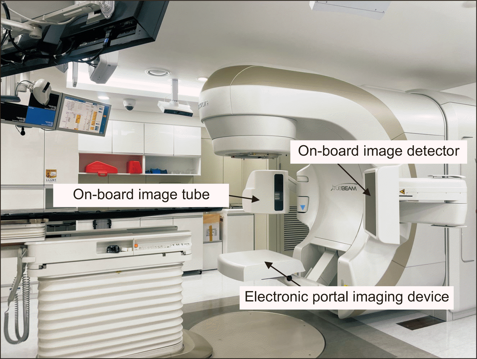
All treatments were administered using the TrueBeam radiotherapy system (Varian Medical Systems) equipped with HyperArc technology. HyperArc is specifically designed for SRS and has been explored for potential applications in SBRT. While some studies have investigated its dosimetric performance in SBRT, further research is needed to establish its routine clinical suitability [5]. The system leverages optimized beam arrangements and automated workflows, including predefined couch and gantry movements, to achieve high target conformity and steep dose gradients. Both PDIP and FractionLab analyze fluence maps. However, PDIP evaluates fluence from the actual beam using an EPID, while FractionLab reconstructs fluence maps based on log files recorded during treatment delivery. In this study, each fluence map—whether from PDIP or FractionLab—was compared to the planned fluence map, and gamma-passing rates were calculated to evaluate the agreement between the planned and delivered dose distributions [15]. This reduced treatment time and improved plan quality for complex cases, such as multiple metastases or small, irregularly shaped tumors [5].
3. Traditional QA method: PDIP
PDIP served as the conventional QA method for comparison, utilizing an EPID to calculate dose distributions delivered during treatment simulations. The system captured 2D dose maps, which were then analyzed using Varian’s PDIP software. Fig. 1 demonstrates the psQA process using EPID within the portal dosimetry framework for IMRT. Portal dosimetry is a widely adopted approach for psQA in advanced radiotherapy techniques, such as IMRT and VMAT, due to its efficiency and accuracy. By eliminating the need for phantom setups, portal dosimetry significantly reduces QA time and is widely used in clinical practice. However, it evaluates fluence maps rather than directly verifying patient dose calculation algorithms, such as pencil beam convolution and Acuros XB. The gamma-passing rate—a metric used to assess the dose distribution accuracy—was calculated based on two criteria: 3%/3 mm and 2%/1 mm. These criteria were chosen based on their widespread acceptance in clinical practice and recommendations by AAPM Task Group No. 218 [7].
4. Log file-based QA using FractionLab
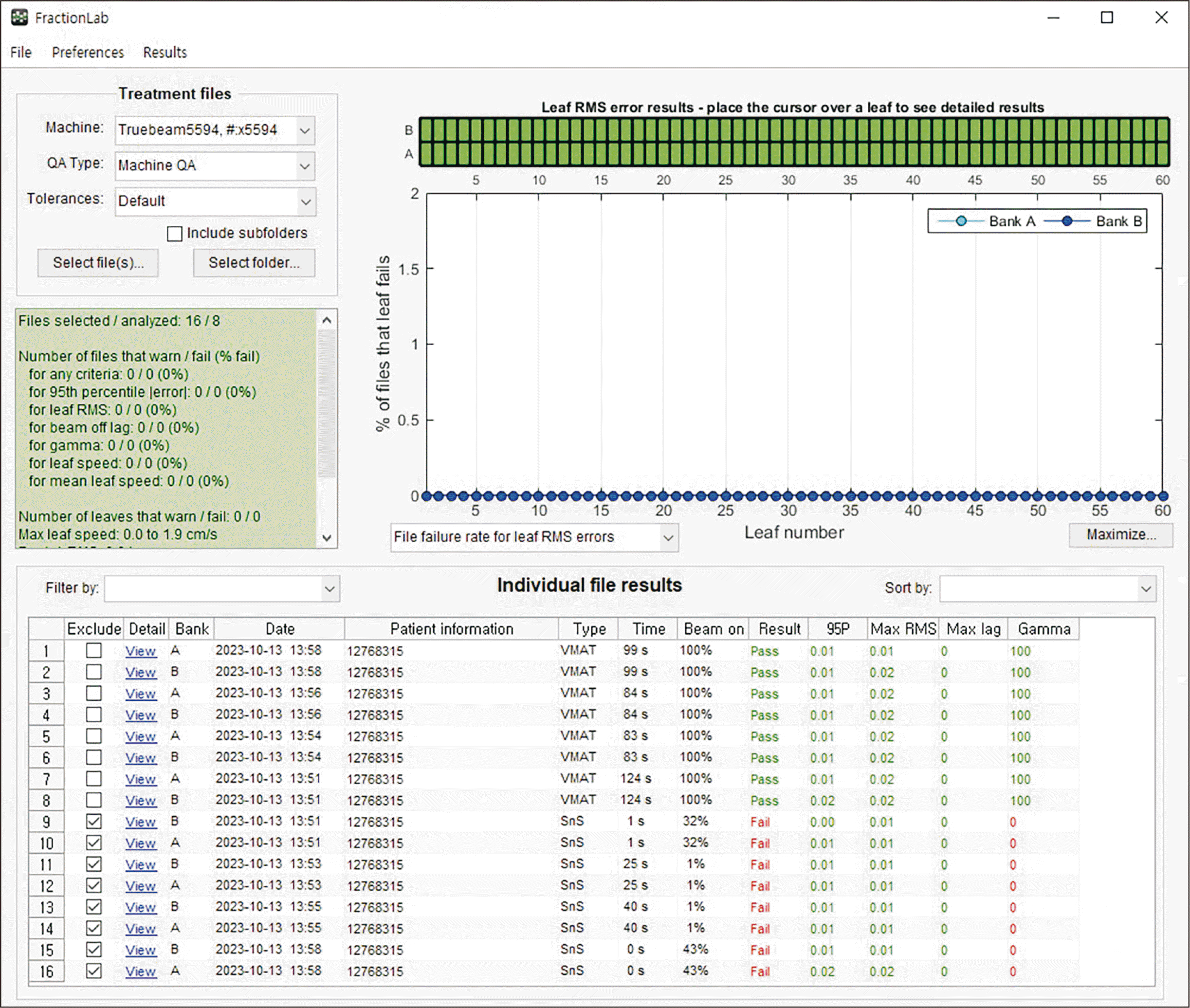
FractionLab was used for the log file-based QA. This system reconstructs fluence maps using log files generated during treatment delivery. The log files contained detailed data, such as MLC positions, gantry angles, and beam outputs [14]. FractionLab was used to compare the reconstructed fluence maps with the planned dose distributions to assess treatment accuracy. Gamma-passing rates were calculated using various criteria, including 3%/3 mm and 2%/1 mm, to validate dose delivery [9,16].
5. Statistical analysis
Statistical analysis was performed using SPSS (version 29.0.2.0, IBM Corp.). Paired t-tests were used to compare gamma-passing rates from FractionLab and PDIP across various gamma criteria. The paired t-test was selected as it is a parametric test suitable for comparing two related measurements within the same sample group, such as gamma-passing rates obtained from the same patients using different QA methods.
Since the sample size (n=43) exceeds the recommended minimum threshold of 30 for the central limit theorem, the distribution of differences can be assumed to be approximately normal, justifying the use of the t-test [8].
To minimize the risk of type I errors from multiple comparisons, a Bonferroni correction was applied, setting a revised significance threshold of P<0.005 to ensure robust statistical conclusions. The significance threshold was adjusted to P<0.005 (0.05/10) to ensure robust statistical conclusions across the 10 gamma criteria.
Additionally, 95% confidence intervals (CIs) for the mean differences in gamma-passing rates between FractionLab and PDIP were calculated to assess the effect size and variability. These CIs were interpreted alongside P-values to evaluate both statistical and clinical significance.
Results with P<0.005 were deemed statistically significant after applying the Bonferroni correction. CIs that excluded zero were interpreted as indicating significant differences in gamma-passing rates, further reinforcing the robustness of the findings [17]. FractionLab’s results were analyzed to identify conditions where it either outperformed or underperformed compared to PDIP [8,11].
Results
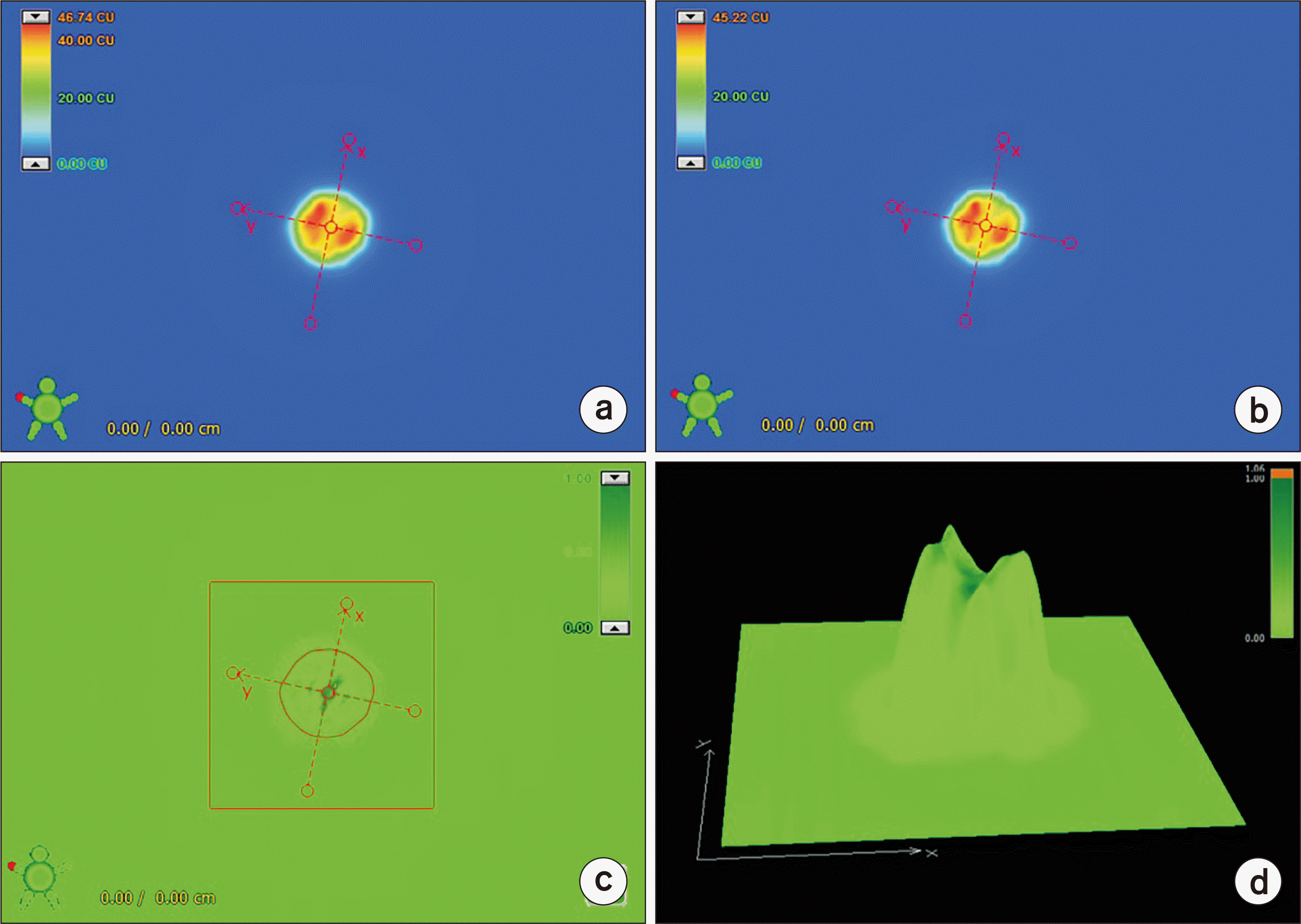
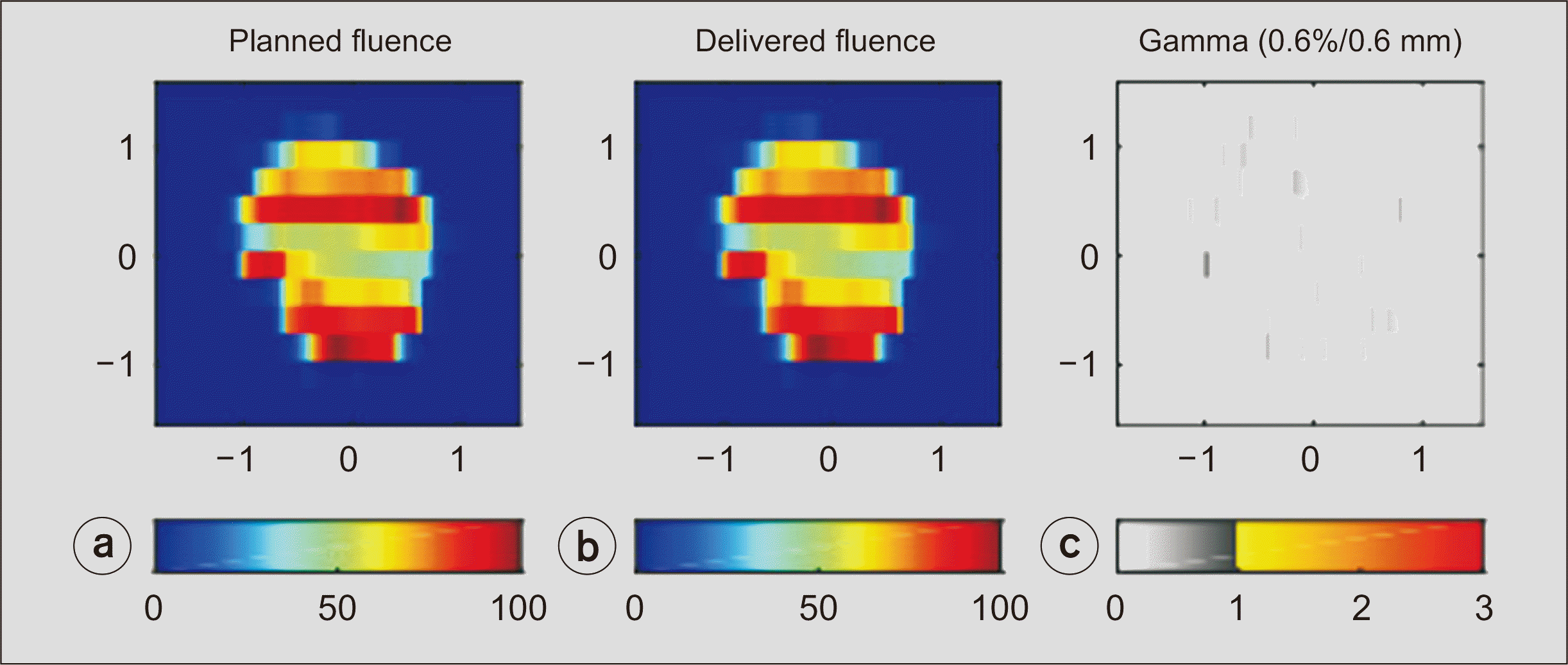
To evaluate their performance under varying conditions, both FractionLab and PDIP were assessed using gamma-passing rate criteria of 3%/3 mm and 2%/1 mm (Table 1). These criteria were used to evaluate each method’s agreement between the planned and delivered dose distributions. For both criteria, FractionLab exhibited a consistent increase in gamma-passing rates as the threshold values were relaxed from 0.1/0.1 to 1.0/1.0. Tables 2 and 3 summarize the mean gamma-passing rates of PDIP and FractionLab for HyperArc-based stereotactic radiosurgery in 43 patients, using various gamma criteria (0.1%/0.1 mm to 1%/1 mm). Table 2 uses a PDIP criterion of 3%/3 mm, while Table 3 uses 2%/1 mm. In this study, patient-specific QA was performed using both portal dosimetry with PDIP and log file–based analysis with FractionLab. An example of QA using PDIP is illustrated in Fig. 2, showing predicted and measured portal dose images and the corresponding gamma evaluation. Additionally, Fig. 3 displays a gamma scatter plot generated by FractionLab using MLC log files, providing visual insight into dose discrepancies across fields. Furthermore, Fig. 4 presents a representative case of FractionLab QA, demonstrating the planned versus delivered fluence distributions and their gamma comparison. In most cases, FractionLab has shown comparable or higher gamma-passing rates than PDIP [13]. However, under the strictest criterion of 2%/1 mm at 0.1/0.1, FractionLab’s passing rate (64.24%) was significantly lower than PDIP’s rate (89.43%), highlighting a limitation under specific conditions [14].
The statistical analysis further reinforced these findings (Table 2). Significant differences were observed under the 3%/3 mm criteria for specific conditions, including 0.1/0.1, 0.2/0.2, 0.3/0.3, and 1.0/1.0, with P-values of <0.001, <0.001, 0.001, and <0.001, respectively. For the 2%/1 mm criteria, significant differences were observed under the strictest (0.1/0.1) and most relaxed (1.0/1.0) conditions, while the intermediate criteria revealed no significant differences (P-values of >0.05). These findings indicate that FractionLab performs comparably to PDIP under less stringent conditions but requires further optimization to meet stricter criteria [9,11,16].
Discussion
The increasing adoption of HyperArc technology has underscored the need for robust QA protocols, particularly to address the complexities of small, irregularly shaped tumors and multiple metastases [4,5,8]. Traditional phantom-based QA methods have been considered the gold standard but are gradually being complemented or replaced by log file-based QA approaches [9]. These methods enhance operational efficiency and enable data-driven evaluations, making them particularly valuable for advanced radiotherapy techniques.
Several studies have confirmed the reliability and potential of log file-based QA methods in standard treatments. Previous research has demonstrated that log file-based QA tools, including FractionLab, achieve gamma-passing rates between 90% and 95% under the 3%/3 mm criteria in general IMRT and VMAT plans, showing strong agreement with conventional measurement-based QA approaches, such as EPID and film dosimetry [18]. Compared to other log file-based QA tools, FractionLab provides distinct advantages in workflow efficiency and data reconstruction.
Mobius3D (Varian Medical Systems) performs automated 3D dose verification and provides comprehensive dose-volume histogram analysis. However, its reliance on computationally demanding algorithms may limit its speed in high-volume clinics.
ArcCHECK (Sun Nuclear Corporation), which integrates log file-based analysis with detector array measurements, improves the spatial resolution but requires extensive setup time.
EPID-based QA methods are widely adopted for their efficiency in real-time fluence map verification. However, they may be less effective in detecting mechanical delivery errors compared to log file-based systems, such as FractionLab.
These comparisons emphasize that FractionLab offers a practical alternative by balancing accuracy, speed, and simplicity, making it particularly advantageous for HyperArc treatments in busy clinical settings [19].
These studies confirm the feasibility of log file-based QA in standard IMRT/VMAT treatments. However, FractionLab’s performance in more complex techniques, such as HyperArc, requires additional considerations. HyperArc’s steep dose gradients and complex beam configurations heighten sensitivity to positional deviations, potentially impacting FractionLab’s gamma-passing rates under stricter criteria. Unlike EPID-based methods, which inherently capture scatter effects and variations in beam modeling, log file-based QA methods like FractionLab rely on reconstructed fluence maps that may not fully account for these complexities. These differences underscore the need for refined QA strategies tailored to specialized treatment techniques, such as HyperArc.
Future studies comparing FractionLab with other log file-based QA systems, such as Mobius3D and ArcCHECK, in HyperArc-specific scenarios will help clarify its performance limitations and define appropriate QA thresholds for clinical adoption. Our study expands upon these findings by specifically evaluating FractionLab’s performance in HyperArc treatments, a specialized VMAT-based technique for SRS. While previous IMRT/VMAT studies have confirmed the general feasibility of log file-based QA, our results highlight its clinical applicability in highly complex, noncoplanar beam deliveries characteristic of HyperArc. By demonstrating comparable or superior performance to PDIP in this setting, our findings contribute to the growing evidence supporting log file-based QA as a robust alternative for psQA in advanced radiotherapy techniques [11,20]. Treatment planning and QA considerations become increasingly complex for patients with multiple metastases and non-single-isocenter plans. Single-isocenter multi-target approaches, commonly used in HyperArc, reduce treatment time by delivering radiation to multiple lesions simultaneously. However, this technique requires highly precise dose conformity to prevent dose spillage into surrounding healthy tissues. In contrast, multi-isocenter approaches offer greater control over individual tumor doses but increase overall setup complexity and potential geometric uncertainties.
When comparing QA methods in these scenarios, PDIP evaluates fluence maps from the actual beam and may be influenced by variations in isocenter alignment, particularly in multiisocenter treatments. Conversely, FractionLab reconstructs fluence maps from log files, enabling direct tracking of deviations in MLC positions and gantry angles across different isocenters. In this study, we observed that for multimetastatic cases using single-isocenter HyperArc plans, FractionLab and PDIP demonstrated comparable gamma-passing rates under the 3%/3-mm criteria. However, for non-single-isocenter treatments, FractionLab exhibited increased sensitivity to MLC positional variations, resulting in slightly lower gamma-passing rates under the strict 2%/1-mm criteria. These findings highlight the importance of tailored QA strategies based on treatment complexity and planning techniques [21,22]. Moreover, log file-based QA methods have demonstrated effectiveness in scenarios involving steep dose gradients and dynamic beam deliveries, confirming their adaptability and precision in complex treatment environments [8]. In this study, FractionLab demonstrated comparable or superior performance to PDIP across most criteria, particularly at 3%/3 mm, where gamma-passing rates were similar for both methods. However, FractionLab’s passing rate was lower under stricter conditions (2%/1 mm, 0.1/0.1). To ensure a fair comparison, the performance was analyzed using the same gamma criterion for each method.
Stricter gamma criteria were applied to FractionLab due to its ability to provide higher-resolution tracking of mechanical parameters, such as MLC positioning and gantry angle variations. Unlike PDIP, which evaluates fluence from the actual beam and is inherently affected by factors like scatter and detector noise, FractionLab reconstructs fluence maps from log files with sub-millimeter precision, making it more sensitive to minor deviations in machine performance. Due to this increased sensitivity, tool-specific criteria should be considered when interpreting the gamma-passing rates. Based on the results of this study, FractionLab’s performance should primarily be evaluated using the 2%/2 mm criteria to reflect its finer detection capabilities, while PDIP remains more suitable for evaluations at 3%/3 mm due to its broader detection range. These adjustments ensure fair and clinically relevant comparisons between the two QA tools [15,20].
Despite its promise, this study has several limitations. The small patient cohort and limited time frame have restricted the generalizability of the findings, emphasizing the need for larger, multicenter studies. Moreover, the limited literature on FractionLab’s application in HyperArc treatments underscores the need for further research. Future studies should explore the long-term outcomes of log file-based QA, its impact on workflow efficiency, and its role in diverse clinical settings. This will help establish its broader applicability and effectiveness, as well as explore the integration of FractionLab with Monte Carlo-based tools to enhance dosimetric accuracy, particularly in small-field SRS treatments [8,12].
Conclusions
This study demonstrated that FractionLab, a log file-based QA tool, performs comparably to or better than PDIP under the 3%/3-mm criteria while maintaining clinical feasibility under stricter criteria. Its consistent accuracy and operational efficiency highlight its potential as a valuable tool for HyperArc psQA in clinical practice.
By offering rapid, data-driven assessments, FractionLab has the potential to improve radiotherapy QA workflows while ensuring treatment accuracy. Its ability to reduce QA time while maintaining precision makes it particularly beneficial for high-volume radiotherapy centers. These advantages position FractionLab as an effective solution for improving operational efficiency in complex treatment environments, such as HyperArc.
However, since log file-based QA does not directly measure the patient dose, further investigations are required to evaluate FractionLab’s long-term reliability and safety in clinical practice. Log files provide detailed mechanical data. However, they may not fully capture factors, such as patient positioning errors, anatomical changes during treatment, or beam attenuation through immobilization devices. To mitigate these concerns, clinics implementing FractionLab should incorporate periodic measurement-based verification. This should be done alongside routine log file analysis to ensure comprehensive QA coverage.
Future research should focus on improving FractionLab’s sensitivity under stricter gamma criteria by refining its fluence reconstruction algorithms and improving its mechanical error detection capabilities. Additionally, integrating FractionLab with Monte Carlo-based tools may improve its dose calculation accuracy, particularly in regions with steep dose gradients. Investigating FractionLab’s feasibility in other complex treatments, such as SBRT for lung, liver, or other extracranial tumors, would offer valuable insights into its broader clinical applicability.
By combining operational efficiency with enhanced precision, FractionLab offers a practical and effective solution for streamlining QA workflows, reducing clinical workload, and improving patient safety in modern radiotherapy practices. As a result, FractionLab offers a practical and effective solution for enhancing QA protocols in modern radiation oncology practices.

 XML Download