



 PDF
 Citation
 Print


INTRODUCTION
The pituitary gland, known as the “master gland,” is crucial in regulating hormonal activities throughout the human body. The anterior pituitary gland, or adenohypophysis, produces six key hormones that regulate various endocrine functions, directly or indirectly influencing other endocrine glandular activities.1
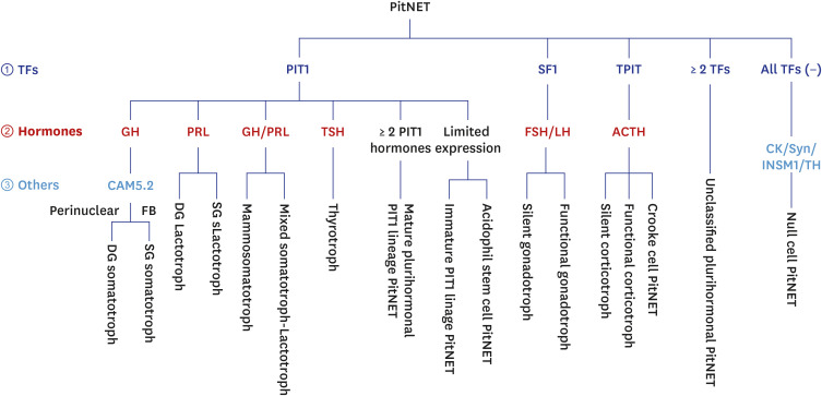
Embryologically, the adenohypophysis originates from Rathke’s pouch, giving rise to progenitor cells that differentiate into specific cell lineages under the influence of pituitary transcription factors (TFs) and signaling molecules.1 Three primary pituitary TFs govern cell lineage specification: pituitary-specific positive transcription factor 1 (PIT1), also known as POU1F1, encoded by POU1F1 (POU domain Transcription Factor Family 1) gene, Steroidogenic factor 1 (SF1), encoded by Nuclear Receptor Subfamily 5 Group A Member 1 (NR5A1), and T-box pituitary transcription factor (TPIT), encoded by TBX19.
PIT1 regulates the growth hormone (GH), prolactin (PRL), and thyroid-stimulating hormone (TSH) production in somatotrophs, lactotrophs, and thyrotrophs, respectively. SF1 controls the expression of follicle-stimulating hormone (FSH) and luteinizing hormone (LH) in gonadotrophs, while TPIT governs the production of adrenocorticotrophic hormone (ACTH) in corticotrophs by regulating the proopiomelanocortin (POMC) gene. Pituitary neuroendocrine tumors (PitNETs) arise from these lineage-specific cell types.2
Pituitary tumors represent a significant proportion of intracranial neoplasms, with their prevalence among primary CNS tumors was 16.5% during 2011–2015 and 17.2% during 2016–2020, according to the Central Brain Tumor Registry of the United States (CBTRUS).234 Among them, PitNETs are the most common tumor.
The classification of anterior pituitary tumors has evolved significantly over time. Historically it was based solely on hormone production, the 2017 WHO update introduced a system considering pituitary cell lineage and tumor hormone production.5 The 5th edition of WHO classification of the CNS tumors and endocrine and neuroendocrine tumors (WHO5) further defined this approach, renaming these tumors as PitNETs and updating classifications based on TF expression and hormone production. This new terminology reflects their epithelial and neuroendocrine differentiation of PitNET, characterized by the expression of cytokeratin (CK) and neuroendocrine markers, such as synaptophysin, insulinoma-associated protein 1 (INSM1), chromogranin A, and CD56.5678 Therefore, PitNET is different from the non-epithelial hormone-producing endocrine tumors, derived from the sympathetic and parasympathetic autonomic nervous system, which are CK-negative but synaptophysin-positive.
Further subclassification of PitNETs requires additional immunohistochemical (IHC) and genetic studies. Corticotrophs, somatotrophs, and lactotrophs are further subtyped based on their secretory granule abundance as densely or sparsely granulated.5
Unique subtypes include Crooke cell corticotroph PitNETs with Crooke’s hyaline change and acidophil stem cell PitNET with acidophilic or oncocytic cytoplasm.9 Molecular profiling and multi-omics data enable more precise subtyping, potentially guiding targeted therapeutic approaches.
The new classification significantly reduced the proportion of nonfunctioning tumors from 20–30% to less than 5%.910 Null cell tumors, defined as TF-negative and hormone-negative tumors, exhibit more aggressive clinical behavior compared to functional or silent PitNETs.101112 Tumors expressing multiple TFs are classified as unclassified plurihormonal PitNET according to the WHO5 classification of CNS tumors. The expression of multiple TFs or multiple lineage pituitary hormones characterize Plurihormonal PitNETs. This subtype represents an important category in the WHO5 new classification system, reflecting the complex nature of some pituitary tumors.
Changes in terminology and diagnostic criteria further exemplify the evolution of PitNET classification. The previous WHO classifications used “atypical pituitary adenomas” and “pituitary carcinoma.” Atypical adenomas are characterized by high mitoses (≥ 3/10 HPFs) and a high Ki-67 proliferating index (> 3%). However, this ‘atypical’ category was excluded from the current classification due to insufficient correlation with aggressive clinical behavior.
The definition of pituitary carcinomas has also evolved. Previously, they were defined as pituitary adenomas with metastatic spread to lymph nodes or discontinuous areas. In the updated WHO5 classification, these tumors are now termed “Metastatic PitNET” to more accurately reflect and provide a more precise classification based on behavior rather than histological features alone.”9
A notable change in the International Classification of Diseases for Oncology (ICD-O) has shifted the behavior code for PitNETs from “0” (benign) to “3” (malignant). This modification, while acknowledging the potential for malignant behavior in some PitNETs, fails to account for the benign characteristics exhibited by the majority of these tumors. The universal application of the ICD-O code 3 to PitNET may lead to inappropriate clinical management.13
Given the relative scarcity of research on the clinicopathological characteristics of PitNET among Korean patients, our study aimed to address this gap by analyzing surgically resected PitNETs.14 We focused on key demographic and clinical parameters, including gender distribution, age range, tumor size, and the prevalence of PitNETs subtypes. Additionally, we explored the diagnostic challenges associated with these tumors. This research is to enhance our understanding of PitNET and consequently improve their clinical management.
METHODS
Case selection
This study analyzed 210 pathologically confirmed cases of surgically resected PitNETs treated at Seoul National University Hospital (SNUH) between 2021 and 2023. All cases underwent trans-sphenoidal resection, and comprehensive data were collected by reviewing medical records, radiological findings, and archival information from SNUH.
Histopathological examination and immunohistochemical analysis
Histopathological examination, IHC studies, and serological hormone level assessments were performed to characterize the clinicopathological features of the surgically resected PitNETs.
Neutral formalin-fixed paraffin-embedded tumor tissues were sectioned at 3 μm thickness and stained with hematoxylin and eosin for histopathological examination. IHC staining was performed using the BenchMark ULTRA system (Ventana-Roche, Manheim, Germany) with a comprehensive panel of monoclonal and polyclonal antibodies targeting various TFs and pituitary hormones, and markers (Table 1). These include ACTH (1:500, Clone 02A3, DAKO, Glostrup, Denmark), ERα (1: 50, Clone 6F11, Novocastra, Newcastle, UK), FSH (1:200, Clone C10, DAKO), GATA3 (1: 3000, Clone L50-823, Cell Marque, Rocklin, CA, USA), GH (1:4,000, polyclonal, DAKO), Ki67 (1:100, clone MIB-1, DAKO), TPIT (1:700, clone CL6251, Abcam, Cambridge, MA, USA), TSH (RTU, clone 0042, DAKO), LH (1:400, C93, DAKO), PIT1 (1:500, clone EPR23555-203, Abcam), PRL (1:200, polyclonal, Ventana, Export, PA, USA), pHH3 (1:100, polyclonal, Cell Marque), P53 (1:100, Cone DO7, DAKO), SF1 (1:500, Clone EPR19744, Abcam).
Table 1
The primary antibodies used in this study
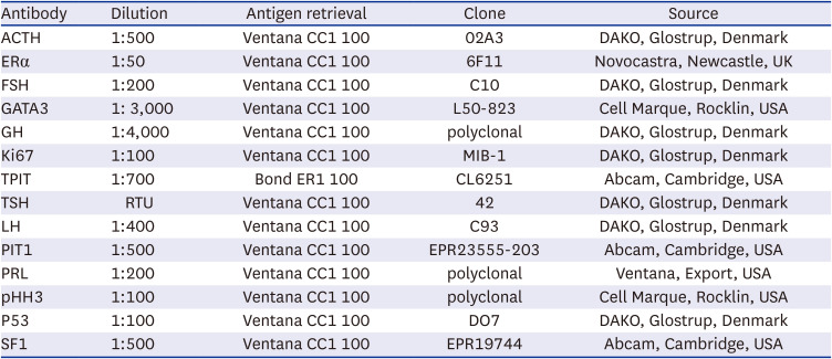
ACTH = adrenocorticotrophic hormone, FSH = follicle-stimulating hormone, GH = growth hormone, TPIT = T-box pituitary transcription factor, TSH = thyroid-stimulating hormone, LH = luteinizing hormone, PIT1 = pituitary-specific positive transcription factor 1, PRL = prolactin, SF1 = steroidogenic factor 1.
Pre-operative serum hormone levels were evaluated to distinguish between functional and non-functional PitNETs. This crucial step aids in identifying tumors that may be secreting hormones at clinically significant levels. IHC evaluation was performed on tumor tissue samples, focusing on the expression of six pituitary hormones. This comprehensive analysis serves two primary purposes: identification of silent PitNETs, characterized by the hormone expression in tumor tissue but lack of clinically relevant hypersecretion, and hormone profile to determine the specific hormone expression by the tumor tissue, if any. Mitotic figures were enumerated on slides with phosphohistone H3 (pHH3) IHC and the Ki-67 labeling index was determined by a computerized Ki-67-positive cell counting algorithm, which analyzed the area of the highest proliferative activity on digitalized images.
PitNET grading
This study employed a French five-tiered grading system, designed specifically for PitNETs, which is currently the sole grading system for these neoplasms.131516 Because p53 IHC expression does not indicate proliferation activity, we exclude p53 IHC evaluation to access proliferation activity.
The French grading system employs a multifaceted approach to PitNETs classification, integrating three key parameters: mitotic rate, Ki-67 proliferation index, and tumor invasiveness. Tumor invasion is determined through histopathological examination and radiological evidence, specifically cavernous or sphenoid sinus involvement. Proliferative PitNETs are characterized by the mitotic rate of ≥ 3/10 high-power fields and a Ki-67 index of > 3%. Based on these parameters, PitNETs are stratified into five distinct grades: Grade 1a: Non-invasive and non-proliferative tumors, Grade 1b: Non-invasive and proliferative tumors, Grade 2a: Invasive but non-proliferative tumors, Grade 2b: Both invasive and proliferative tumors, and Grade 3: Metastatic tumors with cerebrospinal or systemic metastases.
RESULTS
Clinical and radiologic features of PitNET
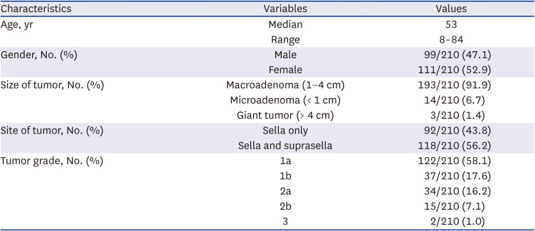
The study cohort comprised 210 patients with a median age of 53 years (range, 8–84 years), with a male-to-female ratio of 1:1.1 (Table 2). Magnetic resonance imaging revealed enhancing masses on T2-weighted sequences. The median tumor diameter was 2.5 cm (range, 0.1–6.5 cm). Macroadenoma (1–4 cm) accounts for 91.9% (193/210), microadenoma (< 1 cm) for 6.7% (14/210), and giant tumors (> 4 cm) for 1.4% (3/210) of cases. Tumor extension was observed in 56.2% (118/210), involving both sella and suprasella regions, while 43.8% (92/210) were confined to the sella.
Table 2
Clinicopathological summary of patients with PitNET
PitNET classification
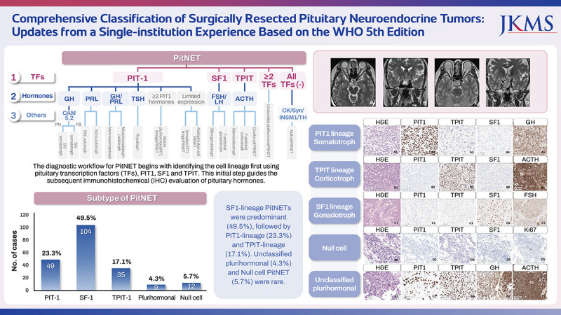
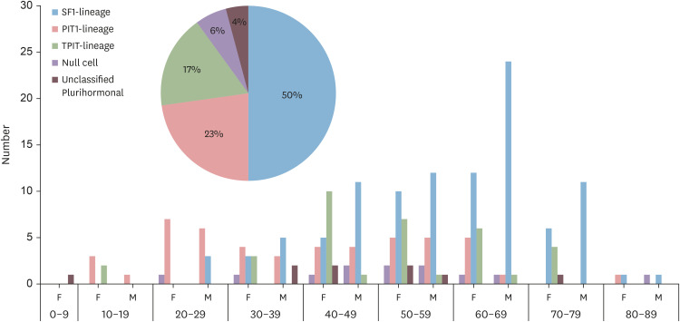
IHC analysis revealed that SF1-lineage PitNETs were the most prevalent (49.5%, 104/210), followed by PIT1 lineage (23.3%, 49/210) and TPIT-lineage (17.1%, 36/210). Null cell tumors (5.7%, 12/210) and unclassified plurihormonal PitNET (4.3%, 9/210) were rare (Table 3).
Table 3
Summary of distribution of PitNET subtypes
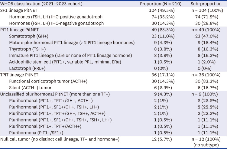
+ = positive, − = negative, PitNET = pituitary neuroendocrine tumor, SF1 = steroidogenic factor 1, FSH = follicle-stimulating hormone, LH = luteinizing hormone, GH = growth hormone, PIT1 = pituitary-specific positive transcription factor 1, TSH = thyroid-stimulating hormone, PRL = prolactin, ACTH = adrenocorticotrophic hormone, TPIT = T-box pituitary transcription factor.
Among PIT1 lineage PitNETs, somatotroph PitNETs were the most common (47.0%), followed by mature plurihormonal PIT1-lineage (18.4%), immature PIT-lineage (16.3%), and thyrotrophs (16.3%), while lactotroph tumors were absent.
SF1-family PitNETs predominantly affected men in their 60s and TPIT lineage tumors were most prevalent among women in their 40s–70s, while PIT1-lineage tumors occurred in men and women in their 20s–60s (Fig. 1). The remaining tumors occurred across a range of age groups.
Fig. 1
Age distribution of patients with a subtype of pituitary neuroendocrine tumor.
SF1 = steroidogenic factor 1, PIT1 = pituitary-specific positive transcription factor 1, TPIT = T-box pituitary transcription factor.
Using the French 5-tiered grading system,16 the distribution was as follows: grade 1a: 58.1% (122/210), grade 1b: 17.6% (37/210), grade 2a: 16.2% (34/210), grade 2b: 7.1% (15/210), and grade 3: 1.0% (2/210) (Table 2).
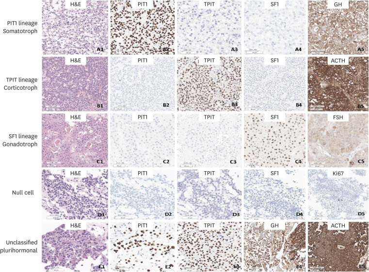
Histopathologically, PitNETs displayed a loss of normal lobular architecture, featuring sheets of monotonous cells with round nuclei (Fig. 2A1, B1, C1, D1, and E1). Fig. 2 illustrates the characteristic IHC staining patterns of TFs and hormones in key subtypes of PitNETs.
Fig. 2
Histopathological and immunohistochemical features of PitNET subtype. (A1-A5) Somatotroph PitNET: sparsely granulated somatotroph tumor with slightly eosinophilic cytoplasm (A1), positive for PIT1 (A2) and GH (A5), negative for TPIT (A3) and SF1 (A4). (B1-B5) Corticotroph PitNET: densely granulated corticotroph tumor with dense basophilic granular cytoplasm (B1), positive for TPIT (B3) and ACTH (B5), negative for PIT1 (B2) and SF1 (B4). (C1-C5) Gonadotroph PitNET: tumor forms sheets and contains focal perivascular pseudorosettes (C1), positive for SF1 (C4) and FSH (C5), negative for PIT1 (C2) and TPIT (C3). (D1-D5, E1-E5) Null cell tumor (D1) and plurihormonal (GH+ACTH) PitNET (E1) show sheet of monotonous cells with round nuclei and loss of normal lobular patterns. Null cell tumor do not express anterior pituitary cell lineage markers and is positive for ki-67 in 1.2% (D2-D5). Plurihormonal (GH+ACTH) PitNET shows positive for PIT1, TPIT, GH and ACTH (E2-E5).
H&E = hematoxylin and eosin, PIT1 = pituitary-specific positive transcription factor 1, TPIT = T-box pituitary transcription factor, SF1 = steroidogenic factor 1, GH = growth hormone, ACTH = adrenocorticotrophic hormone, PitNET = pituitary neuroendocrine tumor.
Functional and silent PitNET
Serological hormone level in each PitNET subtype revealed varying proportions of functional and non-functional tumors: Somatotrophs: 78% functional and 22% nonfunctional, Thyrotrophs: 13% functional and 87% nonfunctional, Plurihormonal PIT1 lineage PitNETs: 50% functional, 50% non-functional, Gonadotrophs; 21% functional and 79% nonfunctional, Corticotrophs: 47% functional and 53% nonfunctional, and Unclassified plurihormonal: 56% functional and 44% nonfunctional (Fig. 3). Among unclassified plurihormonal PiNET, 44% (4/9) were PIT1 and SF1 double positive cases (Somato-gonadotroph PitNET).
Fig. 3
Functional and non-functional tumors were classified based on serum hormone levels, with the following proportions across subtypes; 78% functional and 22% nonfunctional in somatotrophs, 13% functional and 87% nonfunctional in thyrotrophs, evenly split at 50% each in plurihormonal PIT1 lineage PitNETs, 21% functional and 79% nonfunctional in gonadotrophs, 47% functional and 53% nonfunctional in corticotrophs, and 56% functional and 44% nonfunctional in unclassified plurihormonal PitNETs. Immunohistochemical analysis revealed hormone IHC-positive or hormone IHC-negative subtypes within lineages: 71.2% gonadotrophins (FSH and LH or both)-positive and 28.8% gonadotrophs-negative in SF1 lineage, 83.3% ACTH-expressing and 16.7% ACTH-negaitve corticotrophs in the TPIT lineage.
PitNET = pituitary neuroendocrine tumor, IHC = immunohistochemical, PIT1 = pituitary-specific positive transcription factor 1, FSH = follicle-stimulating hormone, LH = luteinizing hormone, SF1 = steroidogenic factor 1, ACTH = adrenocorticotrophic hormone, TPIT = T-box pituitary transcription factor.
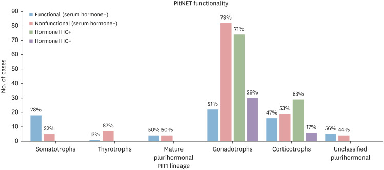
However, hormone (either FSH, LH or both) IHC expression was observed in 71.2% of gonadotroph PitNET (Fig. 3). Therefore, 50.0% of SF1 lineage tumors were silent gonadotrophs [IHC hormone-positive (71%) but no hormone hypersecretion (21%)]. This result represents hormone producing but not secreting PitNET, which is particularly relevant in the context of gonadotroph tumors, which often present as clinically non-functioning (79% in our cohort) despite their potential for hormone production (71.2% in our cohort).
The TPIT lineage revealed 83.3% ACTH-expressing and 16.7% ACTH-negative. Within the PIT1 lineage, the silent tumors may represent immature PIT1 lineage (formerly triple-negative pituitary adenomas).
Clinicopathological features of grade 3 PitNETs by French classifications
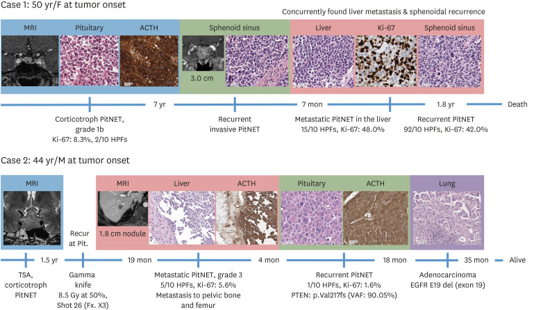
Our cohorts were composed of PitNETs with relatively short follow-up periods, thus, we could not find the survival difference between grades, however, two cases of grade 3 tumors show shorter progression-free survival. The clinicopathological features of these two cases are summarized in Fig. 4.
Fig. 4
Chronological progression of two cases of metastatic PitNETs.
ACTH = adrenocorticotrophic hormone, PitNET = pituitary neuroendocrine tumor, VAF = variant allele frequency.
Case 1
A 50-year-old woman was initially diagnosed with a grade 1b PitNET at the sella and suprasella area, 1 cm in diameter, without cavernous sinus involvement. Its Ki-67 index was 8.3% and mitotic rate was 2/10 HPFs. The tumor recurred after 7 years, invading the nasal cavity. Metastases to the liver and bones followed 7 months later. Liver metastasis showed aggressive features, nuclear atypia, 15 mitoses/10 HPFs and a Ki-67 index of 48.0%. Concurrently, a recurrent PitNET was identified in the sphenoid sinus. She was treated with electrochemotherapy with dacarbazine. Resection of liver and paranasal sinus mass with radiotherapy was given. She died after the latest radiotherapy. Her progression-free survival was 7 years and overall survival was 9.5 years after the initial detection of PitNET.
Case 2
A 44-year-old man initially diagnosed with corticotroph adenoma, 3.1 × 3.6 × 2.4 cm, through transsphenoidal resection at an outside hospital. The tumor recurred 18 months later, treated with gamma knife surgery (8.5 Gy, 50%, 26 times with 3 fractions). Liver and pelvic bone metastases occurred 19 months later, showing 5 mitoses/10 HPFs and a Ki-67 proliferation index of 5.6%. The liver mass was treated with stereotactic ablative radiotherapy (60 Gy) and pelvic bone metastasis was treated by RT (32 Gy). Cisplatin and electrochemotherapy with decarbazine were also given to the patient.
Four months after the metastasis diagnosis, the primary tumor was recurred in the pituitary site, which revealed only 1 mitosis/10 HPFs and a Ki-67 index of 1.6%, classifying it as histologically benign despite its metastatic behavior. Germline pathogenic PTEN (c.649delG, p.Val217fs) mutation was detected in this sellar recurrent PitNET with a high variant allele frequency of 90.05%. The patient developed lung adenocarcinoma (pathologic stage: pT1bN0) 22 months after metastatic PitNET diagnosis. Video-assisted thoracoscopic lobectomy was performed. He has been disease-free for 53 months since the last pituitary tumor recurrence and 8.0 years since her first PitNET episode.
DISCUSSION
The incidence of PitNET tends to increase with age, as reported by several studies.171819 CBTRUS reported that pituitary neoplasms accounted for 16.5–17.2% of surgically resected primary CNS neoplasms during 2011–2020 with the majority being PitNETs.34 The remainder included craniopharyngioma, posterior pituitary neoplasms (pituicytoma, granular cell tumor of sellar region, and spindle cell oncocytoma), and pituitary blastomas. Certain PitNET subtypes exhibit a higher prevalence in women, but there is no overall gender predilection. PitNETs are rare in pediatric population, although approximately 5% of patients are diagnosed before the age of 20 years.920 Consistent with previous reports9212223 our study found that macroadenomas (> 1 cm, < 5 cm) were the most common type, comprising 91.9% of all cases.
PitNET, formally known as pituitary adenoma, are generally categorized into three types as on their function: functional, non-functional, and null cell adenomas. Functioning PitNETs cause symptoms due to hormone excess, were reported to constitute 43% of PitNETs.9 The functional tumors comprised of somatotroph tumors being the most prevalent (18%), followed by lactotroph tumors (12%), corticotroph (5%), gonadotroph (5%), and mixed somatotroph-lactotroph tumors (1%), and thyrotroph (1%).923
Non-functioning tumors constituted 57.0% of all PitNETs.9 These tumors are characterized by the absence of clinically significant hormone related symptoms.9 Non-functional tumors can be further categorized into silent and null cell adenomas.
Silent tumors represent a subset within non-functional tumors. They can synthesize hormones but do not secrete them into the bloodstream at clinically detectable levels. Silent tumors account for approximately 15% of non-functional PitNETs and 10% of all PitNETs.24
Null cell adenomas are now defined by the absence of both hormone expression and pituitary-specific TFs. This new, stricter definition has dramatically reduced their prevalence from 30% of non-functioning adenomas to less than 5%.
Our study highlights the discrepancies between functional status based on serum hormone levels and IHC hormone expression profiles. This is particularly evident in gonadotroph PitNETs, which may express gonadotrophins immunohistochemically without causing clinically significant hormone elevations.
Contrary to various reports, our study revealed a unique distribution of PitNET subtypes: predominance of SF1 lineage PitNETs (49.5%), followed by PIT1 lineage (23.3%), TPIT lineage (17.1%), null-cell tumors (5.7%), and unclassifiable plurihormonal PitNETs (4.3%). This classification is based on WHO5 criteria, utilizing IHC analysis of TFs and pituitary hormones. Non-functional tumors can be categorized based on three features: asymptomatic, those without elevated blood hormone levels, and IHC hormone-negative.
In SF1 lineage PitNETs, the majority of tumors were clinically and serologically non-functional, with a functional to non-functional ratio of 2:8 at the tumor tissue level, most expressed FSH or LH, yielding a functional to non-functional ratio of 7.1:2.9. SF1 lineage PitNET also expresses additional markers, GATA3 or ERα.2526 GATA3-only expressing SF1 lineage tumors are considered the least differentiated and distinct from null cell tumors.12 TPIT lineage, corticotrophs were predominantly functional (ratio 8.3: 1.7). PIT1 lineage PitNET exhibited diverse subtypes: somatotroph (47%), mature PIT1 lineage (18.4%), immature PIT1 lineage (16.3%), thyrotroph (16.3%) and acidophilic stem cell (2.0%) (Table 3).
Hormone-negative tumors constituted 23% (49/210) of our cohort, including gonadotrophs (14.3%), corticotrophs (2.9%), thyrotrophs (0.5%) and null cell tumors (5.7%). Before the 2017 pituitary adenoma classification update, the terms “non-functioning” and “null cell” were often used interchangeably for “hormone-negative tumors based on IHC. However, The WHO5 criteria redefined this definition of null-cell PitNETs, requiring negativity for TFs and hormones. Additional testing with markers like tyrosine hydroxylase and CK may be necessary to rule out paraganglioma.57
This refined classification system significantly reduced the prevalence of null-cell PitNETs in our cohort from 42.4% to 5.7%, aligning with previous studies reporting varying functionality rate among PitNET subtype.1427 The diagnostic workflow for PitNET subtyping is shown in Fig. 5.
Fig. 5
The diagnostic workflow for PitNET begins with identifying the cell lineage first using pituitary TFs, PIT1, SF1 and TPIT. This initial step guides the subsequent IHC evaluation of pituitary hormones. If PIT1 is positive, the tumor belongs to PIT1 lineage, the corresponding hormones (GH, PRL, and TSH) should be examined. If SF1 is positive, the tumor is of gonadotroph lineage, necessitating FSH and LH IHC. If TPIT is positive, the tumor is corticotroph and ACTH staining is required for subtyping. The specific PitNET subtype is determined by the combination of TF expression and corresponding hormone IHC. If all TFs are negative, null cell PitNET should be considered. However, before confirming a null cell diagnosis, paraganglioma should be ruled out using TH IHC. To confirm the neuroendocrine tumor of pituitary, pan-neuroendocrine markers, like synaptophysin and INSM1, should be evaluated. PitNET should be negative for TH. Additionally, CK IHC can aid in distinguishing PitNETs from non-neuroendocrine tumors and DG and SG somatotroph. DG somatotrophs display perinuclear staining patterns, whereas SG somatotrophs exhibit intracytoplasmic FB in CK IHC.
PitNET = pituitary neuroendocrine tumor, TF = transcription factor, IHC = immunohistochemical, GH = growth hormone, PRL = prolactin, TSH = thyroid-stimulating hormone, CK = cytokeratin, TH = tyrosine hydroxylase, DG = densely granulated, SG = sparsely granulated, FB = fibrous bodies.
Our study cohort lacked lactotroph PitNETs, reflecting current best practices in pituitary tumor management. This absence is primarily due the preference for medical therapy over surgery as the first-line treatment for lactotroph PitNETs.72829
The management strategy for lactotroph PitNET considers factors such as tumor size, hormone levels, symptoms, and patient age. Dopamine agonists, particularly cabergoline and bromocriptine, are the preferred initial treatment. These medications effectively reduce prolactin secretion and often cause significant tumor shrinkage.
Cabergoline is generally preferred due to its superior efficacy and better tolerability. Medical therapy is especially advantageous for patients with microadenomas, individuals wishing to preserve fertility, or those seeking to avoid surgical intervention.
Certain PitNET subtypes have been identified as having a higher propensity for early recurrence and treatment resistance. The WHO 2017 classification identified several subtypes as “high-risk,” including male lactotroph tumors, silent corticotrophs, Crooke cell PitNETs, sparsely granulated somatotroph tumors, and silent plurihormonal PIT1 lineage tumors, acidophilic stem cell tumors, Null cell, lactotroph in men, and metastatic PitNETs.9 Each subtype exhibits unique histopathological and clinical features that complicate their management.
Sparsely granulated somatotroph PitNETs are more aggressive, larger at diagnosis, and resistant to somatostatin analogs. Silent corticotroph PitNETs tend to have a more aggressive course, later-stage diagnosis, and a higher recurrence rate. Crooke cell corticotroph PitNETs are rare, aggressive, and resistant to conventional treatment. Plurihormonal PIT1-positive PitNETs can produce multiple hormones and aggressive growth, complicating diagnosis and management. Lactotroph PitNETs in men tends to present later, are larger and more invasive, more aggressive, and resistant to dopamine agonists. Metastatic PitNETs, formerly called pituitary carcinomas, are difficult to treat due to their metastatic potential and lack of reliable predictive histopathological features.
These extended observations would provide more robust data for prognosis and treatment planning, ultimately improving patient care and outcomes. A multidisciplinary approach involving endocrinologists, neurosurgeons, and oncologists is often required to optimize the outcomes of these aggressive PitNETs.
In a retrospective analysis across multiple centers, including data from Lyon, grade 1a (noninvasive and non-proliferative) tumors were most prevalent, accounting for 47.3% to 51.2% of cases. Conversely, grade 2b tumors (invasive and proliferative) were less common, ranging from 7–8%.13
Our results revealed a higher proportion of non-invasive tumors compared to the previous study by Raverot et al.30 Specifically, grades 1a and 1b collectively represented 75.7% (grades 1a: 58.1% and 1b: 17.6%) of our cases, in contrast to 58.9% in Raverot et al.’s study.30 We observed a lower percentage of invasive tumors, particularly grade 2a (16.2% of our cohort vs. 32.3% of Raverot et al.’s cohort).30 The proportion of grade 2b tumors was comparable between studies (7.1% vs. 8.8%). Notably, we identified two case (1.0%) of grade 3 tumor, which was absent in Raverot et al.’s cohort,30 highlighting the potential for aggressive behavior and metastasis even in conventionally classified PitNETs.
Most PitNET in our study demonstrated a relatively benign clinical course, aligning with the generally indolent nature of most pituitary tumors. However, this observation should be interpreted cautiously due to the limited follow-up period of the study.
Previous studies have demonstrated the prognostic value of this grading system, showing significant differences in recurrence-free survival rates across tumor grades (P < 0.001). Their study showed 5-year recurrence-free survival rates of 87.1%, 84.5%, 55.6%, and 31.6% for grades 1a, 1b, 2a, and 2b, respectively.30 Tumor invasiveness emerged as a significant predictor of recurrence (P < 0.001).
We emphasize the importance of long-term follow-up for all PitNET patients, even those initially classified as lower grade. Comprehensive molecular profiling may provide valuable insights into factors contributing to aggressive behavior and metastatic potential.
Long-term studies are needed to fully validate the prognostic value of the French grading system and establish definitive correlations with patient outcomes.
In conclusion, this study provides a comprehensive analyses of surgically resected PitNETs using the new WHO5 classification system. Key findings include the predominance of SF1-lineage tumors, particularly gonadotroph PitNETs, and a notable absence of lactotroph PitNETs in our cohort. A significant finding was the discrepancy between serologically non-functional and immunohistochemically gonadotroph-positive tumors among SF1-lineage PitNETs. This study also applied the French five-tiered grading system, but noted that its prognostic value needs further verification through longer-term follow-up studies with more cases.
The research represents an important step in translating the WHO5 classification into clinical practice and understanding its implications for patient care in pituitary tumor management. Future research should focus on long-term follow-up studies and the integration of molecular markers to enhance the accuracy of tumor classification and improved prognostication in PitNET cases.
 XML Download