



 PDF
 Citation
 Print


INTRODUCTION
Inherited retinal degeneration (IRD) comprises a group of heterogeneous diseases characterized by the progressive loss of photoreceptors and/or retinal pigment epithelial cells. These diseases differ clinically as well as genetically and are caused by anomalies in various cell structures and pathways, including phototransduction cascades, retinal transcription factor-related pathways, RNA splicing machinery, retinal metabolism, retinal cell structure, and ciliary structure and function.12 Retinitis pigmentosa (RP) is considered the most prevalent and well-known IRD, affecting over 1 million patients worldwide and one in 4,000 people.3
Before the era of genetic diagnosis, IRDs were primarily diagnosed based on clinical findings. Phenotypes, including locations of main lesions, affected cell types, disease progression patterns, and systemic involvement, were considered.4 However, rapid progress in genetic examinations led to a phase during which many previous clinical diagnoses were revealed to be genetically heterogeneous, calling into question previous diagnostic categorizations. A single phenotype can be caused by multiple genes (genotypic heterogeneity), and several variants in a single gene result in various phenotypes (phenotypic heterogeneity). Genetic testing has become an essential tool in diagnosing IRD, including incomplete penetrance and unclear inheritance.
As molecular genetic testing is not a widely accessible form of examination readily available in all medical institutions, patients tend to undergo genetic testing after being clinically diagnosed. In many cases, patients decide to undergo genetic testing many years or even decades after the clinical diagnosis for several reasons: they are often afraid to face the results, are misinformed, or the tests are costly. In some cases, the final diagnosis is changed according to genetic testing results.
We analyzed a Korean cohort of patients clinically diagnosed with RP using whole-exome sequencing (WES). The aim of this study was to determine the mutation spectrum and allele frequency and to observe the final diagnoses in this cohort after sequencing.
METHODS
Study population
This observational, open-label cohort study was conducted at a single tertiary hospital (Soonchunhyang University Hospital, Bucheon). Patients referred to the Soonchunhyang University Hospital Bucheon with a clinical diagnosis of RP who underwent WES between January 2017 and February 2018 were enrolled. WES was performed on 145 patients from 100 families.
DNA extraction, libraries construction, and sequencing
WES procedures, including exome capture and sequencing, were performed by Oneomics Co., Ltd. (Gimpo, Korea). Libraries were prepared using the IDT xGen Exome Hyb Panel v2 Library Preparation Kit (Integrated DNA Technologies, Coralville, IA, USA) according to the manufacturer’s instructions.
DNA integration was assessed using gel electrophoresis and a qubit 2.0 fluorometer (Thermo Fisher Scientific, Waltham, MA, USA) for DNA degradation, contamination, and concentration. High-quality genomic DNA (1 μg) from each sample was used for the Agilent SureSelect Human All Exon v5 capture process (Agilent Technologies, Santa Clara, CA, USA). Randomly fragmented DNA was end-repaired, subjected to polyA tailing at the 3′ end, ligated with an indexing-specific paired-end adapter, and amplified according to the manufacturer’s protocol (Agilent Technologies). Qualified exome-captured libraries were sequenced with a throughput of at least 100× coverage using a HiSeq 4000 according to the manufacturer’s protocol (Illumina, San Diego, CA, USA).
Mapping, filtering, calling joint variants, filtering variants, and annotation
Several quality checks were performed before calling the variants to avoid false-positives. Furthermore, several quality checks were performed using FastQC, including assessment of the sequencing data quality, guanine-cytosine content, adapter sequence residues, overrepresented sequences, and duplication levels to detect sequencing errors, polymerase chain reaction (PCR) duplication, and adapter contamination. The remaining adapters were trimmed using CutaDapt software. FastQC and Cutadapt were processed using the QC analysis pipeline, TrimGalore (https://github.com/FelixKrueger/TrimGalore). Single-nucleotide polymorphism and insertion-deletion calling was performed by following the GATK4 pipeline introduced by the Broad Institute.56
Reads were mapped to the human reference genome, hg19, using Burrows-Wheeler Aligner–MEM software, and PCR-duplicated reads were eliminated using the Genome Analysis Toolkit (GATK, version 4.4.0.0) MarkDuplicates. After eliminating duplicate reads, base quality score recalibration was performed to adjust the base quality score of the misaligned reads using the GATK BaseRecalibrator. For recalibrated reads, variants were called using HaplotypeCaller in GATK. Hard filtering of variants was performed separately for single-nucleotide polymorphisms and insertions-deletions using GATK VariantFiltration based on their respective types. The filter-passed variants were annotated using SnpEff and SnpSift in various human databases.
For further analysis, the open-source tool TAPES was employed to predict and transform annotated variants according to the categorical criteria established by the American College of Medical Genetics and Genomics (ACMG).7 After prediction, we filtered the variants using the following criteria to focus on inherited retinal diseases: only variants that affected the protein structure were selected and filtered from the Retinal Information Network database. Variants with a frequency greater than 0.01 in the gnomAD (version 2.1.1) East Asian population were removed. The filtered variants were evaluated using PolyPhen-2, SIFT, CADD, FATHMM, MutationAssessor, and MutationTaster to predict their pathogenicity.
Bioinformatic analysis
All identified variants were classified according to the guidelines of the ACMG and Association for Molecular Pathology (AMP) as follows: pathogenic, likely pathogenic, variants of uncertain significance, likely benign, or benign.8 The pathogenicity of variants of unknown significance in patients were re-evaluated according to known clinical features and inheritance patterns.
Variants were considered putative causative when one of the following criteria was met: 1) the mutation was classified as pathogenic or likely pathogenic according to ACMG/AMP guidelines; 2) variants of uncertain significance were as putative causative variants when the allele frequency was below 0.1% across all populations; more than half of the six pathogenicity prediction tools including PolyPhen2,9 SIFT,10 CADD,11 FATHMM,12 Mutation Assossor,13 and Mutation Taster14 indicated deleterious effects; and the clinical presentation was consistent. Variants incompatible with known phenotypes or inheritance patterns were excluded from analysis. Familial WES results were used to determine inheritance patterns when available.
Patients were determined to have causative variant(s) when 1) one pathogenic variant was present in a gene with autosomal dominant or X-linked inheritance, and 2) two heterozygous variants or one homozygous pathogenic variant was present in a gene with autosomal recessive inheritance. For cases with autosomal recessive traits, segregation analysis was performed when possible, and segregation was additionally inferred using co-occurrence of variants when available.15 Cases that did not meet these criteria were classified as unsolved.
RESULTS
Demographic characteristics
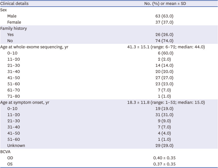
We recruited 145 patients from 100 families who underwent WES. We analyzed 100 unrelated Korean probands clinically diagnosed with RP at other institutions (Table 1). Of the 100 probands, 63 were male (63.0%) and 37 were female (37.0%). The mean age at genetic testing was 41.3 ± 15.1 years (range: 6–72; median: 44.0). The mean age at symptom onset was 18.3 ± 11.8 years (range: 1–52; median: 15.0). Initial symptoms most often occurred in the age range of 11–20 years (31/100, 31.0%), followed by 0–10 years (19/100, 19.0%). Twenty-six (26.0%) probands had a family history of RP.
Table 1
Baseline characteristics and clinical details of 100 Korean patients with clinically diagnosed retinitis pigmentosa after molecular diagnosis
Identification of pathogenic variants
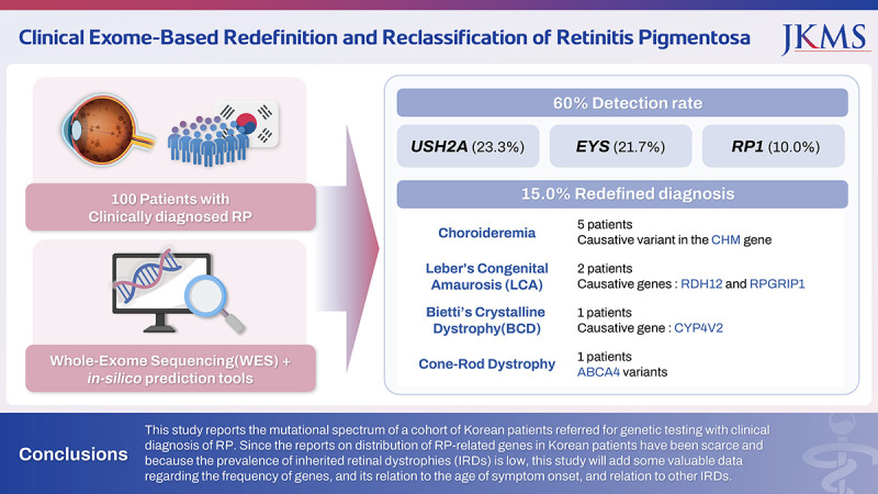
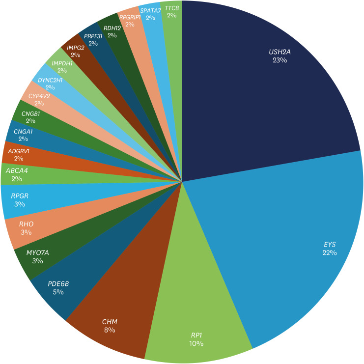
We detected 104 pathogenic variants in 21 known IRD-related genes in 60/100 (60.0%) patients (Supplementary Tables 1 and 2).1617181920212223242526272829303132333435363738394041424344 Among these variants, 86/104 (82.7%) were previously reported to be associated with IRD and 18/104 (17.3%) were novel variants in defined IRD genes. Among the defined pathogenic variants, 39/104 (37.5%) were missense variants, 32/104 (30.8%) were frameshift deletions or insertions, 22/104 (21.6%) were stop-gain variants, and 11/104 (10.6%) were predicted to affect splice regions. USH2A variants were the most common (31/104, 29.8%), followed by those in EYS (24/104, 23.1%) and RP1 (10/104, 9.6%).
Definite causative genes were detected in 60/100 patients (60.0%) (Fig. 1). Of these 60 cases, USH2A was the most common causative gene (14/60, 23.3%), followed by EYS (13/60, 21.7%) and RP1 (6/60, 10.0%). Of the five patients with RP with autosomal dominant inheritance, RHO (2/5, 40.0%) was the most common causative gene. Among 48 RP patients with autosomal recessive inheritance, USH2A (14/48, 29.2%) and EYS (13/48, 27.1%) were the most common causative variants. Patients with autosomal recessive with RP1 as causative variants were revealed to carry an Alu insertion (case 21).2045 Of the seven patients with X-linked inheritance, five were re-diagnosed with choroideremia (5/7, 71.4%), and the other two patients with X-linked RP carried RPGR as the causative gene (2/7, 28.6%).
Mutational spectrum according to age of symptom onset
The frequency of causative genes in each age group was analyzed based on the age at which initial symptoms developed. In the group aged < 10 years, the causative gene was detected in 8 of 19 probands, with a detection rate of 42.1%. The most common gene in this group was PDE6B (2/8, 25.0%). In the group aged 11–20 years, the gene was detected in 20 of 31 probands (64.5%), and the most common gene was USH2A (6/20, 30.0%), followed by EYS (4/20, 20.0%). In the group aged between 21 and 30 years, 8 of 9 probands had the causative gene detected in 8 of 9 probands (88.9%), with the most common gene being EYS (4/8, 50%). In the group aged 31–40 years, the causative gene was detected in 3 of 7 probands (42.9%); the most common gene was USH2A (2/3, 66.7%). In the group aged > 41 years, two of five patients had the causative gene with the lowest detection rate of 40.0%; these genes were CHM and RP1 (1/2 and 50%).
Mutational spectrum according to family history of RP
The frequency of causative genes according to the family history of RP was analyzed. The causative gene detection rate was 69.2% (18/26) when the proband had a family history of RP. The most common gene was USH2A (6/18, 33.3%), followed by EYS (3/18, 16.7%) and CHM (2/18, 11.1%). In patients with no known family history of IRD, the detection rate was lower at 56.8% (42/74), with the most common gene being EYS (10/42, 23.8%), followed by USH2A (8/42, 19.0%) and RP1 (5/42, 11.9%).
Redefining clinical diagnosis based on genetic diagnosis
After confirming the causative genes, clinical diagnoses were redefined based on the genetic findings (Table 2). Five of the 60 probands (8.3%) carried the causative variant of CHM, and the clinical diagnosis was redefined as choroideremia (Table 3). Leber congenital amaurosis was diagnosed in 2/60 probands (3.3%), with RDH12 and RPGRIP1 as the causative genes in each patient. One patient (1/60, 1.7%) was diagnosed with Bietti’s crystalline dystrophy after CYP4V2 was identified as the causative gene. In another patient (1/60, 1.7%), ABCA4 variants were detected with clinical findings suggestive of cone-rod dystrophy.
Table 2
Redefined clinical diagnosis of 60 of 100 Korean patients with clinically diagnosed retinitis pigmentosa after molecular diagnosis
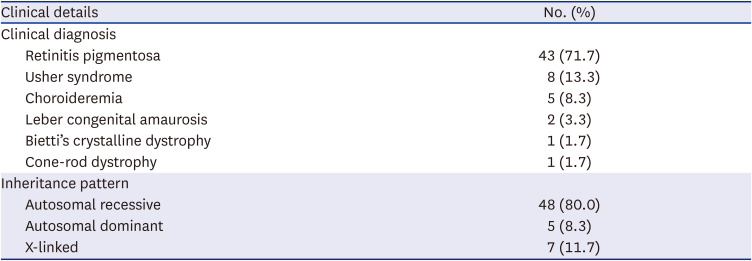
Table 3
Causative variants of patients with redefined diagnosis other than retinitis pigmentosa
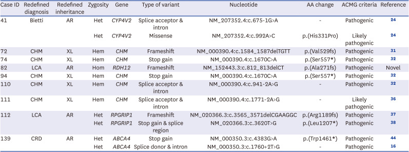
| Case ID | Redefined diagnosis | Redefined inheritance | Zygosity | Gene | Type of variant | Nucleotide | AA change | ACMG criteria | Reference |
|---|---|---|---|---|---|---|---|---|---|
| 41 | Bietti | AR | Het | CYP4V2 | Splice acceptor & intron | NM_207352.4:c.675-1G>A | - | Pathogenic | 24 |
| Het | CYP4V2 | Missense | NM_207352.4:c.992A>C | p.(His331Pro) | Likely pathogenic | 24 | |||
| 72 | CHM | XL | Hem | CHM | Frameshift | NM_000390.4:c.1584_1587delTGTT | p.(Val529fs) | Pathogenic | 31 |
| 74 | CHM | XL | Hem | CHM | Stop gain | NM_000390.4:c.1670C>A | p.(Ser557*) | Pathogenic | 32 |
| 82 | LCA | AR | Hom | RDH12 | Frameshift | NM_152443.3:c.812_813delCT | p.(Ala271fs) | Pathogenic | Novel |
| 94 | CHM | XL | Hem | CHM | Stop gain | NM_000390.4:c.1670C>A | p.(Ser557*) | Pathogenic | 32 |
| 110 | CHM | XL | Hem | CHM | Splice acceptor & intron | NM_000390.4:c.941-2A>G | - | Pathogenic | 32 |
| 111 | CHM | XL | Hem | CHM | Splice acceptor & intron | NM_000390.4:c.1771-2A>G | - | Likely pathogenic | 36 |
| 112 | LCA | AR | Het | RPGRIP1 | Frameshift | NM_020366.3:c.3565_3571delCGAAGGC | p.(Arg1189fs) | Pathogenic | 37 |
| Het | RPGRIP1 | Stop gain & splice region | NM_020366.3:c.3620T>G | p.(Leu1207*) | Pathogenic | 38 | |||
| 139 | CRD | AR | Het | ABCA4 | Stop gain | NM_000350.3:c.4383G>A | p.(Trp1461*) | Pathogenic | 44 |
| Het | ABCA4 | Splice donor & intron | NM_000350.3:c.1760+2T>G | - | Pathogenic | 16 |
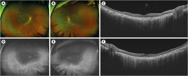
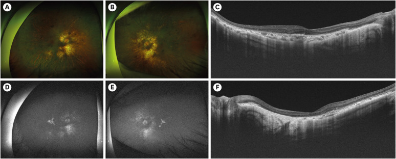
There were three representative redefined clinical diagnosis cases and a case with RP with a rare variant. Case 41 was initially diagnosed with RP due to diffuse photoreceptor, retinal pigment epithelium, and choriocapillaris degeneration, although bony spicules were not prominent (Fig. 2). WES identified CYP4V2 as the causative gene, which led to a revised diagnosis of Bietti’s crystalline dystrophy. Despite not being initially recognized, some shiny yellow deposits in the posterior pole, consistent with Bietti’s crystalline dystrophy, were observed in the fundus and red-free photography.
Fig. 2
Clinical phenotype of case 41 whose diagnosis was redefined as Bietti’s crystalline dystrophy. (A, B) Wide-field fundus photography. (C, F) Optical coherence tomography B-scans including foveal center. (D, E) Fundus autofluorescence photography.
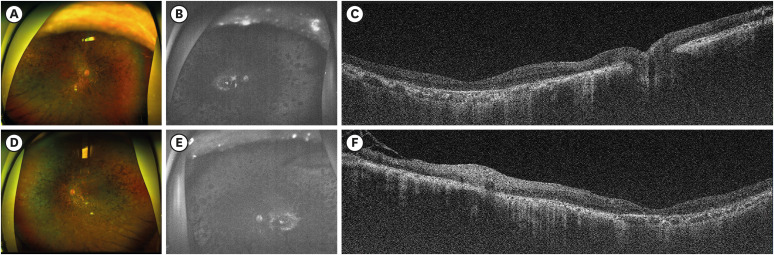
In case 76, the patient was clinically diagnosed with RP 7 years before molecular testing. His elder brother and sister were also diagnosed with RP, making his younger brother the only sibling without a clinical diagnosis of RP. His fundus photos showed extensive bony-spicule pigmentation with near full destruction of the outer segment, sparing only a portion of macular center (Fig. 3). A novel homologous variant of TTC8 was identified using WES.
Fig. 3
Clinical phenotype of case 76 in which a novel homologous variant of TTC8 was identified as the causative variant of retinitis pigmentosa. (A, B) Wide-field fundus photography. (C, F) Optical coherence tomography B-scans including foveal center. (D, E) Fundus autofluorescence photography.
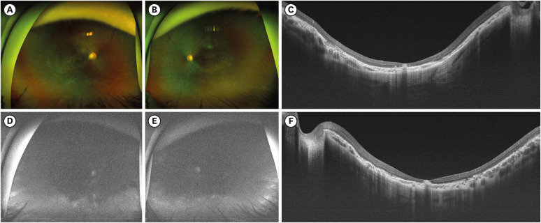
The patient in case 82 developed symptoms immediately after birth, including nystagmus, and was clinically diagnosed with RP because of diffuse photoreceptor degeneration with diffuse bony spicule pigmentation (Fig. 4). However, this patient did not undergo genetic testing. WES revealed pathogenic variants of RPGRIP1, which led to a redefined Leber congenital amaurosis diagnosis.
Fig. 4
Clinical phenotype of case 82 whose diagnosis was redefined as Leber’s congenital amaurosis. (A, B) Wide-field fundus photography. (C, F) Optical coherence tomography B-scans including foveal center. (D, E) Fundus autofluorescence photography.
In case 110, RP was clinically diagnosed based on advanced photoreceptor, retinal pigment epithelium, and choriocapillaris degeneration, sparing only a portion of the macular center with diffuse bony spicule pigmentation (Fig. 5). A pathogenic variant of CHM was identified using WES, and the clinical diagnosis was redefined as choroideremia.
DISCUSSION
In the present study, 100 unrelated probands underwent WES to identify suspected clinical RP causative mutations. The overall diagnostic yield was 60.0%, with USH2A (23.3%), EYS (21.7%), and RP1 (10.0%) as the most frequent causative genes. Additionally, the mutational spectrum was diverse and included various genes. The results of some molecular diagnoses led to a change in the previous clinical diagnoses, with choroideremia being the most notable.
The detection rate of causative genes in this study (60.0%) was comparable or slightly higher than that of previous studies, which ranged from 37% to 57.7%.18212533464748 There have been several reports on the varying genetic spectra of IRDs, including RP, among different regions, even in countries that are geopolitically close of East Asia.18253346 Previous studies showed that EYS variants account for the largest portion of causative genes in Japan, with prevalence ranging from 23.5% to 51.0%.464950 In contrast, USH2A variants are the most common causative mutations in European, Chinese, and Taiwanese populations, with a prevalence of 20–40%.151525354 Reports from South Korea have been mixed regarding whether EYS and USH2A is the most common among causative genes, with one study reporting EYS as the most common (8.2%), followed by USH2A (6.8%) and another study reporting USH2A as the most common (22.4%), followed by EYS (10.3%).1825 Our results support the possibility of USH2A being the most common causative gene in the Korean population, although the proportions of USH2A and EYS mutations in the spectrum may not differ widely. This result also suggests a unique mutational spectrum of patients with RP in Korea, which differs from that of both Japan and China.
The prevalence of third most common mutation, RP1, was 10.0%, which is higher than that in Japanese and Chinese patients with RP (3–4%).4653 This result is in accordance with a previous report of causative variants of Korean patients with IRD, which reported a prevalence of 5.8% for the RP1 mutation, ranking fourth after USH2A, ABCA4, and EYS.18
The genetic distribution varied among age groups at symptom onset. In the group aged < 10 years, PDE6B variants were the most common, accounting for 25.0% of causative genes, whereas in the group aged 11–20 years, USH2A (30.0%) and EYS (20.0%) were the most common genes. EYS was the most common causative gene in patients aged 21–30 years, accounting for 50% of all cases. These results were in line with the clinical characteristics of RP caused by variants of each gene; patients with PDE6B variants tend to experience initial symptoms before age 10 years, whereas patients with USH2A and EYS variants experience the first symptoms in late adolescence or adulthood.25515355
Patients with a family history had a higher rate of causative gene detection (69.2%) than those without a family history (56.8%). This rate was in accordance with previous results showing lower detection rates in sporadic cases than in familial cases. This result may have been obtained because many genes causing sporadic RP have yet to be discovered, with the possibility of non-genetic factors affecting sporadic cases.5657
In 40.0% (40/100) of cases, no causative variant was confirmed, possibly because of the innate limitations of WES. Approximately 85% of known causative mutations occur in exonic regions that encode proteins, indicating that WES cannot identify the remaining 15% of causative mutations. In addition, some genetic regions have low or no coverage in the exome sequencing approach.18 Patients may have shown clinical characteristics closely resembling RP because of non-genetic causes. Further studies involving WES and an accurate clinical diagnosis are warranted to resolve these cases.
This study had several limitations. First, this was a single-institution-based retrospective study and could have been influenced by selection bias. Most patients were referred from other institutions for genetic testing and therefore could not be followed from the time of initial symptom onset or clinical diagnosis. Although genetic testing of family members was conducted when possible, it was not performed in all patients. Thus, we relied on patients’ self-reports to evaluate the presence of a family history in some cases. Further analysis including copy number variation analysis was not available. Systemic evaluations were not performed, including a hearing test for Usher syndrome.
In conclusion, we report the mutational spectrum of a cohort of Korean patients referred for genetic testing with a clinical diagnosis of RP. As reports on the distribution of RP-related genes in Korean patients are scarce and the prevalence of IRDs is low, this study adds valuable data regarding the frequency of genes, their relation to the age of symptom onset, and their relation to other IRDs.
 XML Download