



 PDF
 Citation
 Print


INTRODUCTION
Clonorchis sinensis Looss, 1907 is one of common human infecting zoonotic trematodes in Family Opisthorchiidae, Class Trematoda. Since McConnell first discovered adult worms of C. sinensis from an infected Chinese man in 1874 and published his recovery in The Lancet in 1875,1 it has been targeted of research for biology, epidemiology, pathology, and clinical studies. Clonorchiasis, human infection by C. sinensis, is transmitted by eating raw freshwater fish, and it has been endemically transmitted in East Asia including China, Korea, East Russia, Japan, and northern Vietnam. Although clonorchiasis was eliminated in Japan, over 15 million infected population is still estimated in the endemic areas until present.2
The World Health Organization (WHO) is running many programs to control or eliminate neglected tropical diseases (NTDs) under the discipline of the United Nation’s Sustainable Development Goals (SDGs) by 2030.3 WHO enlists 20 target NTDs of priority by 2030 including food-borne trematodes, such as fascioliasis, clonorchiasis, opisthorchiasis, and paragonimiasis. According to the SDGs strategy, clonorchiasis is one of target NTDs for control or elimination by 2030. At present, clonorchiasis is the only one food-borne trematodiasis for which mass drug administration is recommended in the areas where its prevalence is > 10% by WHO Western Pacific Region.4
One evolutionary study reviewed DNA evidence and speculated that human liver flukes, such as Clonorchis, Fasciola, Opisthorchis, and Dicrocoelium, spread to human from animals in early prehistoric era.5 Several archaeology studies reported that clonorchiasis was widely and heavily endemic over thousand years in East Asia and Korea.6789
In Korea, clonorchiasis had been widely prevalent in the past under favorite natural and social epidemiology environment. Total 8 national status surveys were made in Korea for intestinal parasites from 1971 to 2012 and reported egg positive rates of C. sinensis from 1.4% to 4.6% by fecal examination.10 The status of intestinal parasites in Korea has been characterized by prevailing endemicity of soil-transmitted helminths during 1970s, rapid declining during 1980s, and elimination of disease in 1990s.11 The soil-transmitted helminths, especially Ascaris, had been successfully eliminated over the country in Korea thanks to national efforts,12 but C. sinensis remained highly endemic by 2010s.
In 1994, the International Agency for Research on Cancer (IARC) classified C. sinensis in Group 2A as a probable biological carcinogen to human with limited evidence while put Opisthorchis viverrini in the Group 1. Later in 2009, the IARC Monograph Working Group for Volume 100B discussed the issue and reclassified C. sinensis in the Group 1 biological carcinogen.13 The reclassification in 2009 meant that scientific evidence for carcinogenesis by C. sinensis was sufficient and facilitated research on mechanism of the carcinogenesis. The present review article updates basic knowledge on clonorchiasis and study findings of carcinogenesis by C. sinensis.
In other words, O. viverrini, the taxonomically nearest fluke in the Family Opisthorchiidae, was grouped in the Group 1 already in 1994 with sufficient research evidence for its carcinogenesis of human cholangiocarcinoma (CCA). After the grouping in 1994, O. viverrini has been investigated actively for the pathogenesis and carcinogenic mechanism by several Thai and international researchers in Thailand.14
C. SINENSIS AND CLONORCHIASIS
Biology
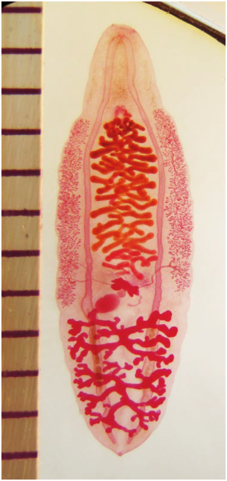
C. sinensis is a slender, leaf-shaped digenetic trematode in the Family Opisthorchiidae, Class Trematoda. Mature worms measure 10–15 mm long, 4–5 mm wide, and 1 mm thick (Fig. 1). The anterior body, from ventral sucker to oral sucker, is sharp and slender, while the posterior end is round. The anterior part is moving actively while they are alive. The worms live in the intrahepatic or extrahepatic bile duct of mammals such as humans, dogs, cats, pigs, and rats. A few worms may be found in the intestine. Experimentally, rabbits and guinea pigs are highly susceptible.15 The worms may live in the bile duct of human about 30 years16 and one adult worm produces about 4,000 eggs a day.17 A rough estimate based on these data suggests that one single mature worm produces over 30 million eggs throughout its life.
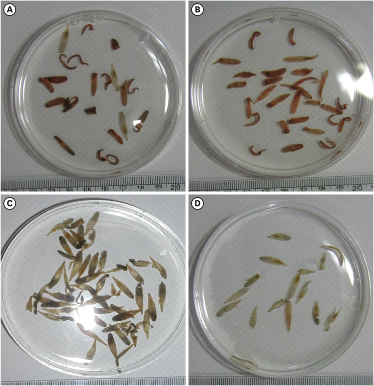
The C. sinensis worms recovered from humans after medication appeared in 3 different colors: red, black, and pale white (Fig. 2).1718 Red worms are young and actively producing eggs, with the uterus filled with fresh eggs giving them their red color. Black worms are middle-aged, with lipofuscin pigmentation. The pigments make worms appear grossly black.19 Pale white worms are older with few eggs, atrophied reproductive organs, and depigmentation. The white worms are looking almost unable to reproduce. Some humans are infected with worms of same color but some heavily infected humans often have worms of mixed colors, which reflects different stages of infection (Fig. 2).
Fig. 2
Adult worms of C. sinensis from 4 infected humans after praziquantel medication. (A) Twenty-two worms recovered from a man, mostly red worms and a few dark or white worms. (B) Twenty-eight worms from a man with mostly red worms. (C) Thirteen pale white worms from a man. (D) Forty-eight worms from a man with mostly dark and a few red worms. The red worms are young and actively producing eggs; dark worms are some aged and with lipofuscin pigments in their posterior part; pale white worms are in senile change, grossly pale with few eggs in the uterus and depigmented. Fig. 2C and Fig. 2D are reproduced from reference 18 and 17, respectively.
When eggs are discharged through feces and drained into nearby water bodies, they may have a chance of continuing life cycle. It is difficult to figure out what proportion of the eggs successfully complete life cycle and produce next generation, but the high egg productivity is likely a major factor contributing survival of the fluke. The eggs in water are ingested by freshwater snails, the first intermediate host. In the snail tissue, a miracidium hatches out of the egg shell’s operculum and metamorphoses into a sporocyst. The sporocyst produces 20–30 rediae, and each mature redia produces about 40–50 cercariae, which are shed from the snail into the water. Ultimately, the larval stages of C. sinensis in the snail host allow one egg to amplify to 1,000 to 1,500 cercariae.20 The snail hosts are mainly Parafossarulus and Bithynia species, which are commonly and widely distributed in rivers, small streams, and reservoirs in East Asia.20
The cercariae swim freely in the water and find the susceptible second intermediate host, mostly freshwater fish of the Family Cyprinidae.20 When a cercaria encounters a suitable fish, it penetrates the skin beneath the scales and encysts in the muscle, becoming a metacercaria. A total of 15 freshwater fish species in 10 Families have been listed as the second intermediate hosts of C. sinensis in Korea.21 The metacercaria infects the final hosts when the fish is ingested.
In the final hosts, the metacercaria excysts rapidly in the duodenum through the action of trypsin and endogenous cysteine proteinases.22 The excysted juvenile worm migrates through the common bile duct and finally reaches the intrahepatic bile duct. An animal experiment demonstrated that it took 9–10 minutes for the worms to migrate from the duodenum to the intrahepatic bile duct in rabbits by bile chemotaxis.23 When the small size of the juvenile worms is considered, the initial migration into the intrahepatic bile duct is significantly rapid, taking only 10 minutes.
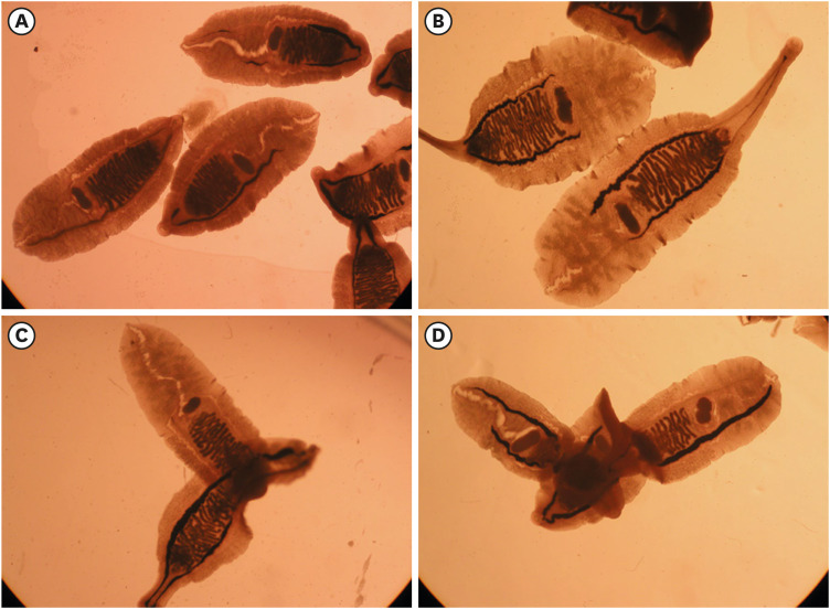
In the bile duct of the final host, the juvenile worms grow and maturate rapidly. Two or more worms make close contacting and overlapping their bodies, when fresh adult worms were incubated in culture media (Fig. 3).24 This body contact may be a process of sperm exchange between mature worms. In mature worms, fertilized ova, which are commonly called eggs, are accumulated in the gravid uterus and the miracidium is maturated within the intrauterine eggs. The eggs with mature miracidium are discharged through the uterine pore into the bile flow. Finally, the eggs in the bile pass out the host body mixed in feces. The eggs first appear in feces after 26 days from the metacercarial infection, which is the incubation period of clonorchiasis. The infected bile duct undergoes pathological changes due to both mechanical and chemical stimulation of the worms, and eventually the worms reshape the bile duct into a nest that is suitable for their survival and reproduction.25
Fig. 3
Live worms of C. sinensis cultured in media.24 (A) Mature worms with a dark-looking uterus by eggs. (B) Worms are moving actively by extending their anterior end. (C, D) Some worms are overlapped actively.
Epidemiology
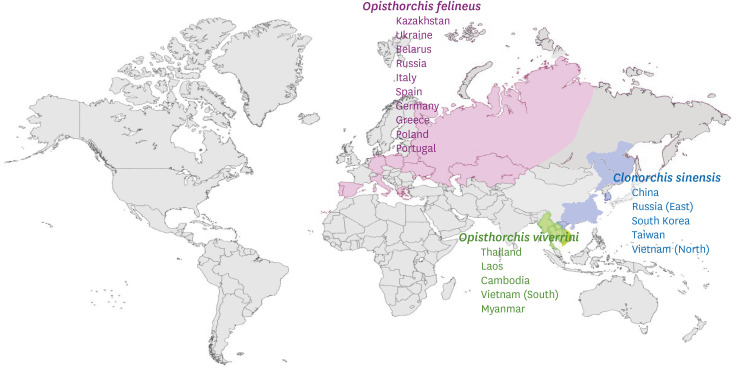
Clonorchis and Opisthorchis are sister genera in the Family Opisthorchiidae. C. sinensis causes clonorchiasis, while O. viverrini and Opisthorchis felineus cause opisthorchiasis. These are human liver fluke diseases transmitted by fish. Clonorchiasis is primarily found in East Asia, from East Russia to Northern Vietnam, while O. viverrini is found in the Indochina Peninsula and O. felineus has a wide distribution, from Siberian Russia to Europe (Fig. 4).2 The three flukes do not overlap in their distribution, and this segregated distribution reflects different biological characteristics among them. In Korea, only clonorchiasis is transmitted in areas near rivers, streams, and lakes.
In the endemic areas, men in their 50s or 60s tend to have the highest and heaviest infection.25 Humans become infected with C. sinensis by eating raw freshwater fish which determines the epidemiological characteristics of clonorchiasis. Men are more likely to fish and eat raw fish while drinking alcohol, making them more likely infected than women in all endemic areas. In most endemic areas, older men or women are resistant to changing their behavior of eating raw fish. Many infected people in endemic areas have been infected for a lifetime. The life span of C. sinensis in human body may be quite long as about 30 years.16 Therefore, both the host and the parasite age together, and older individuals in endemic areas tend to have a higher prevalence and heavier worm burdens due to accumulated infections over their lifetimes.
The accumulated infection means that new worms are superimposed on preexisting worms in the same host. In endemic communities, C. sinensis worms of different ages have been recovered from a single human host (Fig. 2).1718 The co-infection of different aged worms in one host is indirect evidence of ineffective host immune responses in humans, meaning the body does not protect reinfection or superinfection. The host immune response to existing worms has no protective effect against newly infecting worms in humans. The host immune response has been shown to vary depending on the host species, but in rats and rabbits, it has been observed to provide protection against reinfection or superinfection of C. sinensis.26
In Korea, a total of 8 national surveys had been conducted to assess the status of intestinal parasitic infections.10 These surveys monitored the prevalence of soil-transmitted helminths, food-borne trematodes, and cestodes as national statistics. Based on the survey data, the WHO endorsed the elimination of soil-transmitted helminthiases in Korea in 2000.12 The first national status survey of parasitic infections, conducted in 1971, reported a 4.6% egg positive rate for C. sinensis.10 The population-based cross-sectional survey estimated at least 2.5 million individuals were infected with clonorchiasis throughout the country in 1970s, when no effective anthelmintics for clonorchiasis were available. After the successful elimination of soil-transmitted helminthiases in 1980s, clonorchiasis showed the highest egg positive rates among intestinal helminthiases in subsequent national surveys from the 1990s through 2012. The egg positive rates of C. sinensis in the national surveys were 4.6% in 1971, 1.8% in 1976, 2.6% in 1981, 2.7% in 1986, 2.2% in 1992, 1.4% in 1997, 2.4% in 2004, and 1.9% in 2012.10
Seo et al.27 investigated prevalence of food-borne trematodes among 13,373 residents along 7 river basins in Korea and demonstrated that all of the major river basin areas were endemic for clonorchiasis with an overall prevalence rate of 21.5%, estimating 830,000–890,000 infected individuals in these river basin areas. In 1981, the overall prevalence was 2.7% by the 3rd national survey,10 estimating 1.4 million cases of clonorchiasis in general population. The two datasets combined estimated 2.2 to 2.3 million individuals with clonorchiasis in Korea in 1981.
Although praziquantel was introduced for treatment in 1980s, clonorchiasis remained endemic along the river basins in southern Korea, and an overall prevalence of 1.9% in 2012.10 The Ministry of Health and Welfare implemented a community program of screening and chemotherapy for clonorchiasis at river basin areas in southern Korea from 2011 to 2020.28 The program examined 23,000–42,000 residents annually and observed a slow decrease of the egg positive rates, from 11.1% in 2011 to 3.8% in 2020. Continuous screening, chemotherapy, and health education significantly reduced the prevalence of clonorchiasis < 5% in most endemic areas, serving as a good control model of clonorchiasis at the community level.28 These updated data suggest that Koreans with clonorchiasis must be far less than one million at present.
China is a large country but clonorchiasis was mainly distributed in two regions; southeastern region along the Pearl River in Guangdong and Guangxi provinces, and northeastern region along the Songhuajiang River and Nen River in Heilongjiang and Jilin provinces.2930 In 2016, a surveillance of clonorchiasis was implemented in 301 surveillance points in 30 provinces in China.30 The survey detected 6,226 cases (2.04%) infected with C. sinensis among the 305,081 screened people, from 70 counties in 15 provinces. However, 89.4% of the infected people were distributed in Jilin, Heilongjiang, Guangdong, and Guangxi provinces. Qian et al.2 made a meta-analysis and estimated that 10.82 million individuals have clonorchiasis in China. A study in a community in Guangxi province traced individuals with clonorchiasis and observed 40.6% of them were reinfected within one year. Of the reinfected individuals, 75.1% were reinfected once, 23.2% twice, and 1.7% 3 times.31 Many infected individuals are frequently reinfected in most endemic areas because they live in the liver fluke environment that is formed by both biological ecology and sociocultural tradition.
A meta-analysis of food-borne trematodiases across the Great Mekong Subregion recorded prevalence of opisthorchiasis as 21.11% (95% confidence interval [CI], 17.74–24.47%) in Thailand, Laos, Cambodia, and Vietnam while clonorchiasis had a prevalence of 25.33% (95% CI, 18.32–32.34%) in China and Vietnam.32 In Northern Vietnam, the prevalence of clonorchiasis was estimated at 20.30% (95% CI, 9.13–31.47%). About 20 provinces in Northern Vietnam are known to be endemic of clonorchiasis, and Nam Dinh and Ninh Binh are heavily endemic, with prevalence rates over 20%.2
Additionally, several thousand residents with clonorchiasis are estimated along the Amur river in Russia, downflow basin of the Songhuajiang in China.2 In Japan, clonorchiasis was endemic in many regions of Honshu, northeast Shikoku, and north Kyushu but not in Hokkaido in the past. Industrialization and agricultural modernization after the World War II drastically changed freshwater ecology, making the first intermediate host vulnerable throughout Japan. Eventually, clonorchiasis rapidly disappeared from former endemic regions and no new cases have been reported recently.33
Clinical manifestations
Most people with clonorchiasis have few symptoms or signs, especially when only a small number of flukes are involved, causing minimal tissue damage. A few flukes typically trigger little or no noticeable subjective symptoms because they live in the lumen of the hypertrophic bile duct. However, heavy infection leads to more severe manifestations. The symptoms, signs, and pathological findings in clonorchiasis depend on the duration of infection, number of infecting worms, the frequency of reinfection, and any associated microbial infections.25 Individuals with chronic and heavy infections may experience epigastric pain, feeling of indigestion, fever, and loose stools. Overtime, they may develop serious complications, such as gallstone formation, liver abscess, cholangitis, cholecystitis, pancreatitis, and CCA.25 A cross-sectional study in an endemic community in southeastern China investigated correlation between C. sinensis infection and hepatobiliary morbidity.34 The prevalence and infection intensity of C. sinensis were higher in male, elder people, and the individuals consuming alcohol, same as found in Korea. C. sinensis infection was associated with the increase of diarrhea (adjusted odds ratio [aOR], 2.2, 95% CI, 1.1–4.5) and with the increase of fatty liver (aOR, 2.7, 95% CI, 1.4–5.2). Moderate C. sinensis infection was associated with the increase of gallbladder stone (aOR, 3.0, 95% CI, 1.1–8.6), while moderate and heavy infections with the increase of intrahepatic bile duct dilatation (aOR, 2.2, 95% CI, 1.0–4.9 and aOR, 4.3, 95% CI, 1.9–9.9, respectively) and the development of periductal fibrosis (aOR, 3.2, 95% CI, 2.1–4.9).34
Pathogenesis
When a human host swallows metacercariae, the juvenile C. sinensis worms excyst in the intestine and first crawl into the intrahepatic bile duct, where they grow rapidly. The worms stimulate the bile duct mucosal wall, leading to adenomatous hyperplasia of the mucosa.3536373839 The hyperplastic bile duct becomes wider and tortuous, and eventually transforms into a comfortable nest, nurturing the fluke. Over time, the thin and narrow bile duct dilates up to 10 times in its original diameter, from 0.8 mm to 8 mm.40 The volume of the dilated intrahepatic bile duct may increase by about 1,000-times, providing more space for the worms in the liver. The volume expansion of the intrahepatic bile duct may create pressure on surrounding liver tissue. As the worm burden increases, the volume pressure can damage the periductal liver parenchyma.
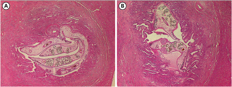
Histopathological findings demonstrate that the worms in the duct lumen are surrounded by and in close contact with hyperplastic bile duct epithelial cells, mixed with many metaplastic mucin-producing cells. The hyperplastic epithelial cells showed a 15-fold increase of cell division activity.40 The thick adenomatous hyperplastic cell layer, interspersed with mucin-secreting cells, helps minimize damage to the duct wall by the worms (Fig. 5). The worms feed on blood and epithelial cells in the bile duct and survive for a long time.20 Inflammatory changes are most evident at the duct wall and periductal tissue, with subsequent fibrosis gradually progressing around the infected duct.3637383940 Dysplastic cells were noted among the hyperplastic cells.38 When the infected host is treated with chemotherapy, the pathological changes in the bile duct significantly resolve after the worms are eliminated. However, the ductal dilatation and periductal fibrosis persist in mild degree over one year.41 The remaining mild degree pathology of the intrahepatic bile duct can lead to false-positive results of the ultrasound scanning images of the liver after clonorchiasis has been treated. 424344
Fig. 5
Adult worms of C. sinensis live in the intrahepatic bile duct inducing the duct into a nurturing nest in an infected cat. (A, B) The infected bile ducts show dilation, adenomatous hyperplasia, mucin-secreting metaplasia, inflammation, and fibrosis. Original magnification ×40, hematoxylin-eosin stained.
CARCINOGENESIS BY CLONORCHIASIS
Notice of correlation between C. sinensis and human CCA
The first patient of CCA associated with C. sinensis was reported in China in 1919,1 and after then several studies reported causal correlation between CCA and C. sinensis. In Korea, Mr. Ju Shik Lee, an officer of Ministry of Health and Social Affairs, claimed that there were many patients with liver cancer and related diseases of jaundice and ascites in Busan, Korea in 1948.45 He investigated those patients and noticed that many of them were heavily infected with C. sinensis. Some surveys and hospital data confirmed that C. sinensis was highly prevalent with egg positive rate 50–70% in Busan and neighboring localities along the Nakdong-gang river basin. However, his work was stopped by the Korean War in 1950 and could not continue thereafter. His finding suggested possible correlation between clonorchiasis and CCA in endemic areas of clonorchiasis.
Epidemiological studies
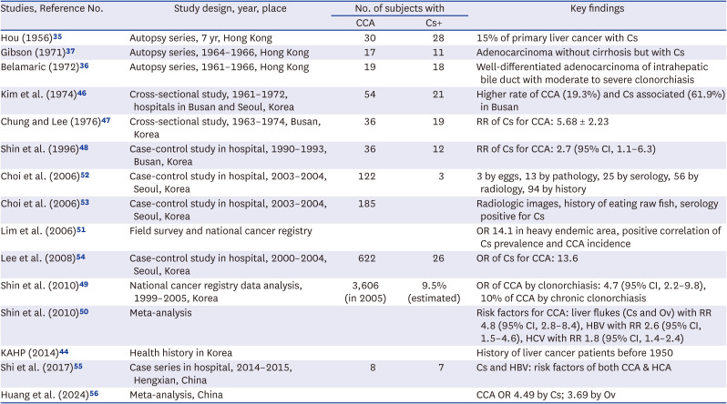
There are several reports from hospital case studies and epidemiological studies that report a correlation between clonorchiasis and CCA. In Hong Kong, studies based on autopsy series from the 1950s and 1960s described pathological observations of CCA without cirrhosis in association with C. sinensis.353637 These articles reported a high prevalence of clonorchiasis in Hong Kong and described pathological characteristics of the intrahepatic bile duct in cases of CCA with clonorchiasis. The tumor masses spread along the bile duct in the liver causing bile duct obstruction. Histopathologically, the bile ducts were dilated with adenomatous hyperplasia of the epithelium and periductal fibrosis. Most cases showed multiple metastatic tumors. These case series studies described autopsy findings of CCA patients describing liver pathology of human clonorchiasis and suggesting causal relation with clonorchiasis in Hong Kong.
In Korea, Kim et al.46 compared hospital data from patients with primary cancer and CCA in Seoul and Busan, where Seoul was non-endemic but Busan was endemic for clonorchiasis. The proportion of primary liver carcinoma was 26.7% in Seoul and 27.5% in Busan, showing no significant difference. However, CCA accounted for 8.6% of the primary liver carcinomas in Seoul but 19.3% in Busan, which was a significant difference. The prevalence rate of clonorchiasis was 38.9% in CCA patients, compared to 8.0% in hepatocellular carcinoma (HCA) patients. The study revealed a higher incidence of CCA in clonorchiasis endemic area compared to the non-endemic area, as well as a higher infection rate of clonorchiasis in CCA patients than that in HCA patients. Another study in Busan demonstrated that the relative risk of clonorchiasis for CCA was 5 times higher compared to HCA.47 The two clinical studies described a highly probable association between CCA and clonorchiasis in Korea.
Later, Shin et al.48 investigated the epidemiological correlation between CCA and clonorchiasis in Busan, suggesting a relative risk of 2.7 (95% CI, 1.1–6.3) for CCA due to clonorchiasis. Shin et al.49 estimated the odds ratio (OR) of CCA attributable to clonorchiasis at 4.7 (95% CI, 2.2–9.8), and found that approximately 10% of CCA cases in Korea were associated with chronic clonorchiasis. A meta-analysis identified risk factors for CCA, revealing that liver flukes (both C. sinensis and O. viverrini), hepatitis B virus, and hepatitis C virus had relative risks (95% CI) 4.8 (2.8–8.4), 2.6 (1.5–4.6), and 1.8 (1.4–2.4), respectively.50 The study also demonstrated that past history of clonorchiasis is a significant risk factor of CCA, alongside the current infection.
Lim et al. (2006)51 surveyed the prevalence of clonorchiasis in non-endemic (2.1%), lightly endemic (7.8%), and moderately endemic (31.3%) areas and compared the mortality and incidence rates of CCA in these regions using the national mortality data and cancer registry database in Korea. The age-standardized CCA mortality rates were 1.2, 1.1, and 2.6/100,000 persons and the incidence rate of CCA was 0.3, 1.8, and 5.5/100,000 persons in the matched non-endemic, lightly endemic, and moderately endemic areas, respectively. The OR of clonorchiasis was 14.7 (95% CI, 7.90–25.00) in the moderately endemic area compared to the non-endemic area. The proportion of CCA among primary liver cancers was 1.4%, 8.9%, and 13.2% in the non-endemic, lightly endemic, and moderately endemic areas, respectively.51
Choi et al.52 collected data from patients with CCA and clonorchiasis and analyzed radiologic images of the liver. They observed a strong etiological correlation of clonorchiasis and CCA and reviewed patients’ imaging data. Most clonorchiasis patients showed dilation of the intrahepatic bile ducts and dilation of the distal duct from the mass in CCA. These combined imaging features were considered pathognomonic for clonorchiasis-associated CCA.52
In a later study, retrospective analysis of clinical data from a tertiary hospital in Seoul confirmed that radiologic images of C. sinensis, positive history of eating raw freshwater fish, and positive serology were significant risk factors for CCA.53 The ORs for these risk factors were OR, 8.615 (95% CI, 5.045–16.062), OR, 2.385 (95% CI, 1.527–3.832), and OR, 2.272 (95% CI, 1.147–4.811), respectively. Another clinical study identified several significant risk factors for intrahepatic CCA, with clonorchiasis being one of the risk factors, showing an OR of 13.6 in a tertiary hospital in Korea.54 These findings of human CCA provided clear evidence of the significant causal correlation between clonorchiasis and CCA in human population, which contributed to the reclassification of C. sinensis from Group 2A to Group 1 biological carcinogen by the IARC in 2009.13
In China, Shi et al.55 analyzed 20 patients with primary liver cancer, including 8 with CCA and 11 with HCA. Seven of the 8 CCA patients were infected with C. sinensis and 2 of them were co-infected with hepatitis B virus. Among the 11 HCA patients, 10 were infected with C. sinensis and 4 were co-infected with hepatitis B virus. They suggested a high risk of both CCA and HCA due to co-infection of C. sinensis and hepatitis B virus. Shin et al.50 also proposed that hepatitis B and C viruses are oncogenic in relation to CCA. The synergistic oncogenic effect of C. sinensis and hepatitis viruses or other microbial pathogens, may warrant further research. One meta-analysis in China reported ORs of human liver flukes with CCA: 4.49 (95% CI, 3.43–5.87, P < 0.001) for C. sinensis and 3.69 for O. viverrini. The OR of clonorchiasis with cholelithiasis was 3.69.56 These studies are summarized in Table 1.
Table 1
Studies on human clonorchiasis and CCA
| Studies, Reference No. | Study design, year, place | No. of subjects with | Key findings | |
|---|---|---|---|---|
| CCA | Cs+ | |||
| Hou (1956)35 | Autopsy series, 7 yr, Hong Kong | 30 | 28 | 15% of primary liver cancer with Cs |
| Gibson (1971)37 | Autopsy series, 1964–1966, Hong Kong | 17 | 11 | Adenocarcinoma without cirrhosis but with Cs |
| Belamaric (1972)36 | Autopsy series, 1961–1966, Hong Kong | 19 | 18 | Well-differentiated adenocarcinoma of intrahepatic bile duct with moderate to severe clonorchiasis |
| Kim et al. (1974)46 | Cross-sectional study, 1961–1972, hospitals in Busan and Seoul, Korea | 54 | 21 | Higher rate of CCA (19.3%) and Cs associated (61.9%) in Busan |
| Chung and Lee (1976)47 | Cross-sectional study, 1963–1974, Busan, Korea | 36 | 19 | RR of Cs for CCA: 5.68 ± 2.23 |
| Shin et al. (1996)48 | Case-control study in hospital, 1990–1993, Busan, Korea | 36 | 12 | RR of Cs for CCA: 2.7 (95% CI, 1.1–6.3) |
| Choi et al. (2006)52 | Case-control study in hospital, 2003–2004, Seoul, Korea | 122 | 3 | 3 by eggs, 13 by pathology, 25 by serology, 56 by radiology, 94 by history |
| Choi et al. (2006)53 | Case-control study in hospital, 2003–2004, Seoul, Korea | 185 | Radiologic images, history of eating raw fish, serology positive for Cs | |
| Lim et al. (2006)51 | Field survey and national cancer registry | OR 14.1 in heavy endemic area, positive correlation of Cs prevalence and CCA incidence | ||
| Lee et al. (2008)54 | Case-control study in hospital, 2000–2004, Seoul, Korea | 622 | 26 | OR of Cs for CCA: 13.6 |
| Shin et al. (2010)49 | National cancer registry data analysis, 1999–2005, Korea | 3,606 (in 2005) | 9.5% (estimated) | OR of CCA by clonorchiasis: 4.7 (95% CI, 2.2–9.8), 10% of CCA by chronic clonorchiasis |
| Shin et al. (2010)50 | Meta-analysis | Risk factors for CCA: liver flukes (Cs and Ov) with RR 4.8 (95% CI, 2.8–8.4), HBV with RR 2.6 (95% CI, 1.5–4.6), HCV with RR 1.8 (95% CI, 1.4–2.4) | ||
| KAHP (2014)44 | Health history in Korea | History of liver cancer patients before 1950 | ||
| Shi et al. (2017)55 | Case series in hospital, 2014–2015, Hengxian, China | 8 | 7 | Cs and HBV: risk factors of both CCA & HCA |
| Huang et al. (2024)56 | Meta-analysis, China | CCA OR 4.49 by Cs; 3.69 by Ov | ||
Animal models
Several animal experiments have investigated immunopathology, host-parasite interactions, genetic alterations, and CCA pathogenesis by C. sinensis infection. C. sinensis-induced CCA is characterized continuous excessive remodeling of the bile duct cells through persistent stimulation and injury by the worms.
The tumor-associated hepatic microenvironment consists of hepatocytes, hepatic sinusoidal endothelial cells, hepatic stellate cells, cholangiocytes, Kupffer cells and recruited immune cells, including bone-marrow-derived macrophages.57 Adult C. sinensis worms cause mechanical injury and inflammation on the bile duct wall, adenomatous hyperplasia, and metaplasia of mucin-producing cells in the biliary mucosa, as described above. In addition to these histopathological findings, changes in the bile duct epithelium in response to stimuli in acute and chronic clonorchiasis include infiltration of Kupffer cells, macrophages and cyst formation in mice. These pathological changes in mice tend to correlate with the longevity of C. sinensis infection, the worm burden, and the susceptibility of the host.58 For pathogen recognition, Toll-like receptor 2 (TLR2) and TLR4 were reportedly upregulated in a mouse model of clonorchiasis during C. sinensis infection, and TLR4 upregulation induced tumor necrosis factor (TNF)-α secretion in excretory-secretory product (ESP)-stimulated biliary epithelial cells in mice.596061
Kim et al.62 reported that Kupffer cells most likely served as antigen-presenting cells and showed a 70-fold increase during the primary infection of C. sinensis. The recovered Kupffer cells produced significantly higher levels of cytokines, including TNF-α, interleukin (IL)-6, IL-10, and IL-13, in C. sinensis-infected FVB mice.63
However, the effects of persistent TNF production by Kupffer cells and cholangiocytes upon chronic liver injury and high levels of reactive oxygen species (ROS) remain unclear. Kupffer cells were concentrated in the periductal tissues and near the veins. Additionally, the livers of mice with cirrhosis showed infiltration of Kupffer cells around the portal areas.64 The number of recruited Kupffer cells increased in regions characterized by tissue remodeling during the cirrhotic stage of infection. Kupffer cells are activated by different components and can be differentiated into M1 (classical) or M2 (alternative) macrophages in the liver. M1 macrophages suppressed tumor growth, whereas M2 macrophages contributed to cancer progression.64 The fibrotic and cirrhotic stage following C. sinensis infection markedly increased the proportion of Arg-1-producing macrophages, the M2 phenotype, which modulate fibrosis and tissue repair, and also promote neoplastic transformation of bile duct cells.62
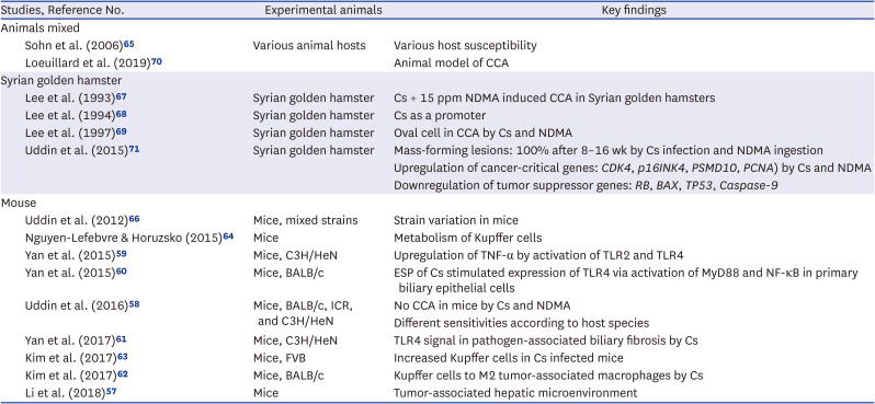
Different host animals exhibit varying susceptibility to C. sinensis infection, but hamsters, guinea pigs, and rabbits are considered good experimental laboratory animal hosts.156566 Among them, the Syrian golden hamster is the suitable model for studies of CCA.67686970 Previous studies have reported that C. sinensis infection acts a promoter, and the initiator, such as N-nitrosodimethylamine (NDMA), is required to induce CCA.6768 Carcinogens, including NMDA, initiate tumorigenesis through DNA methylation and induce a genotoxic effect, leading to structural changes in DNA.
No definite neoplastic changes of CCA were observed in BALB/c, ICR, B6, and C3H/He mice infected with C. sinensis and exposure to NDMA.58 Instead of CCA, C3H/He mice developed extensive fibrosis when challenged with C. sinensis, NDMA, and dicyclanil together. Interestingly, adenomas developed only in FVB mice, accompanied cystic dilatation of the infected bile duct.58 No other animals developed CCA through experimental infection with C. sinensis.
Features of hamster model
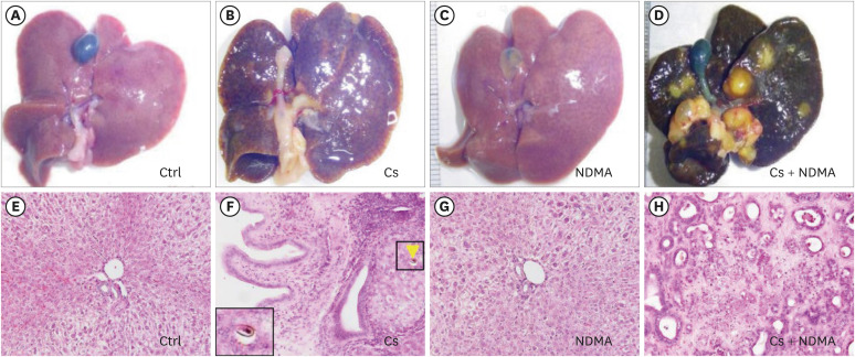
Mass-forming lesions (MFLs) are a major pathologic finding of CCA induced by C. sinensis and NDMA in the Syrian golden hamster model, with 100% prevalence of MFLs in the C. sinensis + NDMA group of hamsters.71 Histological progressions of CCA occurred sequentially from well-differentiated CCA, followed by poorly differentiated CCA in a time-dependent manner, beginning at the 8th week of infection (Fig. 6).
Fig. 6
Experimental carcinogenesis in Syrian golden hamsters induced by Cs and NDMA. Gross morphology of the livers: Ctrl (A), Cs only (B), NDMA only (C), Cs + NDMA (D). Histopathology of the livers: Ctrl (E), Cs only, boxes marking eggs in the bile duct (F), NDMA only (G), Cs + NDMA (H). Original magnification ×200 (E-H), hematoxylin-eosin stained. Reproduced from the reference 71.
Ctrl = control, Cs = C. sinensis, NDMA = N-nitrosodimethylamine.
Another study using the Syrian golden hamster model reported that continuous mechanical and chemical irritation by C. sinensis and NDMA upregulated cancer-critical genes, including PSMD10, CDK4, and PCNA and downregulated tumor suppressor gene TP53, as well as the protein RB, caspase-9, and BAX.71 Downregulation of BAX and caspase-9 may further facilitate the survival of transformed cells with denatured DNA and those undergoing uncontrolled proliferation to form a CCA mass. PCNA overexpression can be detected in tumor tissue as a marker of CCA in tumor necrosis. The animal experiments on carcinogenesis are summarized in Table 2.
Table 2
Animal studies on Cs and CCA
| Studies, Reference No. | Experimental animals | Key findings | |
|---|---|---|---|
| Animals mixed | |||
| Sohn et al. (2006)65 | Various animal hosts | Various host susceptibility | |
| Loeuillard et al. (2019)70 | Animal model of CCA | ||
| Syrian golden hamster | |||
| Lee et al. (1993)67 | Syrian golden hamster | Cs + 15 ppm NDMA induced CCA in Syrian golden hamsters | |
| Lee et al. (1994)68 | Syrian golden hamster | Cs as a promoter | |
| Lee et al. (1997)69 | Syrian golden hamster | Oval cell in CCA by Cs and NDMA | |
| Uddin et al. (2015)71 | Syrian golden hamster | Mass-forming lesions: 100% after 8–16 wk by Cs infection and NDMA ingestion | |
| Upregulation of cancer-critical genes: CDK4, p16INK4, PSMD10, PCNA) by Cs and NDMA | |||
| Downregulation of tumor suppressor genes: RB, BAX, TP53, Caspase-9 | |||
| Mouse | |||
| Uddin et al. (2012)66 | Mice, mixed strains | Strain variation in mice | |
| Nguyen-Lefebvre & Horuzsko (2015)64 | Mice | Metabolism of Kupffer cells | |
| Yan et al. (2015)59 | Mice, C3H/HeN | Upregulation of TNF-α by activation of TLR2 and TLR4 | |
| Yan et al. (2015)60 | Mice, BALB/c | ESP of Cs stimulated expression of TLR4 via activation of MyD88 and NF-κB in primary biliary epithelial cells | |
| Uddin et al. (2016)58 | Mice, BALB/c, ICR, and C3H/HeN | No CCA in mice by Cs and NDMA | |
| Different sensitivities according to host species | |||
| Yan et al. (2017)61 | Mice, C3H/HeN | TLR4 signal in pathogen-associated biliary fibrosis by Cs | |
| Kim et al. (2017)63 | Mice, FVB | Increased Kupffer cells in Cs infected mice | |
| Kim et al. (2017)62 | Mice, BALB/c | Kupffer cells to M2 tumor-associated macrophages by Cs | |
| Li et al. (2018)57 | Mice | Tumor-associated hepatic microenvironment | |
IN VITRO EXPERIMENTS WITH ESP OF C. SINENSIS
The ESP is a mixture of various functional molecules secreted from the liver fluke. O. viverrini is known to secrete granulin-like growth factor, which stimulates angiogenesis in the host tissue, promoting rapid wound healing and accelerating neoplastic pathways.72 Additionally, thioredoxin from O. viverrini downregulates apoptosis of cholangiocytes, supporting its induction of CCA.73 Various proteinases, including granulin, have been observed from C. sinensis,7475 and the ESPs of C. sinensis were suggested to be related with CCA,7677 but the oncogenic molecules in the ESPs of C. sinensis still require further investigation.
Several in vitro studies have cultured epithelial cells stimulated with ESPs of C. sinensis and investigated changes of tumor related intracellular molecules and genes. The stimulated cells exhibited various changes of the cell characteristics. The findings from these cell studies may help explain the mechanisms behind cellular changes that lead to CCA transformation due to C. sinensis infection.
Cancer-associated microenvironment
Several studies have reported that oxidative stress potentially mediates C. sinensis-associated carcinogenesis.777879 Bile duct inflammation induces endogenous oxidative stress through ROS and reactive nitrogen species, leading to oxidative/nitrative DNA damage in cholangiocytes.77 In response to oxidative stress in hepatocytes, TNF-producing Kupffer cells are activated via JNK and ROS. TNF from Kupffer cells then induces JNK-mediated cholangiocyte proliferation and oncogenic transformation.80 Specifically, the levels of peroxiredoxin (Prdx) isomers (Prdx2, 3, and 6) and thioredoxin1, which are potential carcinogens, were found very high, similar to the levels of intracellular ROS in ESP-exposed HuCCT1 cells.80 Nam et al.77 and Pak et al.81 demonstrated that C. sinensis ESPs significantly induced matrix metalloproteinase-9 expression and activity by enhancing the activity of nuclear factor kappa B (NF-κB) and subsequent induction of IκBα. Through this signal pathway, C. sinensis ESPs regulate the migration and invasion of various CCA cell lines.82
Won et al.83 developed a three-dimensional (3D) in vitro model to understand the microenvironment by culturing HuCCT1 cells and normal H69 cholangiocytes together with C. sinensis ESP. This 3D system consisted of a type I collagen extracellular matrix, HuCCT1 cells treated with ESP, and quiescent biliary ductal plates formed by H69 cholangiocytes. Ultimately, the expression of local and cell-cell adhesion proteins such as vinculin, paxillin, and MMPs (1, 9 and 13) was increased in ESP-stimulated HuCCT1 cells within the 3D system. HuCCT1 cells migrated to the direction of the ESP by a gradient of ESP. Consequently, both IL-6 and tumor growth factor (TGF)-β1 levels were elevated in normal H69 cholangiocytes when they were stimulated with C. sinensis ESP, and a cadherin switch (decrease in E-cadherin/increase in N-cadherin expression) was observed in HuCCT1 cells. Won et al.84 also reported that HuCCT1 cells underwent epithelial-mesenchymal transition-like changes in response to ESP stimulation, promoting their migration through the interaction between cancer cells and normal cholangiocytes. These findings suggest that C. sinensis ESP promotes cholangiocytes to undergo neoplastic changes.
Cell division cycle
ESPs are highly immunogenic and may exert cytotoxic effects on the biliary epithelium, stimulating inflammation, promoting proliferation, and suppressing apoptosis.8586 Kim et al.86 reported that C. sinensis ESPs increased cell proliferation and suppressed apoptosis in the human HEK293T cells. Furthermore, normal epithelial cells actively proliferated after treatment with ESP. Specifically, when normal epithelial cells (HEK293T, human kidney epithelium cell line; H69, human bile duct epithelium cell line) were treated together with ESP and NMDA, the G2/M phase of the cell cycle became dominant, while most cells remain at the G1/S phase in the control. In addition to the changes in cell population, cell cycle-associated molecules, including CDK2, cyclin B, Rb, pRb, and E2F1 were significantly upregulated.858687 Co-stimulation with ESP and NDMA induced overexpression of E2F1 and downregulation of pRb, indicating the involvement of the Rb-E2F1 pathway.86 E2F1 expression is correlated with tumor proliferation, and overexpression of E2F1 potentially results in aggressive tumors with a high proliferation rate and suppressed apoptosis.87 The abnormal distribution of cells in the division cycle suggests that these cells may receive stress for division and acquire neoplastic characteristics.
Cancer related molecules
Studies using C. sinensis-infected mouse liver tissues reported that COX-2 and 5-LOX expression, along with increased 8-oxodG nuclear accumulation in inflammatory cells, were detected in the inflammatory nidus.78 Oxidative stress-mediated liver lesions were observed following C. sinensis infection of mice. COX-2 is known to be overexpressed in various inflammatory diseases and in CCA.88 In primary pancreatitis, biliary epithelial cells exhibited COX-2 upregulation, similar to carcinoma cells. At the intracellular level, combined treatment of normal H69 cholangiocytes with ESP of C. sinensis and NDMA significantly upregulated COX-2.89 Kim et al.89 reported that normal H69 cholangiocytes stimulated by C. sinensis ESP and NDMA overexpressed Cy19, Ki-67, and COX-2. These molecular changes support the idea that normal H69 bile duct cells can undergo neoplastic transformation when stimulated by ESP of C. sinensis and NDMA.
C. sinensis was found to secrete C. sinensis granulin (CsGRN), a growth factor-like protein in the ESP. The CsGRN was localized in the testes and tegument of adult worms.74 The in vitro study with hepatocytes and biliary epithelial cells found enhancement of cell migration, upregulation of vimentin, N-cadherin, β-catenin, MMP2, and MMP9, and downregulation of ZO-1 in PLC-GRN/RBE-GRN cells. When the CsGRN was introduced into normal human hepatocytes, the cells underwent cell arrest and malignant transformation by regulating epidermal growth factor receptor-mediated RAS/MAPK/ERK and PI3K/Akt signaling pathways, which suggested that CsGRN could serve as a novel oncoprotein.75
ESPs of C. sinensis induce pro-inflammatory responses in primary biliary epithelial cells by upregulating TLR4 and its downstream signaling pathways, including MyD88-dependent IκB-α degradation, and NF-κB activation.5960 A DNA modification may be potentially triggered by increased levels of proinflammatory cytokines and NF-κB, which regulate COX-2 and inducible nitric oxide synthase (iNOS) and disrupt homeostasis of oxidants/anti-oxidants and DNA repair enzymes.77 Additionally, free radicals generated enzymatically by C. sinensis ESPs can cause NF-κB-mediated inflammation in HuCCT1 cells.77 The HuCCT1 cells exposed to ESP of C. sinensis induce free radical production through the activation of NOX, XO, and iNOS, and xanthine oxidase leading to increased expression of the NF-κB-regulated proinflammatory genes such as IL-1β and IL-6. The ESP-induced stimulation in HuCCT cells was mediated through the TLRs, leading to production of free radicals that can damage DNA.90
Cancer related genes
Neoplastic transformation of cells begins at the genetic level, and several studies have provided evidence of genetic changes. Using miRNA microarray chips containing 135 cancer-related miRNAs, 13 miRNAs were upregulated and 3 were downregulated in human HuCCT1 CCA stimulated by C. sinensis ESP.91 These dysregulated miRNAs were involved in cell proliferation, inhibition of tumor suppressor genes, neoplasia, cell migration, and invasion. Among these, 6% of miRNAs were associated with DNA methylation, an important factor in carcinogenesis. In particular, tumor-suppressor miRNA including let-7a, let7i, and miR-124a were downregulated in both cancerous and non-cancerous cells via ESP stimulation.
Analysis of gene expression profiles across the three developmental stages of C. sinensis may provide insight into their pathogenesis and carcinogenesis. Yoo et al.92 reported that gene expression profiles in the C. sinensis transcriptome contained several contigs encoding proteins associated with inhibition and regulation of apoptosis and cell proliferation, such as granulin, epithermal growth factor, TGF interacting proteins, oncogenic laminins, JNK, cyclin-dependent kinase, and transcription factors in adult C. sinensis. These results suggest that apoptosis inhibitors and regulators identified in the C. sinensis transcriptome promote neoplastic transformation by preventing the death of DNA-damaged cells.
Gap junction molecules and connexins (Cxs)
In the homeostatic condition of the tissue, cells are networked tightly with gap-junction molecules, mainly Cxs.93 NF-κB may influence the production of Cx 43, a gap-junction protein, in liver cirrhosis.94 The formation and nitrosation of nitric oxide (NO) can contribute to C. sinensis-associated carcinogenesis. Induction of iNOS under inflammatory conditions suggests that NO is involved in Cx 43 upregulation.95 Among gap junction proteins, Cx 43 plays a key role in almost all steps of the inflammatory response of cells, inflammatory cell migration, and wound healing.939495 Cx 43 upregulation has been shown to increase tumor cell invasion in melanoma, playing a key role in carcinogenesis.96 Expression profiles of Cx are altered in various cancers.979899
The gap-junction proteins Cx 43 and Cx 26 were upregulated, while Cx 32 was downregulated in H69 cholangiocytes stimulated by C. sinensis ESP and NDMA.89 The study confirmed that silencing of Cx 43 reduced ESP- and NDMA-induced cell proliferation and COX-2 overexpression to control levels (Fig. 7). Consequently, exposure to ESPs of C. sinensis combined with low level NDMA promotes carcinogenesis in normal bile duct epithelial cells through dysregulated intercellular communication, mediated by altered gap-junction molecules.
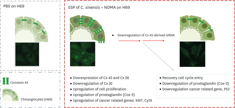
Summary of in vitro experiments
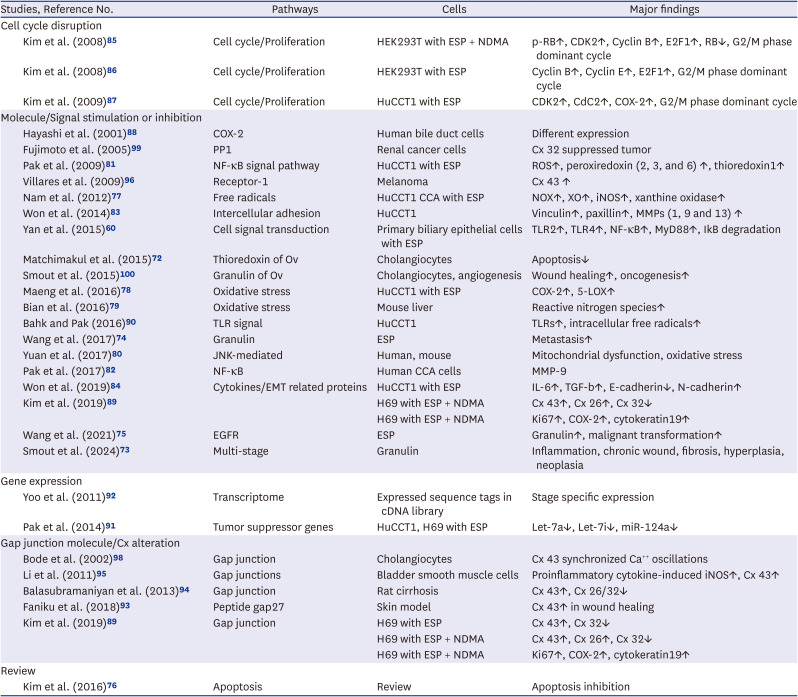
The carcinogenetic mechanism of CCA associated with C. sinensis is multistage, beginning with initial stimulation of normal cholangiocytes to proliferate due to the worm and endogenous or exogenous radicals. The stimulation is followed by DNA damage, overexpression of oncogenes, downregulation of apoptosis inhibitory genes, disrupted communication between cells, and uncontrolled proliferation and migration of the damaged cells. Smout et al.100 suggested mechanistic insights that secretions of the liver flukes, such as Ov-GRN-1 granulin growth factor, combined with aberrant inflammation and repeated cycles of chronic wounding at the parasitic sites, promote biliary hyperplasia, fibrosis, and ultimately malignant transformation. The in vitro cell experiment studies are summarized in Table 3.
Table 3
In vitro or in vivo studies for carcinogenesis with ESPs of C. sinensis
| Studies, Reference No. | Pathways | Cells | Major findings | |
|---|---|---|---|---|
| Cell cycle disruption | ||||
| Kim et al. (2008)85 | Cell cycle/Proliferation | HEK293T with ESP + NDMA | p-RB↑, CDK2↑, Cyclin B↑, E2F1↑, RB↓, G2/M phase dominant cycle | |
| Kim et al. (2008)86 | Cell cycle/Proliferation | HEK293T with ESP | Cyclin B↑, Cyclin E↑, E2F1↑, G2/M phase dominant cycle | |
| Kim et al. (2009)87 | Cell cycle/Proliferation | HuCCT1 with ESP | CDK2↑, CdC2↑, COX-2↑, G2/M phase dominant cycle | |
| Molecule/Signal stimulation or inhibition | ||||
| Hayashi et al. (2001)88 | COX-2 | Human bile duct cells | Different expression | |
| Fujimoto et al. (2005)99 | PP1 | Renal cancer cells | Cx 32 suppressed tumor | |
| Pak et al. (2009)81 | NF-κB signal pathway | HuCCT1 with ESP | ROS↑, peroxiredoxin (2, 3, and 6) ↑, thioredoxin1↑ | |
| Villares et al. (2009)96 | Receptor-1 | Melanoma | Cx 43 ↑ | |
| Nam et al. (2012)77 | Free radicals | HuCCT1 CCA with ESP | NOX↑, XO↑, iNOS↑, xanthine oxidase↑ | |
| Won et al. (2014)83 | Intercellular adhesion | HuCCT1 | Vinculin↑, paxillin↑, MMPs (1, 9 and 13) ↑ | |
| Yan et al. (2015)60 | Cell signal transduction | Primary biliary epithelial cells with ESP | TLR2↑, TLR4↑, NF-κB↑, MyD88↑, IkB degradation | |
| Matchimakul et al. (2015)72 | Thioredoxin of Ov | Cholangiocytes | Apoptosis↓ | |
| Smout et al. (2015)100 | Granulin of Ov | Cholangiocytes, angiogenesis | Wound healing↑, oncogenesis↑ | |
| Maeng et al. (2016)78 | Oxidative stress | HuCCT1 with ESP | COX-2↑, 5-LOX↑ | |
| Bian et al. (2016)79 | Oxidative stress | Mouse liver | Reactive nitrogen species↑ | |
| Bahk and Pak (2016)90 | TLR signal | HuCCT1 | TLRs↑, intracellular free radicals↑ | |
| Wang et al. (2017)74 | Granulin | ESP | Metastasis↑ | |
| Yuan et al. (2017)80 | JNK-mediated | Human, mouse | Mitochondrial dysfunction, oxidative stress | |
| Pak et al. (2017)82 | NF-κB | Human CCA cells | MMP-9 | |
| Won et al. (2019)84 | Cytokines/EMT related proteins | HuCCT1 with ESP | IL-6↑, TGF-b↑, E-cadherin↓, N-cadherin↑ | |
| Kim et al. (2019)89 | H69 with ESP + NDMA | Cx 43↑, Cx 26↑, Cx 32↓ | ||
| H69 with ESP + NDMA | Ki67↑, COX-2↑, cytokeratin19↑ | |||
| Wang et al. (2021)75 | EGFR | ESP | Granulin↑, malignant transformation↑ | |
| Smout et al. (2024)73 | Multi-stage | Granulin | Inflammation, chronic wound, fibrosis, hyperplasia, neoplasia | |
| Gene expression | ||||
| Yoo et al. (2011)92 | Transcriptome | Expressed sequence tags in cDNA library | Stage specific expression | |
| Pak et al. (2014)91 | Tumor suppressor genes | HuCCT1, H69 with ESP | Let-7a↓, Let-7i↓, miR-124a↓ | |
| Gap junction molecule/Cx alteration | ||||
| Bode et al. (2002)98 | Gap junction | Cholangiocytes | Cx 43 synchronized Ca++ oscillations | |
| Li et al. (2011)95 | Gap junctions | Bladder smooth muscle cells | Proinflammatory cytokine-induced iNOS↑, Cx 43↑ | |
| Balasubramaniyan et al. (2013)94 | Gap junction | Rat cirrhosis | Cx 43↑, Cx 26/32↓ | |
| Faniku et al. (2018)93 | Peptide gap27 | Skin model | Cx 43↑ in wound healing | |
| Kim et al. (2019)89 | Gap junction | H69 with ESP | Cx 43↑, Cx 32↓ | |
| H69 with ESP + NDMA | Cx 43↑, Cx 26↑, Cx 32↓ | |||
| H69 with ESP + NDMA | Ki67↑, COX-2↑, cytokeratin19↑ | |||
| Review | ||||
| Kim et al. (2016)76 | Apoptosis | Review | Apoptosis inhibition | |
ESP = excretory-secretory product of C. sinensis, NDMA = N-nitrosodimethylamine, Cx = connexin, NF-κB = nuclear factor kappa B, ROS = reactive oxygen species, MMP = matrix metalloproteinase, IL = interleukin, TGF = tumor growth factor, Ov = O. viverrini, TLR = Toll-like receptor, CCA = cholangiocarcinoma, EMT = epithelial-mesenchymal transition, EGFR = epidermal growth factor receptor, iNOS = inducible nitric oxide synthase.
CONCLUSION
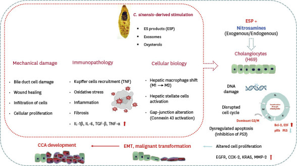
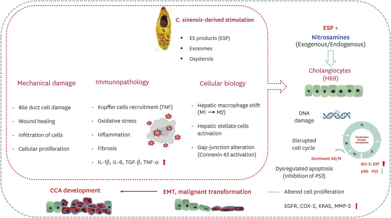
There is substantial evidence supporting the carcinogenesis of C. sinensis from both epidemiological and experimental studies. Epidemiological research has shown an overall OR of 4.7 for CCA associated with clonorchiasis, and 10% of Korean CCA patients linked to the infection. The carcinogenic factors include ESPs with granulin of the parasite, a diet rich in nitrosamines, co-infections with microbes, and endogenous radicals produced by local chronic inflammation. These stimulants lead to expression of oncogenes and suppression of apoptosis. Eventually, oncogenic intracellular molecules accumulate, and the altered gap-junction proteins disrupt intercellular communication in the bile duct epithelial cells. These neoplastic cells then proliferate forming CCA masses. Fig. 8 draws the theoretical process of carcinogenesis by C. sinensis. Since C. sinensis was classified as a biological carcinogen for human CCA, clonorchiasis poses a significant public health threat in East Asia, where it still remains prevalent. To eliminate the disease globally, changes in diet habits, such as avoiding raw fish, are essential, along with mass praziquantel therapy, in streamlining with the WHO guidelines.
Fig. 8
Carcinogenic mechanism of CCA induced by C. sinensis.
CCA = cholangiocarcinoma, ESP = excretory-secretory product of C. sinensis, TNF = tumor necrosis factor, IL = interleukin, TGF = tumor growth factor, EMT = epithelial-mesenchymal transition, EGFR = epidermal growth factor receptor, MMP = matrix metalloproteinase.
 XML Download