Abstract
One of the main parameters in the analysis of skeletal remains in forensic anthropological cases is the estimation of age. This study aimed to investigate the correlation between age and the fusion status of the sternal junction. This cross-sectional study was carried out on 184 sterna from 94 females and 90 males obtained from known-age cadavers in the Thai population. By direct observation, the fusion stage of the manubrio-sternal and sterno-xiphoidal junctions was studied and divided into unfused and fused joints. The results showed that a large proportion of the sterna remain unfused throughout adulthood, with fusion observed in both young and old cadavers. Insignificant differences in the rate of fusion, the sexes and ages were observed. None of the sterna under 30 years of age in females and 32 years of age in males showed fusion of the manubrio-sternal and sterno-xiphoidal junctions. Based on the variability of the sternal fusions observed in this study, we highlighted a very limited role of the sternum alone in the estimation of age in the Thai population.
Age is one of the biological profiles that is particularly useful in narrowing the number of missing persons compared to the profile of the deceased [1]. In adults, age estimation focuses not only on bone degeneration but also the appearance and fusion of the ossification centers of the bones [2]. Several regions of the body have been studied to make the most accurate age estimation, such as pubic symphysis, auricular surface, cranial suture, sternal end of the rib, and medial end of the clavicle [3-8]. In the Thai population, the sternum has been used for estimating stature [9]. However, this bone may be a reliable source of information and can offer useful information for age estimation in some cases with this bone.
The sternum has been considered a potential application in forensic age estimation in young adult and adult skeletons. Most of the authors studied direct observation of the fusion of manubrio-sternal and sterno-xiphoidal junctions in different population groups [10-22]. All of these studies found that the manubrio-sternal and sterno-xiphoidal junctions fused at different times. Studies in this regard have been carried out in the Chinese [11], Indian population [12-16], South African [17], Turkish [18, 19], Egyptian [20], Spanish [21], Japanese [22] and mixed population (mainly British) [10]. For the Thai population, Monum et al. [23] developed a model for age estimation in the Thai male population using radiological analysis of chest plate ossification. However, the question arises of whether the results obtained from the radiological examination can be compared with those from direct observation. To the best of our knowledge, there was no study of age estimation using direct observation of the sternum in a Thai population. The results from the non-Thai population may be unreliable with the Thai population due to variations in genetics and environment.
Therefore, the aim of this study was to examine the sterna of a Thai population for the fusion of manubrio-sternal and sterno-xiphoidal junctions using the direct observation method, and to determine the applicability of this method for age-at-death estimation.
This study has been approved by the Research Ethics Committee of the Faculty of Medicine Siriraj Hospital, Mahidol University, Bangkok, Thailand (Protocol No. 950/2564 (IRB2) on December 1st, 2021). Informed consent was obtained from the legal heirs before collecting the sterna. Between December 5th, 2021 and November 10th, 2022, the intact sterna were removed from the deceased who underwent autopsy in the Forensic Pathology Unit, Department of Forensic Medicine, Faculty of Medicine Siriraj Hospital, Mahidol University. All of the deceased were Thai nationals, 20 years of age or older. Because it is generally recognized that the sternal body segments develop fully and unite at the age of 20–25, the age of 20 was chosen [24-26]. The age and nationality were approved by identification documents issued by the Thai government. The deceased with pathological sterna or injuries at any location in the examined area was excluded from this study.
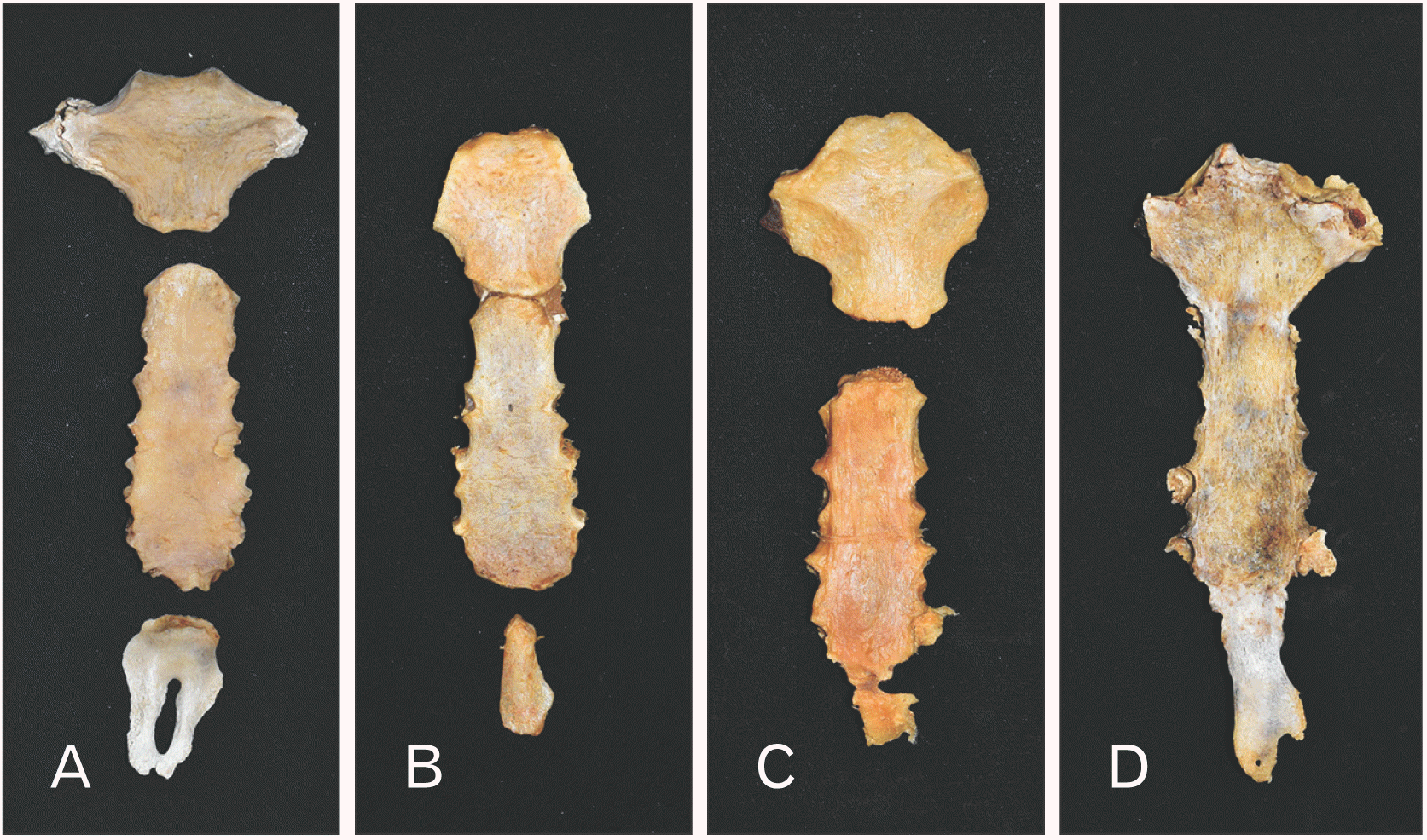
A total of 184 sterna were included, consisting of 94 females (20 to 83 years) and 90 males (20 to 84 years). The mean age of females and males in this study population was 44.62 (standard deviation [SD]=17.20) years and 49.07 (SD=16.59) years, respectively. After manual maceration of the surrounding muscles and soft tissues, the sterna were allowed to macerate in warm water for 4 weeks. All residual soft tissue was removed from the bones using a gentle brush and running water to prevent any damage to the bone surface. The bones were allowed to dry at room temperature. The sterna were studied with a naked eye examination and the fusion status of their junctions was observed. The manubrio-sternal and sterno-xiphoidal junctions were classified as unfused or fused (Fig. 1). Fusion was considered when it was discovered that a part of the joint space was fused. It was deemed nonfusion if none of the surfaces was found to be fused. The observers did not know the age and sex of the sample during their examination. Additionally, a blind test was carried out with a sample of known age.
All statistical analyses were performed with SPSS statistical software, version 20.0 (SPSS Inc.). A P-value of less than 0.05 was considered statistically significant. Descriptive statistics was used to illustrate the occurrence of fusion throughout the sample and binary logistic regression tests were used to find variations between the sexes, ages and the fusion rates. To clarify the correlations between the fusion rates and the true age, point-biserial correlation coefficient was carried out. The association between age above and below 45 and the state of fusion of the manubrio-sternal and sterno-xiphoidal junctions was also evaluated using an odd ratio. The age of 45 was chosen as the halfway point between data entries in the sample of this study and the midpoint between the various reports of fusion in the literature [17].
Tables 1 and 2 demonstrate the fusion of the manubrio-sternal and sterno-xiphoidal junctions in various age groups among females and males. Direct observation of the manubrio-sternal junction revealed that 46.74% (n=86) of the sterna had a fusion of the joint. The earliest fusion of manubrio-sternal junction was reported at 30 years of age in females and 36 years in males, while nonfusion was observed until the age of 83 years in females and 84 years in males. In female, the manubrio-sternal junction fused at a mean age of 54.94 years (SD=13.91), while males showed a mean age of fusion of 55.33 years (SD=13.02).
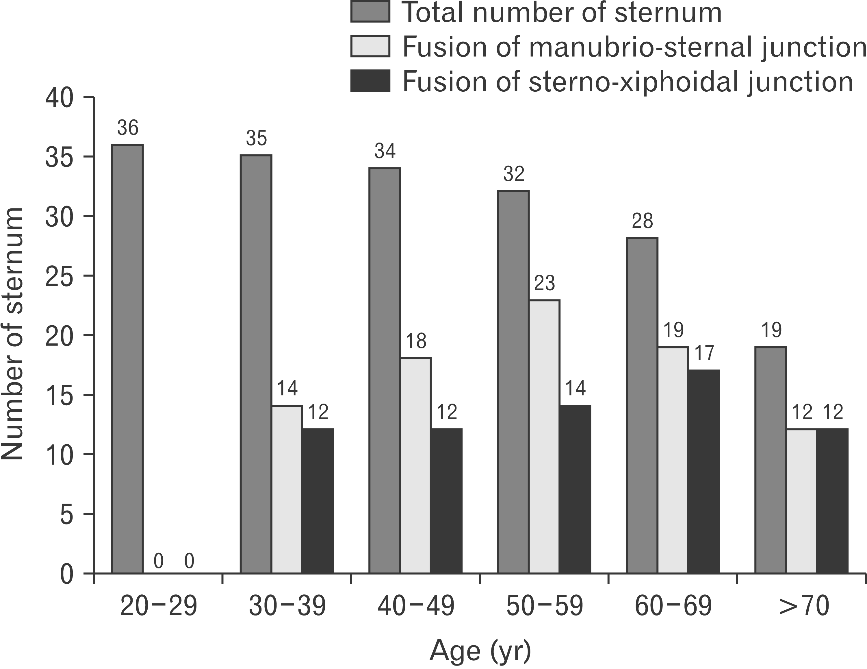
A total of 67 (36.4%) sterna demonstrated a fused sterno-xiphoidal junction, consisting of 33 (35.1%) female sterna and 34 (37.8%) male sterna. The oldest sterna with unfused sterno-xiphoidal junction were 76 years old in females and 78 years old in males, whereas the youngest sterna with complete fusion were 30 years old in females and 32 years old in males. In female, the sterno-xiphoidal junction fused at a mean age of 57.09 years (SD=12.92), while males showed a mean age of fusion of 55.59 years (SD=15.47). It was noted that, with advancing age, more males and females had sterna with fused manubrio-sternal and sterno-xiphoidal junction. Number of sterna with fusion of the manubrio-sternal and sterno-xiphoidal junctions in each age group are illustrated in Fig. 2.
Binary logistic regression was used to analyze the relationship between age, sex and the rate of sternal joint fusion. It was found that statistical insignificance was observed both from the manubrio-sternal (OR, 1.811; 95% CI, 0.937–3.499; P=0.077) and sterno-xiphoidal junctions (OR, 0.656; 95% CI, 0.333–1.294; P=0.224). A moderate correlation was observed for the correlation between age and fusion stages of sternal elements (Rpb=0.547, P<0.001). An odd ratio was calculated to assess the association between either above and below the age of 45 years and the fusion state of the manubrio-sternal and sterno-xiphoidal junctions. Statistically significant differences were observed between the age of fusion above and below 45 years for the manubrio-sternal and sterno-xiphoidal junctions (P<0.001). Females and males with age over 45 years were 4.14 and 4.44 times more likely to have the fusion of manubrio-sternal junction than those with age below 45 years, respectively. In addition, females and males over the age of 45 had fusions of the sterno-xiphoidal junctions at rates of 5 and 4.14 times higher than those under 45 years of age.
Age estimation is one of the four essential components needed to aid in personal identification, and therefore, a number of skeletal regions are analyzed to enhance the age estimation methods and accuracy rates. This study sought to determine whether there was a correlation between age and the fusion of manubrio-sternal and sterno-xiphoidal junctions in a Thai population. The present study showed that the fusion of the manubrio-sternal and sterno-xiphoidal junctions increased with advancing age. Significant variations were observed from person to person and the fusion of the manubrio-sternal and sterno-xiphoidal junctions might not occur at all. Throughout adulthood, most sterna remain unfused, and fusion is seen in both young and old people. Therefore, the fusion rate of both the manubrio-sternal and sterno-xiphoidal junctions was not a reliable age indicator in Thai population. Previous studies have reported similar results on the high variability in the fusion rate of the manubrio-sternal and sterno-xiphoidal junctions [12-23]. Table 3 summarizes the studies about the fusion of manubrio-sternal and sterno-xiphoidal junctions. The studies focusing on age estimation from the sternum have yielded contradictory results. The possible reason for such differences may be the genetic difference of the population included in the study. Furthermore, it is crucial to note that there is confusion regarding the definition of fusion in the literature. The assessment of fusion may be affected by the differences between the methodological approach used (radiological or direct observations).
In this study, an insignificantly statistical difference between sexes and ages was observed in the fusion rate of the manubrio-sternal and sterno-xiphoidal junctions. Fusion of the manubrio-sternal junction occurred proportionately more frequently in females (55.3%) than in males (37.8%). A greater proportion of sterna in different age groups of females revealed a fusion of the manubrio-sternal junction than the sterno-xiphoidal junctions. These findings were inconsistent with previous studies by Singh and Pathak [13] and Bacci et al. [17], who found a higher number of fused sterna in males than in females. This could be a result of unrecognized alterations in bone metabolic turnover and related biochemical changes in the joint [17]. In addition, males showed an earlier and, much higher number of fused sterno-xiphoidal junctions in all age ranges compared to females. This may be due to differences in breathing patterns between sexes, with males showing a more active movement of the abdomen and possibly more mechanical stress on the xiphoid process because several breathing muscles attach to this structure [27, 28]. This finding also indicates that the age estimation from the fusion rate of sterno-xiphoidal junctions is less reliable in males than in females.
In this study, the fusion of the manubrio-sternal junction occurs in all age groups after the age of 30 years in females and 36 years in males. Fusion at the manubrio-sternal junction can occur through primary or matrical fusion (not age-related) and secondary or sclerotic fusion (probably age-related) [10, 12, 17, 18]. The primary cartilaginous joint between the manubrium and mesosternum can be obliterated during early life and this process is thought to be the cause of the matrical fusion. On the other hand, sclerotic fusion is assumed to be the outcome of pathological processes because it occurs in late adulthood and involves the obliteration of a secondary cartilaginous joint between the manubrium and mesosternum [10]. Thus, the matrical fusion is taken into account as a potential cause of the high variance in the fusion time of the manubrio-sternal junction. Previous literature recommended using radiographic investigation for the analysis of manubrio-sternal junction because this technique is more effective to distinguish between matrical and sclerotic fusion [12, 17, 26]. Direct observation of the fusion of the manubrio-sternal junction should be interpreted cautiously and should not be directly compared to the results of the radiographic examination. In this study, the fusion status of the sternum was evaluated using the direct observation approach, which is most often used in forensic anthropological examination in developing countries because it is more cost-effective and suits field work. Due to funding limitations and inadequate facilities, a radiographic examination could not be performed on the sternum in the present study. This may be considered a limitation of this study. Examinations of the unfused state should be considered more reliable than those of the fusion state when using direct observation to estimate the age of the sternum.
The results of this study showed decreases in fusion rates among females aged over 70 and males aged 60–69. Therefore, these findings indicate that there is limited correlation between the incidence of the fusion of manubrio-sternal and sterno-xiphoidal junctions and chronological age of elderly Thai people. The factor which may contribute to the discrepancy in these results is the generation difference. The impact of secular change cannot be overlooked as there is an obvious difference in socio-economic and nutritional status between the two generation [29]. As a result, the fusion of manubrio-sternal and sterno-xiphoidal junctions is not useful for forensic age estimation in elderly Thai population.
Most earlier investigations discovered that the age at which different sternal elements fused was variable, making age estimation based on sternal elements unreliable. However, the fusion of the manubrio-sternal and sterno-xiphoidal junctions in this study may have some potential to differentiate between young people and older adults. This study attempted to express a proportion of the likelihood of age between fusion and nonfusion of the manubrio-sternal and sterno-xiphoidal junctions. The results showed that females or males with fused sternal elements are approximately 4 times more likely to be older than those without fused sternal elements. Furthermore, the fusion of the sterno-xiphoidal junctions starts as early as 30 years in females and 32 years in males, whereas the earliest fusion of the manubrio-sternal junction was reported at 30 years in females and 36 years in males. This is useful for estimating the age of the Thai population. For example, if an unknown sternum is discovered in a forensic anthropological setting, it can be assumed that an unidentified female or male was no younger than 30 years if she or he has fused sternum. All techniques have validity, but the best strategy for estimating age is one that recognizes and considers all available evidence [30, 31]. We believe that these values, rather than serving as age range indicators, may be employed in forensic anthropological analysis to support conventionally recognized age estimation techniques.
There is a need for additional studies using large sternum samples or using a combination of direct and radiological examinations among the Thai population. Studies on Thai sub-adult sterna are also required as non-metric traits offer more precise and reliable results for sex and age determination than adult traits.
In conclusion, this study found that the age at which the manubrio-sternal and sterno-xiphoidal junctions fused was highly variable, resulting in a low effectiveness of using this method as a forensic age-at-death estimation in the Thai population. Based on the results of this study, we recommend using the fusion of sternal joints to indicate ages older than 30 years of age in females and 32 years of age in males.
Acknowledgements
We would like to give special thanks to Mr. Suthipol Udompunturak for his assistance with the statistical analyses. We also thank Ms. Maneerat Saithong for her help with the preparation of photographs. We are also indebted to Mr. Mark Simmerman for the English-language editing of this manuscript. Finally, we thank the anonymous reviewers for their helpful suggestions.
Notes
References
1. Buikstra JE, Ubelaker DH. 1994. Standards for data collection from human skeletal remains: proceedings of a seminar at the field museum of natural history (Arkansas Archeological Survey Research Report). 12154th ed. Arkansas Archeological Survey.
2. Franklin D. 2010; Forensic age estimation in human skeletal remains: current concepts and future directions. Leg Med (Tokyo). 12:1–7. DOI: 10.1016/j.legalmed.2009.09.001. PMID: 19853490.

3. Brooks S, Suchey JM. 1990; Skeletal age determination based on the os pubis: a comparison of the Acsádi-Nemeskéri and Suchey-Brooks methods. Hum Evol. 5:227–38. DOI: 10.1007/BF02437238.

4. Mulhern DM, Jones EB. 2005; Test of revised method of age estimation from the auricular surface of the ilium. Am J Phys Anthropol. 126:61–5. DOI: 10.1002/ajpa.10410. PMID: 15386244.

5. Bassed RB, Briggs C, Drummer OH. 2010; Analysis of time of closure of the spheno-occipital synchondrosis using computed tomography. Forensic Sci Int. 200:161–4. DOI: 10.1016/j.forsciint.2010.04.009. PMID: 20451338.

6. Işcan MY, Loth SR, Wright RK. 1984; Age estimation from the rib by phase analysis: white males. J Forensic Sci. 29:1094–104. DOI: 10.1520/JFS11776J. PMID: 6502109.
7. Webb PA, Suchey JM. 1985; Epiphyseal union of the anterior iliac crest and medial clavicle in a modern multiracial sample of American males and females. Am J Phys Anthropol. 68:457–66. DOI: 10.1002/ajpa.1330680402. PMID: 4083337.

8. Chantharawetchakun T, Vachirawongsakorn V. 2021; Age estimation in the Thai male population using epiphyseal union of the medial clavicle. Chiang Mai Med J. 60:149–55. DOI: 10.12982/CMUMEDJ.2021.13.

9. Jeamamornrat V, Monum T, Keereewan W, Mahakkanukrauh P. 2022; Stature estimation using the sternum in a Thai population. Anat Cell Biol. 55:170–8. DOI: 10.5115/acb.22.045. PMID: 35773219. PMCID: PMC9256492.

10. Ashley GT. 1954; The morphological and pathological significance of synostosis at the manubrio-sternal joint. Thorax. 9:159–66. DOI: 10.1136/thx.9.2.159. PMID: 13179129. PMCID: PMC1019362.
11. Sun YX, Zhao GC, Yan W. 1995; Age estimation on the female sternum by quantification theory I and stepwise regression analysis. Forensic Sci Int. 74:57–62. DOI: 10.1016/0379-0738(95)01737-4. PMID: 7665133.

12. Chandrakanth HV, Kanchan T, Krishan K, Arun M, Pramod Kumar GN. 2012; Estimation of age from human sternum: an autopsy study on a sample from South India. Int J Legal Med. 126:863–8. DOI: 10.1007/s00414-012-0752-0. PMID: 22875076.

13. Singh J, Pathak RK. 2013; Sex and age related non-metric variation of the human sternum in a Northwest Indian postmortem sample: a pilot study. Forensic Sci Int. 228:181.e1–12. DOI: 10.1016/j.forsciint.2013.02.002. PMID: 23453187.

14. Chopra M, Singh H, Kohli K, Aggarwal OP. 2014; Age estimation from sternum for age group 25 years onwards. J Indian Acad Forensic Med. 36:340–2.
15. Manoharan C, Dhanalakshmi V, Thangam D, Joe AE. 2016; Estimation of age from human sternum- an autopsy study. Indian J Forensic Community Med. 3:128–32. DOI: 10.5958/2394-6776.2016.00029.1.
16. Sahu MR, Tripathy PR, Mohanty MK, Padhi KS, Naveen A. 2022; Age estimation from sternum of eastern Indian population: autopsy based study. Indian J Forensic Community Med. 9:59–64. DOI: 10.18231/j.ijfcm.2022.014.

17. Bacci N, Nchabeleng EK, Billings BK. 2018; Forensic age-at-death estimation from the sternum in a black South African population. Forensic Sci Int. 282:233.e1–7. DOI: 10.1016/j.forsciint.2017.11.002. PMID: 29195663.

18. Oktay C, Aytaç G. 2022; Evaluation of manubriosternal joint fusion and second costal cartilage calcification: are they useful for estimating advanced age groups? J Forensic Sci. 67:450–9. DOI: 10.1111/1556-4029.14951. PMID: 34893986.

19. Bolatlı G, Ünver Doğan N, Koplay M, Fazlıoğulları Z, Karabulut AK. 2020; Evaluation of sternal morphology according to age and sex with multidetector computerized tomography. Anatomy. 14:29–38. DOI: 10.2399/ana.20.016.

20. Ali MIM, Mosallam W, Mostafa EMA, Aly SM, Ali NM. 2021; Sternum as an indicator for sex and age estimation using multidetector computed tomography in an Egyptian population. Forensic Imaging. 26:200457. DOI: 10.1016/j.fri.2021.200457.

21. Macaluso PJ, Lucena J. 2014; Morphological variations of the anterior thoracic skeleton and their forensic significance: radiographic findings in a Spanish autopsy sample. Forensic Sci Int. 241:220.e1–7. DOI: 10.1016/j.forsciint.2014.05.009. PMID: 24933632.

22. Monum T, Makino Y, Prasitwattanaseree S, Yajima D, Chiba F, Torimitsu S, Hoshioka Y, Yoshida M, Urabe S, Oya Y, Iwase H. 2020; Age estimation from ossification of sternum and true ribs using 3D post-mortem CT images in a Japanese population. Leg Med (Tokyo). 43:101663. DOI: 10.1016/j.legalmed.2019.101663. PMID: 31954957.

23. Monum T, Mekjaidee K, Pattamapaspong N, Prasitwattanaseree S. 2017; Age estimation by chest plate radiographs in a Thai male population. Sci Justice. 57:169–73. DOI: 10.1016/j.scijus.2017.02.003. PMID: 28454625.

24. O'Neal ML, Dwornik JJ, Ganey TM, Ogden JA. 1998; Postnatal development of the human sternum. J Pediatr Orthop. 18:398–405. DOI: 10.1097/01241398-199805000-00024. PMID: 9600571.
25. Bayaroğulları H, Yengil E, Davran R, Ağlagül E, Karazincir S, Balcı A. 2014; Evaluation of the postnatal development of the sternum and sternal variations using multidetector CT. Diagn Interv Radiol. 20:82–9. DOI: 10.5152/dir.2013.13121. PMID: 24100061. PMCID: PMC4463249. PMID: 78dd171619aa4c48957fa983e336a0e9.
26. Cunningham C, Scheuer L, Black S, Liversidge H, Christie A. 2016. Developmental juvenile osteology. 2nd ed. Elsevier Science;p. 235–7. DOI: 10.1016/B978-0-12-382106-5.00003-7.
27. Parreira VF, Bueno CJ, França DC, Vieira DS, Pereira DR, Britto RR. 2010; Breathing pattern and thoracoabdominal motion in healthy individuals: influence of age and sex. Rev Bras Fisioter. 14:411–6. DOI: 10.1590/S1413-35552010000500010. PMID: 21180867.
28. Kaneko H, Horie J. 2012; Breathing movements of the chest and abdominal wall in healthy subjects. Respir Care. 57:1442–51. DOI: 10.4187/respcare.01655. PMID: 22348414.
29. Jordan S, Lim L, Seubsman SA, Bain C, Sleigh A. 2012; Secular changes and predictors of adult height for 86 105 male and female members of the Thai Cohort Study born between 1940 and 1990. J Epidemiol Community Health. 66:75–80. DOI: 10.1136/jech.2010.113043. PMID: 20805198. PMCID: PMC3230828.
30. Saunders SR, Fitzgerald C, Rogers T, Dudar C, McKillop H. 1992; A test of several methods of skeletal age estimation using a documented archaeological sample. Can Soc Forensic Sci J. 25:97–118. DOI: 10.1080/00085030.1992.10757005.

31. Baccino E, Ubelaker DH, Hayek LA, Zerilli A. 1999; Evaluation of seven methods of estimating age at death from mature human skeletal remains. J Forensic Sci. 44:931–6. DOI: 10.1520/JFS12019J. PMID: 10486944.

Fig. 1
Photograph showing the sterna with (A) no fusion at both junctions, (B) fusion at manubrio-sternal junction but no fusion at sterno-xiphoidal junction, (C) no fusion at manubrio-sternal junction but fusion at sterno-xiphoidal junction, and (D) fusion at both junctions.

Fig. 2
Age distribution of the study population and the proportion of sterna with the fusion of manubrio-sternal and sterno-xiphoidal junctions.

Table 1
The fusion of the female sterna in the various age groups
Table 2
The fusion of the male sterna in the various age groups
Table 3
Summary of studies of the fusion of the manubrio-sternal and sterno-xiphoidal junctions
| Study | Nationality | Assessment | Sample size | Manubrio-sternal fusion ages (yr) | Sterno-xiphoidal fusion ages (yr) |
|---|---|---|---|---|---|
| Chandrakanth et al. [12] | South Indian | Gross | 118 | Starts at 31 (F), 35 (M) | Starts at 30 (F, M) |
| Unfused even up to 75 (F), 70 (M) | Unfused even up to 46 (F), 48 (M) | ||||
| Singh and Pathak [13] | North-west Indian | Gross | 343 | Starts at 17 (F), 19 (M) | Almost fused at 46 (F), 50 (M) |
| Almost fused at 38 (F), 42 (M) | |||||
| Chopra et al. [14] | North Indian | Gross | 200 | Starts at 35 (F), 29 (M) | Starts at 26 (F, M) |
| Manoharan et al. [15] | South Indian | Gross | 100 | Highly variable | Starts at 40 (F), 32 (M) |
| All fused at 60 (F, M) | |||||
| Sahu et al. [16] | Eastern Indian | Gross | 102 | Starts at 17 (F), 19 (M) | Starts at 25 (F), 23 (M) |
| Unfused up to 80 (F), 78 (M), highly variable | Unfused up to 85 | ||||
| Bacci et al. [17] | South African | Gross | 461 | Highly variable | Highly variable |
| Oktay and Aytaç [18] | Turkish | CT | 200 | Highly variable | - |
| Bolatlı et al. [19] | Turkish | CT | 700 | Starts at 10–19 (F, M) | Starts at 10–19 (F, M) |
| Ali et al. [20] | Egyptian | CT | 165 | Starts at 25–35 (F, M) | Starts at 25–35 (F, M) |
| Unfused up to 55–65 (F, M) | Unfused up to 55–65 (F, M) | ||||
| Macaluso and Lucena [21] | Spanish | Radiograph | 122 | Starts at 23 (F), 29 (M) | Starts at 22 (F), 29 (M) |
| Unfused up to 91 (F), 88 (M) | Unfused even up to 91 (F), 67 (M) | ||||
| Monum et al. [22] | Japanese | CT | 320 | Highly variable | Highly variable |
| Monum et al. [23] | Thai (male only) | Radiograph | 136 | 15–81 | 15–70 |
| The present study | Thai | Gross | 184 | Starts at 30 (F), 36 (M) | Starts at 30 (F), 32 (M) |
| Unfused even up to 83 (F), 84 (M) | Unfused even up to 76 (F), 78 (M) |


 PDF
PDF Citation
 Print
Print


 XML Download