Journal List > Korean Circ J > v.51(2) > 1146435

Kaneko, Hachinohe, Kobayashi, and Fujita: Snare-Assisted Valve Delivery to Overcome a Severely Calcified Aortic Arch during Transcatheter Aortic Valve Replacement
An 89-year-old woman underwent transfemoral transcatheter aortic valve replacement with a 26-mm Evolut PRO (Medtronic, Minneapolis, MN, USA) bioprosthesis because of severe aortic stenosis.
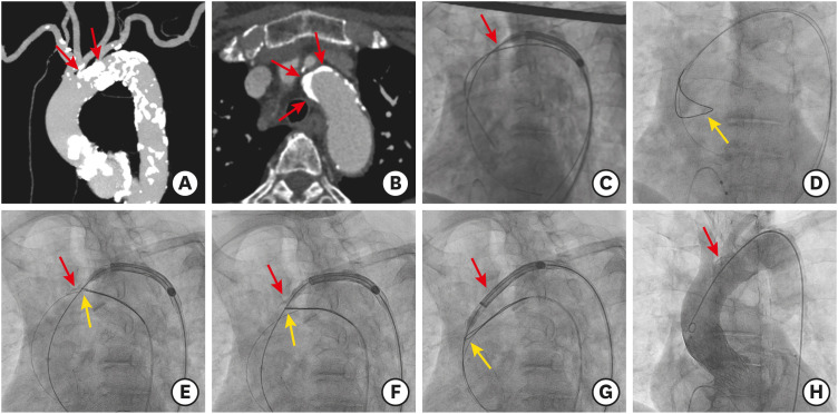
As shown in Figure 1, the delivery catheter failed to pass through the severely calcified aortic arch, because of a dense calcified mass in the outer curvature of the aortic arch (Supplementary Video 1). Eventually, a snare-assisted delivery using a 35-mm loop Amplatz Goose Neck Snare kit (ev3 Endovascular, Inc., Plymouth, MN, USA) was attempted. We tightened the snare to grasp and retain the stiff wire and the nose cone of the delivery catheter. Further, we advanced the delivery catheter while simultaneously pulling the snare catheter downward. Finally, the delivery catheter was passed and the valve implantation was successful without major aortic complications (Supplementary Videos 2 and 3).
Severely calcified aortic arches are a major limitation in the implantation of currently available self-expanding valves due to the rigidity and non-steerable features of these valves, with the risk of delivery failure or fatal complications including aortic dissection or rupture.1) The inflexible and bulky delivery catheter tends to follow the outer curvature of the aortic arch, and massive calcification can obstruct the device passage. The snare technique can direct the wire and delivery catheter away from the aortic wall, leading to successful crossing.
In an extremely calcified aortic valve and a horizontal aorta that blocks the passage of a bioprosthetic valve, a snare catheter can achieve coaxiality with the aortic orifice and successful crossing of the aortic valve.2)

Notes

Funding: The authors received no financial support for the research, authorship, and/or publication of this article.

Conflict of Interest: The authors have no financial conflicts of interest.

Author Contributions:

  • Supervision: Fujita T.

  • Validation: Hachinohe D, Kobayashi K.

  • Writing - original draft: Kaneko U.

References

1. Szlapka M, Michel E, Ricciardi MJ, Malaisrie SC. Valve-in-valve-prosthesis embolization and aortic dissection: single procedure, double complication. Eur J Cardiothorac Surg. 2019; 56:204–205. PMID: 30561574.
crossref
2. Kaneko U, Hachinohe D, Kobayashi K, et al. Evolut self-expanding transcatheter aortic valve replacement in patients with extremely horizontal aorta (aortic root angle ≥ 70°). Int Heart J. 2020; 61:1059–1069. PMID: 32921666.
crossref

SUPPLEMENTARY MATERIALS

Supplementary Video 1

The delivery catheter fails to pass through the severely calcified aortic arch.

Supplementary Video 2

Simultaneous pulling of the snare results in successful passage of the delivery catheter.

Supplementary Video 3

Final aortography confirms no major aortic complications.
Figure 1

Snare-assisted delivery of a self-expanding valve.

(A, B) Baseline computed tomography reveals a severely calcified aortic arch with dense calcified plaques protruding into the lumen. (C) The protruding calcification hampers the passage of a delivery catheter. (D) First, we insert a standard wire inside the snare introduced from the contralateral femoral access. Second, the standard wire inserted in the left ventricle is exchanged with the stiff wire. (E-G) Next, the snare is tightened and the stiff wire and nose cone of the delivery catheter are retained. Simultaneous pulling of the snare results in successful passage of the delivery catheter. (H) Final angiography shows an optimal valve position without aortic complications. Red arrows indicate the dense calcified plaque of the aortic arch, and yellow arrows indicate the snare position.
kcj-51-185-g001
TOOLS
Similar articles